The effect of smoking cessation on posterior ocular
structures
O efeito da suspensão do tabagismo em estruturas oculares posteriores
Bekir Küçük1, Serkan Akkaya2
1. Ophthalmology Department, Bozok University Faculty of Medicine, Yozgat, Turkey. 2. Ophthalmology Department, Kayseri Education and Research Hospital, Kayseri, Turkey.
Submitted for publication: October 30, 2017 Accepted for publication: February 9, 2018
Funding: No specific financial support was available for this study.
Disclosure of potential conflicts of interest: None of the authors have any potential conflict of interest to disclose.
Corresponding author: Bekir Küçük - E-mail: bekirkucuk1983@hotmail.com
Approved by the following research ethics committee: Kayseri Education and Research Hospital (# 50/2016).
ABSTRACT | Purpose: This study evaluated changes in cho roidal and macular thickness in healthy volunteers and chronic smokers. Methods: Thirtythree eyes of 33 chronic smokers (study group) and 33 eyes of 33 healthy controls who had never smoked were prospectively evaluated. Comprehensive ophthalmic assessment included slit lamp biomicroscopy, stereoscopic fundus examination, and intraocular pressure measurement. Spectral domain optical coherence tomography was used to measure choroidal and macular thickness 1 month before smoking cessation (smoking period) and after 3 months of smoking cessation (nonsmoking period). Results:
The mean age of the participants was 41.88 ± 6.52 years (range, 2652), and the average smoking duration was 8.6 ± 2.5 years (range, 516). The thickness of the paracentral choroid (nasal: 1,500 μm, p=0.001 and temporal: 1,500 µm, p=0.001) had significantly decreased after 3 months of smoking cessation. The thicknesses of the subfoveal choroid in the smoking and nonsmoking periods were not significantly different (p=0.194). The mean central macular thickness was 267.21 ± 18.42 µm in the smoking period and 268.42 ± 18.28 μm in the nonsmoking period (p=0.022). Conclusions:
Smoking was associated with statistically significant changes in paracentral choroidal and central macular thickness in healthy volunteers. Pathological studies should be performed to evaluate the effects of smoking on posterior ocular structures.
Keywords: Choroid/anatomy & histology; Macula lutea/anatomy & histology;Smoking cessation;Tomography, optical coherence
RESUMO | Objetivo: Este estudo avaliou as mudanças na espessura da coroide e da mácula em voluntários saudáveis e fumantes crônicos. Métodos: Trinta e três olhos de 33 fumantes crônicos (grupo estudado) e 33 olhos de 33 controles saudáveis
que nunca fumaram foram avaliados prospectivamente. A avaliação oftalmológica abrangente incluiu biomicroscopia de lâmpada de fenda, exame de fundo estereoscópico e medição da pressão intraocular. A tomografia de coerência óptica de domínio espectral foi utilizada para medir a espessura da coroide e da mácula um mês antes da cessação do tabagismo (período de fumar) e após 3 meses da cessação do tabagismo (período de abstinência). Resultados: A idade média dos participantes foi de 41,88 ± 6,52 anos (faixa de 2652 anos) e a duração média do tabagismo foi de 8,6 ± 2,5 anos (faixa, 516 anos). A espessura da coroide paracentral (nasal: 1.500 μm, p=0,001, temporal: 1.500 μm, p=0,001) diminuiu significativamente após 3 meses de cessação do tabagismo. As espessuras de coroide subfoveal nos períodos de tabagismo e nãotabagismo não foram signifi cativamente diferentes (p=0,194). A espessura macular central média foi de 267,21 ± 18,42 μm no períodos de tabagismo e 268,42 ± 18,28 μm no períodos de nãofumantes (p=0,022).
Conclusões: O tabagismo foi associado a mudanças estatis ticamente significativas na espessura paracentral de coroide e macular central em voluntários saudáveis. Estudos patológicos devem ser realizados para avaliar os efeitos do tabagismo nas estruturas oculares posteriores.
Descritores: Coroide/anatomia & histologia; Mácula lútea/ana tomia & histologia; Abandono do hábito de fumar; Tomografia de coerência óptica
INTRODUCTION
Smoking is the leading preventable cause of death worldwide and the most damaging preventable lifestyle factor affecting public health globally and nationally(1).
Smokingrelated causes of death include lung cancer, chronic obstructive pulmonary disease, and cardiovas cular diseases(2). Smoking is also associated with ocular
vascular diseases such as hypertensive retinopathy, agerelated macular degeneration, and anterior ischemic optic neuropathy(3).
by the US Food and Drug Administration as a smoking cessation treatment(4,5). It is a highaffinity partial nico
tinic agonist at the α2β4 nicotinic receptor and a full agonist at the neuronal α7 nicotinic receptor(6). Bupro
pion was initially approved for the treatment of major depressive disorders(7) and subsequently licensed as a
smoking cessation agent. It is currently available in the United States as a firstline treatment to stop smoking(8).
Bupropion acts by inhibiting dopamine and norepine phrine reuptake, and it weakens the stimulatory effects of nicotine on nicotinic acetylcholine receptors(9).
Age(10), axial length(11), chorioretinal diseases(12), severe
myopia(13), and smoking(14) have been shown to affect
choroid and macular thickness. The effects of smoking on the choroidal layer and ocular function have been stu
died(1418), but data on the effects of smoking cessation on
posterior ocular structures are lacking. Spectral domain optical coherence tomography (SDOCT) is a noninvasive, noncontact, highly sensitive modality that provides highresolution ophthalmic images and can be used to measure choroid layer thickness. This study was desig ned to objectively assess the effects of smoking cessation on the retina and choroid using SDOCT.
METHODS
Study population and design
This prospective study was performed at the Depart ment of Ophthalmology and Chest Diseases Hospital, Smoking Cessation Policlinic at, Kayseri Education and Research Hospital, Turkey. The study adhered to the tenets of the Declaration of Helsinki and was approved by the local ethics committee. The participants were given oral and written information about the study, and all provi ded written informed consent. Thirtythree eyes of 33 chronic smokers and 33 eyes of 33 healthy controls who had never smoked were studied. The chronic smokers were at the start of the study and reevaluated at the end of the study after not smoking for 3 months.
Examination protocol and study measurements
One month before smoking cessation, each volunteer was given a comprehensive ophthalmic evaluation that included bestcorrected visual acuity, slit lamp biomi croscopy, dilated stereoscopic fundus examination, in traocular pressure measurement by Goldmann appla nation tonometry, and OCT imaging measurements. The same evaluation was repeated after 3 months of smoking cessation, and the results were compared with those
obtained in the control group. Choroidal images were obtained with a Heidelberg Spectralis SDOCT (Heidelberg Engineering, Heidelberg, Germany) equipped with an enhanced depth imaging module. Choroidal thickness was the vertical distance from the hyperreflective line of Bruch’s membrane to the hyperreflective line of the inner surface of the sclera. The choroidal thickness was measured at the subfovea, temporally at 500, 1,000, and 1,500 µm and nasally at 500, 1000, and 1,500 µm from the center of the fovea. The central foveal thickness was also measured. All OCT scans were performed in the morning to avoid diurnal fluctuations.
Exclusion criteria
Patients with a history of ocular disease that preven ted examination of the cornea and retina, a history of ocular surgery, ocular or systemic diseases, a spherical refractive error >3 D, or a cylindrical refractive error >3 D were excluded.
Statistical analysis
Statistical analysis was performed with the Statisti cal Package for the Social Sciences version 21.0 (SPSS Inc., Chicago, IL, USA). The normality of continuous variable dis tributions normality was checked with the ShapiroWilk test. If the differences in the foveal and choroidal mea surements before and after smoking cessation were normally distributed, they were com pared with the paired ttest. If not, the Wilcoxon signedrank test was used. Differences in the mean values of independent groups were compared with the independent Student’s ttest or MannWhitney U test was used. Analysis of covariance (ANCOVA) was used to evaluate the significance of differences of ini tial and final participant evaluation measurements. The initial choroidal thickness results were covariates in the ANCOVA. Logarithmic transformation of nonparametric data was performed as needed to perform parametric analysis. Results were reported as means ± standard de viation and with 95% confidence intervals p≤0.05 were considered statistically significant.
RESULTS
23 (70%) were men, and 10 (30%) were women. The average smoking duration was 8.6 ± 2.5 years (range, 516). The subfoveal and paracentral choroidal thickness measurements in the study group smoking period and the control group are shown in table 2. The mean cen tral macular thickness was 267.21 ± 18.42 µm in the smoking and 268.42 ± 18.28 µm in the nonsmoking period (p=0.022). The mean subfoveal and paracen tral choroidal thickness values during the study group smoking and nonsmoking periods are shown in table 3. The paracentral choroidal thickness measurements
were significantly lower than the baseline values after 3 months of smoking. The subfoveal choroidal thicknesses in the smoking and nonsmoking periods were not signifi cantly different (p=0.194). The subfoveal and paracen tral choroidal thickness measurements of the control and the study nonsmoking period are shown in table 4.
DISCUSSION
Central foveal thickness increased and parafoveal choroidal thickness decreased after smoking cessation. A change in subfoveal choroidal thickness has not pre viously been reported. Overall, the reported effects of cigarette smoking on choroidal thickness are inconsis
Table 2. Subfoveal and paracentral choroidal thickness of the study group during the smoking period and the control group
Choroidal thickness
Study group (smoking period)
(n=33)
Control group (n=33) p
Subfoveal choroidal thickness (median ± interquartile range) (μm)
354.00 ± 086.00 355.00 ± 06.00 0.990α
Temporal1500 (median ±
interquartile range) (μm)
305.00 ± 077.00 281.00 ± 50.00 0.133α
Temporal1000 (median ±
interquartile range) (μm)
327.00 ± 125.00 303.00 ± 63.00 0.393α
Temporal500 (mean ± SD) (μm) 333.88 ± 093.20 323.24 ± 83.99 0.595β
Nasal1500 (mean ± SD) (μm) 299.52 ± 070.84 281.03 ± 50.32 0.548β
Nasal1000 (mean ± SD) (μm) 315.69 ± 078.14 307.39 ± 67.08 0.607β
Nasal500 (mean ± SD) (μm) 334.36 ± 079.55 314.97 ± 60.49 0.269β
SD= standard deviation.
α= Mann
Whitney U test; β= independent Student’s ttest.
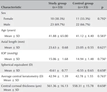
Table 1. Baseline ocular characteristics of the participants
Characteristic
Study group (n=33)
Control group (n=33) p
Sex
Female 10 (30.3%) 11 (33.3%) 0.792a
Male 23 (69.7%) 22 (66.7%)
Age (years)
Mean ± SD 041.88 ± 65.00 041.12 ± 4.40 0.583b
Axial length (mm)
Mean ± SD 023.63 ± 00.68 023.05 ± 0.55 0.621b
IOP (mmHg)
Mean ± SD 015.06 ± 01.68 014.94 ± 1.48 0.756b
Spherical equivalent (D)
Mean ± SD 00.61 ± 00.77 000.55 ± 0.65 0.658b
Average central keratometry (D) Mean ± SD
042.94 ± 01.39 042.78 ± 1.55 0.765b
Central corneal thickness (µm) Mean ± SD
561.36 ± 16.13 558.31 ± 15.78 0.658b
SD= standard deviation; D= diopters; IOP= intraocular pressure.
a= Chi square test; b= Independent Student’s ttest.
Table 3. Subfoveal and paracentral choroidal thickness of the study group participants during the smoking and nonsmoking periods
Choroidal thickness
Study group (smoking periods)
(n=33)
Study group (nonsmoking period) (n=33) p
Subfoveal choroidal thickness (median ± interquartile range) (µm)
354.00 ± 86.00 352.00 ± 88.00 0.194&
Temporal1500 (median ± interquartile range) (µm)
305.00 ± 77.00 287.00 ± 11.00 0.001&
Temporal1000 (median ± interquartile range) (µm)
327.00 ± 12.00 296.00 ± 12.00 0.001&
Temporal500 (mean ± SD) (µm) 333.88 ± 93.20 319.69 ± 76.63 0.044*
Nasal1500 (mean ± SD) (µm) 299.52 ± 70.84 276.58 ± 65.60 0.001*
Nasal1000 (mean ±SD) (µm) 315.69 ± 78.14 300.88 ± 75.14 0.003*
Nasal500 (mean ±SD) (µm) 334.36 ± 79.55 314.36 ± 80.44 0.001*
&= Wilcoxon signedrank test; *= paired ttest.
Table 4. Subfoveal and paracentral choroidal thickness of the control group and study group during the nonsmoking period
Choroidal thickness
Control group (n=33)
Study group (nonsmoking period) (n=33) p
Subfoveal choroidal thickness (median ±
interquartile range) (μm)
355.00 ± 06.00 352.00 ± 88.00 0.783†
Temporal1500 (median ±
interquartile range) (μm)
281.00 ± 50.00 287.00 ± 11.00 0.778†
Temporal1000 (median ±
interquartile range) (μm)
303.00 ± 63.00 296.00 ± 12.00 0.763†
Temporal500 (mean ± SD) (μm) 323.24 ± 83.99 319.69 ± 76.63 0.859€
Nasal1500 (mean ± SD) (μm) 281.03 ± 50.32 276.58 ± 65.60 0.397€
Nasal1000 (mean ± SD) (μm) 307.39 ± 67.08 300.88 ± 75.14 0.713€
Nasal500 (mean ± SD) (μm) 314.97 ± 60.49 314.36 ± 80.44 0.973€
†= MannWhitney U test; €
tent. A study by Kantarci et al.(14) reported that choroidal
thickness in longterm smokers and in healthy partici pants was not significantly different, but other studies found that cigarette smoking caused shortterm(15,16), but
not longterm(16) changes. Others have reported decrea
ses in choroidal thickness in chronic smokers(17,18). A
study by Tamaki et al.(19) found that a decrease in tissue
blood velocity at the optic nerve head and possibly in the choroid, suggesting a significant increase in vascular resistance in those tissues.
An animal study found that increased choroidal vas cular resistance was associated with chronic exposure to tobacco smoke(20). Kaiser et al. reported increased
flow rates in the ophthalmic artery, central retinal artery, and posterior ciliary arteries of chronic smokers(21), but
others have reported decreased blood flow rates in chronic smokers(22,23).
A study of the effects of carbogen breathing revea led abnormal choroidal vascular responses in chronic smokers compared with nonsmokers(24).
Choroidal blood flow is responsive to pCO2(25), but
the cause of vasodilation induced by CO2 is not clear. The reduction of nitrite to NO could lead to vasodila tation because of hypercapnia and reduced pO2(26), but
NOmediated vasodilation is impaired in smokers(27,28).
The evidence suggests that the choroid is affected by smoking because of the presence of a rich capillary network. A recent study found that the central fovea was thinner in active than in passive smokers. In addi tion to affecting the structure of the macula, multifocal electro retinography showed that active smoking redu ced ma cular functional responses (29). However, Kantarci
et al.(14) reported that the central macular thickness did
not differ in longterm smokers and healthy participants. Nicotine replacement therapy, bupropion, and vare nicline are medications indicated for smoking cessation. The US Preventive Services Task Force reported that intervention improved the likelihood of smoking cessa tion within 6 months or more(30), and a metaanalysis by
Cahill et al.(31) reported that varenicline and bupropion
were superior to a placebo in smoking cessation. In this singlecenter study, 33 of 150 chronic smokers with ba seline measurements quit smoking. The study was limited by the small number of participants. They used medications to help stop cigarette smoking, but smoking cessation was selfreported. Despite its weaknesses, this study is valuable because it is the first to examine changes in the central foveal and choroidal thicknesses of patients who stopped smoking. In conclusion, subfo
veal choroidal thickness did not change, central foveal thickness significantly increased, and parafoveal choroi dal thickness was significantly decreased in patients who quit smoking.
REFERENCES
1. Müezzinler A, Mons U, Gellert C, Schottker B, Jansen E, Kee F, et al. Smoking and allcause mortality in older adults: results from the CHANCES consortium. Am J Prev Med. 2015;49(5):e53e63. 2. Hill C. [Tobacco epidemiology]. Rev Prat. 2012;62(3):3279. French. 3. Solberg Y, Rosner M, Belkin M. The association between cigarette smoking and ocular diseases. Surv Ophthalmol. 1998;42(6):53547. 4. Gonzales D, Rennard SI, Nides M, Oncken C, Azoulay S, Billing
CB, Watsky EJ, Gong J, Williams KE, Reeves KR; Varenicline Phase 3 Study Group. Varenicline, an alpha4beta2 nicotinic acetylcholine receptor partial agonist, vs sustainedrelease bupropion and placebo for smoking cessation: a randomized controlled trial. JAMA. 2006;296(1):4755. Comment in: Expert Opin Pharmacother. 2006;7(18):2599603; JAMA. 2006;296(21):2555; author reply 25556; Curr Psychiatry Rep. 2007;9(5):3456; JAMA. 2006;296(1):945. 5. Jorenby DE, Hays JT, Rigotti NA, Azoulay S, Watsky EJ, Williams KE,
Billing CB, Gong J, Reeves KR; Varenicline Phase 3 Study Group. Efficacy of varenicline, an alpha4beta2 nicotinic acetylcholine recep tor partial agonist, vs placebo or sustainedrelease bupropion for smoking cessation: a randomized controlled trial. JAMA. 2006; 296(1):5663. Erratum in: JAMA. 2006;296(11):1355. Comment in: Curr Psychiatry Rep. 2007;9(5):3467; JAMA. 2006;296(1):945; JAMA. 2006;296(21):2555;author reply 25556.
6. Mihalak K, Carroll FI, Luetje CW. Varenicline is a parietal agonist at {alpha]4{beta}2 and a full agonist at {alpha}7 neuronal nicotinic receptors. Mol Pharmacol. 2006;70(3):8015.
7. Fava M, Rush AJ, Thase ME, Clayton A, Stahl SM, Pradko JF, et al. 15 years of clinical experience with bupropion HCl: from bupropion to bupropion SR to bupropion XL. Prim Care Companion J Clin Psychiatry. 2005;7(3):10613.
8. Hurt RD, Sachs DP, Glover ED, Offord KP, Jonhston JA, Dale LC, et al. A comparison of sustainedrelease bupropion and placebo for smoking cessation. N Engl J Med. 1997;337(17):1195202. Comment in: N Engl J Med. 1998;338(9):619; N Engl J Med. 1997; 337(17):12301; N Engl J Med. 1998;338(9):61920.
9. Warner C, Shoaib M. How does bupropion work as a smoking cessation aid? Addict Biol. 2005;10(3):21931.
10. Manjunath V, Taha M, Fujimoto JG, Duker JS. Choroidal thickness in normal eyes measured using Cirrus HD optical coherence tomo graphy. Am J Ophthalmol. 2010;150(3):3259.
11. Goldenberg D, Moisseiev E, Goldstein M, Loewenstein A, Barak A. Enhanced depth imaging optical coherence tomography: choroidal thickness and correlations with age, refractive error, and axial length. Ophthalmic Surg Lasers Imaging. 2012;43(4):296301. 12. Imamura Y, Fujiwara T, Margolis R, Spaide RF. Enhanced depth
imaging optical coherence tomography of the choroid in central serous chorioretinopathy. Retina. 2009;29(10):146973. Comment in: Retina. 2010;30(8):13201.
13. Nishida Y, Fujiwara T, Imamura Y, Lima LH, Kurosaka D, Spaide RF. Choroidal thickness and visual acuity in highly myopic eyes. Retina. 2012;32(7):122936.
15. Ulaş F, Çelik F, Doğan Ü, Çelebi S. Effect of smoking on choroidal thickness in healthy smokers. Curr Eye Res. 2014;39(5):50411. 16. Sızmaz S, Küçükerdönmez C, Pınarcı EY, Karalezli A, Canan H, Yilmaz
G. The effect of smoking on choroidal thickness measured by optical coherence tomography. Br J Ophthalmol. 2013;97(5):6014. 17. Sigler EJ, Randolph JC, Calzada JI, Charles S. Smoking and choroidal
thickness in patients over 65 with earlyatrophic agerelated ma cular degeneration and normals. Eye (Lond). 2014;28(7):83846. 18. Moschos MM, Nitoda E, Laios K, Ladas DS, Chatziralli IP. The im
pact of chronic tobacco smoking on retinal and choroidal thickness in Greek population. Oxid Med Cell Longev. 2016;2016:2905789. 19. Tamaki Y, Araie M, Nagahara M, Tomita K, Matsubara M. The acute
effects of cigarette smoking on human optic nerve head and poste rior fundus circulation in light smokers. Eye (Lond). 2000;14(Pt 1): 6772.
20. Hara K. [Effects of cigarette smoking on ocular circulation chronic effect on choroidal circulation]. Nippon Ganka Gakkai Zasshi. 1991; 95(10):93943. Japanese.
21. Kaiser HJ, Schoetzau A, Flammer J. Blood flow velocity in the extraocular vessels in chronic smokers. Br J Ophthalmol. 1997; 81(2):1335.
22. Steigerwalt RD Jr, Laurora G, Incandela L, Cesarone MR, Belcaro GV, De Sanctis MT. Ocular and orbital blood flow in cigarette smokers. Retina. 2000;20(4):3947.
23. Williamson TH, Lowe GD, Baxter GM. Influence of age, systemic blood pressure, smoking, and blood viscosity on orbital blood velocities. Br J Ophthalmol. 1995;79(1):1722.
24. Wimpissinger B, Resch H, Berisha F, Weigert G, Schmetterer L, Polak K. Response of choroidal blood flow to carbogen breathing in smokers and nonsmokers. Br J Ophthalmol. 2004;88(6):77681. 25. Geiser MH, Riva CE, Dorner GT, Diermann U, Luksch A, Schmet terer L. Response of choroidal blood flow in the foveal region to hyperoxia and hyperoxiahypercapnia. Curr Eye Res. 2000; 21(2):66976.
26. Iadecola C, Zhang F. Nitric oxidedependent andindependent components of cerebrovasodilation elicited by hypercapnia. Am J Physiol. 1994;266(2 Pt 2):R54652.
27. McVeigh GE, Lemay L, Morgan D, Cohn JN. Effects of longterm ci garette smoking on endotheliumdependent responses in humans. Am J Cardiol. 1996;78(6):66872.
28. Butler R, Morris AD, Struthers AD. Cigarette smoking in men and vascular responsiveness. Br J Clin Pharmacol. 2001;52(2):1459. 29. ElShazly AA, Farweez YA, Elzankalony YA, Elewa LS, Farweez
BA. Effect of smoking on macular function and structure in acti ve smokers versus passive smokers. Retina. 2017. doi: 10.1097/ IAE.0000000000001632.
30. Anthenelli RM, Benowitz NL, West R, St Aubin L, McRae T, Lawrence D, et al. Neuropsychiatric safety and efficacy of varenicline, bupro pion, and nicotine patch in smokers with and without psychiatric disorders (EAGLES): a doubleblind, randomised, placebocontrolled clinical trial. Lancet. 2016;387(10037):250720. Comment in: Lancet. 2016;387(10037):24812.