O
r i g i n a la
rt i c l e1 5 5 Arq Bras Oftalmol. 2016;79(3):155-8 http://dx.doi.org/10.5935/0004-2749.20160047
INTRODUCTION
More than 90 million people worldwide are afected by diabetic retinopathy (DR), causing signiicant vision loss(1,2), with macular edema and proliferative retinopathy as the leading causes of visual impairment(2,3). Anti-vascular endothelial growth factor (anti-VEGF) drugs are widely used in the treatment of diabetic macular edema (DME); this is supported by an extensive body of literature, demonstrating substantial improvements in visual and anatomic outcomes(4-10).
The choroid is a vascularized tissue that plays a vital role in pro-viding metabolic support to the outer retina, including the photo-receptors and prelaminar portion of the optic nerve head(11,12). Abnor-mal choroidal blood volume and compromised low may result in
dysfunctioning of the photoreceptor and mortality(13). Choroidal vasculopathy, such as obstruction of the choriocapillaris, vascular de-ge neration, choroidal aneurysms, and neovascularization, may be involved in the pathogenesis of DR(13-15).
Enhanced depth imaging using spectral domain optical cohe-rence tomography (SD-OCT) enables improved visualization of the choroid and assessment of choroidal thickness(16,17).Results of clinical studies have indicated that choroidal thickness may be related to DR severity, and the presence of DME is associated with a signiicant de-crease in choroidal thickness(16). In patients with neovascular age-related macular degeneration (AMD), anti-VEGF drugs have been shown to induce signiicant thinning in choroidal thickness, which may lead to unknown long-term consequences or complications(18).
Effect of intravitreal anti-VEGF on choroidal thickness in patients with diabetic
macular edema using spectral domain OCT
Efeito de injeção intravítrea na espessura de coroide em pacientes com edema macular diabético
utilizando OCT de domínio espectral
Vinicius F. KniggendorF1,2, eduardo a. noVais2, sergio L. KniggendorF1, camiLLa XaVier2, emiLy d. coLe3, caio V. regatieri2,3
Submitted for publication: October 27, 2015 Accepted for publication: February 15, 2016
1 Hospital Oftalmológico de Brasília, Brasília, DF, Brazil.
2 Department of Ophthalmology and Visual Sciences, Escola Paulista de Medicina (EPM), Universidade
Federal de São Paulo (UNIFESP), São Paulo, SP, Brazil.
3 Tufts University School of Medicine, Boston, MA, USA.
Funding: This study was supported in part by CNPq (EAN - Grant number 479648/2012-3) and CAPES foundation.
Disclosure of potential conflicts of interest: None of the authors have any potential conflict of interest to disclose.
Corresponding author: Caio Vinicius Saito Regatieri. Universidade Federal de São Paulo. Depar-tamento de Oftalmologia. Rua Botucatu, 821 - 1o andar - São Paulo, SP - 04023-062 - Brazil
E-mail: caiore@gmail.com
Approved by the following research ethics committee: UNIFESP (#1.093.853).
ABSTRACT
Purpose: To evaluate choroidal thickness (CT ) using spectral domain optical coherence tomography (SD-OCT ) imaging at baseline and 6 months after intravi-treal anti-vascular endothelial growth factor (anti-VEGF) treatment in patients with diabetic macular edema (DME).
Methods: A retrospective chart review was performed to identify patients with DME who underwent intravitreal injection of anti-VEGF (bevacizumab or ranibizu-mab) in a pro re nata (PRN) regimen. Subfoveal choroidal thickness was compared between values obtained at baseline and at 6-month follow-up visits.
Results: Thirty-nine eyes (15 females, 24 males) from 39 patients were enrolled (mean age, 62.43 ± 8.7 years; range, 44-79 years). Twenty-three and 16 eyes were treated with ranibizumab and bevacizumab respectively. The mean number of anti-VEGF injections was 2.28 ± 1.27 (range, 1-5). Mean nasal, subfoveal, and temporal choroidal thickness (CT) measurements at baseline were 234.10 ± 8.63 μm, 246.89 ± 8.94 μm, and 238.12 ± 8.20 μm, respectively, and those at 6 months post-treatment were 210.46 ± 8.00 μm, 215.66 ± 8.29 μm, and 212.43 ± 8.14 μm, respectively. Significant differences in CT were observed between baseline and the 6-month follow-up at all measured points (p=0.0327).
Conclusions: Over a 6-month period, the use of intravitreal anti-VEGF was asso-ciated with significant thinning of the choroid in patients with DME. The clinical significance of a thinner choroid in DME is currently unknown; however, it may contribute to long-term adverse effects on choroidal and retinal function, repre-senting an area requiring future investigation.
Keywords: Angiogenesis inhibitors; Choroid; Diabetic retinopathy; Intravitreal in-jections; Tomography, optical coherence
RESUMO
Objetivos: Avaliar a espessura de coroide pré-tratamento e após 6 meses da in je ção intravítrea de anti-fator de crescimento vascular endotelial (anti-VEGF) em pacientes com edema macular diabético (EMD), utilizando a tomografia de coerência óptica de domínio espectral (SD-OCT ).
Métodos: Análise retrospectiva, com revisão de prontuários, foi realizada para identificação de pacientes submetidos a tratamento com injeções intravítreas de anti-VEGF, no regime pro re nata, para tratamento de EMD. As medidas da es pessu ra de coroide pré-tratamento foi comparada com as medidas após acompa nhamento de 6 meses.
Resultados: Trinta e nove olhos de 39 pacientes (15 femininos, 24 masculinos) foram incluídos, com idade média de 62,43 ± 8,7 anos (variando de 44-79 anos). Trinta e três olhos foram tratados com ranibizumab e 18 com bevacizumab. O número médio de injeções de anti-VEGF foi 2,28 ± 1,27 (variando de 1-5). A medida média pré-tra-tamen to da espessura de coroide nasal, subfoveal e temporal foi 234,10 ± 8,63 μm, 246,89 ± 8,94 μm e 238,12± 8,20 μm, respectivamente. Após acompanhamento de 6 meses as medidas médias da espessura de coroide foram 210,46 ± 8,00 μm, 215,66 ± 8,29 μm e 212,43 ± 8,14 μm. A diferença entre as medidas médias pré e pós trata-mento foi estatisticamente significante (p=0,0327) em todos os pontos medidos. Conclusão: Após um período de 6 meses, o uso de injeções intravítreas de anti-VEGF foi associado com diminuição significante da espessura de coroide nos pa cien tes com EMD. O significado clínico de uma coroide mais fina nos pacientes com EMD é desconhecido mas pode causar eventos adversos a longo prazo para função da coroide e retina, representando uma área para futura investigações.
Ef f E c to fi n t r av i t r E a la n t i-v E G f o nc h o r o i d a l t h i c k n E s si npat i E n t sw i t hd i a b E t i cm a c u l a rE d E m au s i n Gs p E c t r a ld o m a i n o c t
1 5 6 Arq Bras Oftalmol. 2016;79(3):155-8
Currently, intravitreal anti-VEGF injections are the most common treatment for DME. Though anti-VEGF injections improved visual acuity, several studies demonstrated an association with decreased central retinal thickness(18,19). However, there is currently a lack of studies inves-tigating the efect of anti-VEGF injections on the choroid in patients with diabetes. The purpose of the present study was to evaluate the efect of intravitreal anti-VEGF injections on choroidal thickness using SD-OCT in patients treated for DME.
METHODS
This was a retrospective chart analysis conducted at the Federal University of São Paulo (UNIFESP) and Hospital Oftalmológico de Bra-sília, Brazil. The present study was approved by the UNIFESP Institu-tional Review Boards (CEP 1.093.853) and adhered to the tenets of the Declaration of Helsinki. Informed consent was obtained from all subjects after explanation of the nature of the present study.
S
UBJECTSExploratory chart review of data collected from patients with type 2 diabetes previously treated for DME with intravitreal anti-VEGFs between January 2012 and June 2014. Clinically signiicant macular edema (CSME) was assessed by clinical examination and SD-OCT imaging. The diagnosis of CSMA was deined according to the Early Treatment Diabetic Retinopathy Study (ETDRS) protocol. A pro re nata (PRN) protocol was used to treat DME in all patients. Major exclusion criteria included the following: previous anti-VEGF or intravitreous corticosteroid treatment for DME within 6 months of the irst visit; vitreomacular traction syndrome; suspected or conirmed glaucoma; proliferative diabetic retinopathy; previous retinal laser therapy; pre-vious pars plana vitrectomy; high myopia and age-related macular degeneration; and other causes of macular edema.
C
HOROIDALTHICKNESSMEASUREMENTSD-OCT scans were performed using EDI SD-OCT (Spectralis OCT, Heidelberg Engineering, Heidelberg, Germany). The scan pattern used was a high-deinition 1-line raster scanning of 30° consisting of 768 A-scans per frame, followed by an average of 100 frames(20). Mea-surements were performed before and after anti-VEGF treatment. For images to be included in the present study, the signal-to-noise ratio (SNR) had to be at least 20 dB (considered best quality) and should
be taken as close to the fovea as possible (thinnest macular point), with the understanding that slight diferences in positioning afect the measured thicknesses(21). Using the Spectralis linear measurement tool, two independent observers measured CT perpendicularly from the outer edge of the hyper-relective RPE to the inner sclera at 500-mm intervals temporal and nasal to the fovea up to 1,000 mm. Measurements were performed prior to treatment and at a 6-month follow-up (Figure 1).
S
TATISTICALANALYSISData are expressed as means ± standard error of the mean. Statis-tical analyses were performed using a paired t-test for comparing between baseline and follow-up choroidal thickness measurements. The Pearson correlation coeicient was used to evaluate the corre-lation between choroidal thickness and central foveal thickness. A 95% conidence interval with a 5% level of signiicance was adopted; thus, P values of <0.05 were considered to be statistically signiicant. Analysis of variance (ANOVA) with the Bonferroni correction was used to compare groups treated with bevacizumab and ranibizumab. All statistical analyses were performed using Graph Pad Prism 5.0 software for MAC.
RESULTS
Thirty-nine eyes from 39 patients (15 female, 24 male) were in-cluded in the study analysis. The mean age was 62.43 ± 8.7 years, with a range of 44-79 years. Twenty-three eyes were treated with ra-nibizumab and 16 eyes were treated with bevacizumab. The average number of anti-VEGF injections administered was 2.28 ± 1.27 (range, 1-5) over the 6-month follow-up. Central retinal thickness improved in all patients after 6-month anti-VEGF treatment.
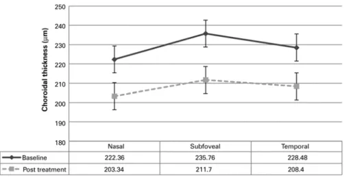
The choroid was noted to be thinnest nasally, thicker temporally, and thickest in the subfoveal region. This pattern was observed at baseline and after the 6-month follow-up, which is consistent with that reported in previous studies(22). Figure 2 shows the mean CT at each location. Choroidal thickness was signiicantly thinner (p=0.0327) at all measured points following anti-VEGF treatment. Mean nasal, subfoveal, and temporal choroidal thickness measurements at base-line were 234.10 ± 8.63 μm, 246.89 ± 8.94 μm, and 238.12 ± 8.20 μm, respectively, and at the 6-month follow-up were 210.46 ± 8.00 μm, 215.66 ± 8.29 μm, and 212.43 ± 8.14 μm, respectively.
A
B
Kn i g g e n d o r f Vf, e t a l.
1 5 7
Arq Bras Oftalmol. 2016;79(3):155-8
In a subgroup analysis of patients who received intravitreal be-vacizumab, mean nasal, subfoveal, and temporal choroidal thickness measurements at baseline were 241.25 ± 14.40 µm, 259.50 ± 13.93 µm, and 249.87 ± 13.30 µm, respectively, with signiicant decreases to 212.75 ± 12.70 μm, 221.12 ± 12.26 μm, and 221.12 ± 12.06 μm, res-pectively, at the 6-month follow-up. In the ranibizumab group, mean nasal, subfoveal, and temporal choroidal thickness measurements at baseline were 229.13 ± 10.83 µm, 238.13 ± 11.56 µm, and 229.95 ± 10.28 µm, respectively, with signiicant decreases to 208.86 ± 10.53 μm, 211.87 ± 11.34 μm, and 206.40 ± 11.00 μm, respectively, at the 6-month follow-up. No signiicant diference (p>0.05) in foveal CT was obser-ved between eyes treated with ranibizumab (211.87 ± 11.34 μm) and those treated with bevacizumab (221.12 ± 12.26 μm). Decreases in central retinal thickness were not correlated with choroidal thinning after 6-month anti-VEGF treatment.
DISCUSSION
The choroid plays an integral role in the metabolic support of the retinal pigment epithelium (RPE), optic nerve, and outer retina; normal choroidal vasculature is essential for appropriate functioning of the retina(11,12). Compromised choroidal blood low may result in dysfunctioning of the photoreceptor and insuicient removal of me tabolites generated by RPE cells, resulting in an accumulation of waste at Bruch’s membrane(13). Previous studies have reported abnor-mal choroidal indings in patients with diabetes such as increased blood vessel tortuosity, focal vascular dilation and narrowing, hyper-cellularity, vascular loops, microaneurysms, areas of non-perfusion, and sinus-like structure formation between the choroidal lobules(14,23). Choroidal thinning, increased thickness of the extracellular matrix, and decreased vessel diameter have been observed on SD-OCT ima-ging in the eyes of patients with diabetes(16,24,25).
Anti-VEGF treatment has demonstrated utility in improving visual and anatomical outcomes in DME(4-10). The efects of anti-VEGF the-rapy on choroidal thickness in various diseases, including AMD and DM, have been reported in several previous studies(18,19). Yiu et al. demonstrated that central choroidal thickness decreases following anti-VEGF therapy for DME after 6 months(19). The present study also found a statistically signiicant decrease in choroidal thickness among patients following anti-VEGF injections.
The results of the present study indicate that intravitreal anti-VEGF therapy may inluence choroidal structure in eyes with diabetic
reti-nopathy treated for DME. This result may be attributable to the indings that both full-length antibody (bevacizumab) and Fab fragment (ranibizumab) drugs are able to penetrate into all retinal layers, reach the choroid, and accumulate in the wall of the choroidal vessel(26-28). As VEGF functions by inducing vessel dilation and increasing ocular blood low, which is mediated by increased nitric oxide production(29), VEGF inhibition by these drugs may cause constriction of the choroi-dal vessels. Choroichoroi-dal thickness is already decreased in patients with diabetes, and the consequences of further thinning of the choroidal vasculature secondary to anti-VEGF administration are unknown. Altered choroidal blood low may result in dysfunctioning of the pho-toreceptor and mortality, contributing to the higher anatomical and functional responses to anti-VEGF treatment in patients with thicker choroids, as demonstrated by a previous report(30).
One of the major limitations of the present study was the retros-pective design. We were unable to determine which anti-VEGFs were administered, and we were unable to identify a control group. Addi-tionally, visual acuity data were not analyzed as the major objective of the present study was to evaluate changes in choroidal thickness.
As anti-VEGF drugs are commonly used in the treatment of DME and other retinal pathologies, a full understanding of the efects of anti-VEGF drugs on the choroidal vasculature is warranted. Further prospective studies are required to completely determine the long--term efects of anti-VEGF therapy on choroidal thickness. Decreased choroidal thickness induced by anti-VEGF administration may have long-term adverse efects on choroidal and retinal functions.
REFERENCES
1. Yau JW, Rogers SL, Kawasaki R, Lamoureux EL, Kowalski JW, Bek T, et al. Global preva-lence and major risk factors of diabetic retinopathy. Diabetes Care. 2012;35(3):556-64. 2. Moss SE, Klein R, Klein BE. The 14-year incidence of visual loss in a diabetic population.
Ophthalmology. 1998;105(6):998-1003.
3. Klein R, Klein BE, Moss SE, Cruickshanks KJ. The Wisconsin Epidemiologic Study of diabetic retinopathy. XIV. Ten-year incidence and progression of diabetic retinopathy. Arch Ophthalmol. 1994;112(9):1217-28.
4. Boyer DS, Hopkins JJ, Sorof J, Ehrlich JS. Anti-vascular endothelial growth factor therapy for diabetic macular edema. Ther Adv Endocrinol Metab. 2013;4(6):151-69. 5. Nguyen QD, Brown DM, Marcus DM, Boyer DS, Patel S, Feiner L, et al. Ranibizumab
for diabetic macular edema: results from 2 phase III randomized trials: RISE and RIDE. Ophthalmology. 2012;119(4):789-801.
6. Michaelides M, Kaines A, Hamilton RD, Fraser-Bell S, Rajendram R, Quhill F, et al. A pros-pective randomized trial of intravitreal bevacizumab or laser therapy in the management of diabetic macular edema (BOLT study) 12-month data: report 2. Ophthalmology. 2010; 117(6):1078-86 e2.
Ef f E c to fi n t r av i t r E a la n t i-v E G f o nc h o r o i d a l t h i c k n E s si npat i E n t sw i t hd i a b E t i cm a c u l a rE d E m au s i n Gs p E c t r a ld o m a i n o c t
1 5 8 Arq Bras Oftalmol. 2016;79(3):155-8
7. Korobelnik JF, Do DV, Schmidt-Erfurth U, Boyer DS, Holz FG, Heier JS, et al. Intravitreal alibercept for diabetic macular edema. Ophthalmology. 2014;121(11):2247-54. 8. Mitchell P, Bandello F, Schmidt-Erfurth U, Lang GE, Massin P, Schlingemann RO, et al.
The RESTORE study: ranibizumab monotherapy or combined with laser versus laser monotherapy for diabetic macular edema. Ophthalmology. 2011;118(4):615-25. 9. Massin P, Bandello F, Garweg JG, Hansen LL, Harding SP, Larsen M, et al. Safety and
eicacy of ranibizumab in diabetic macular edema (RESOLVE Study): a 12-month, ran domized, controlled, double-masked, multicenter phase II study. Diabetes Care. 2010;33(11):2399-405.
10. Nguyen QD, Shah SM, Khwaja AA, Channa R, Hatef E, Do DV, et al. Two-year outcomes of the ranibizumab for edema of the mAcula in diabetes (READ-2) study. Ophthalmo-logy. 2010;117(11):2146-51.
11. McCourt EA, Cadena BC, Barnett CJ, Ciardella AP, Mandava N, Kahook MY. Measu-rement of subfoveal choroidal thickness using spectral domain optical coherence to mography. Ophthalmic Surg Lasers Imaging. 2010;41 Suppl:S28-33.
12. Tan CS, Cheong KX, Lim LW, Li KZ. Topographic variation of choroidal and retinal thicknesses at the macula in healthy adults. Br J Ophthalmol. 2014;98(3):339-44. 13. Cao J, McLeod S, Merges CA, Lutty GA. Choriocapillaris degeneration and related
pathologic changes in human diabetic eyes. Arch Ophthalmol. 1998;116(5):589-97. 14. Hidayat AA, Fine BS. Diabetic choroidopathy. Light and electron microscopic
obser-vations of seven cases. Ophthalmology. 1985;92(4):512-22.
15. Shiragami C, Shiraga F, Matsuo T, Tsuchida Y, Ohtsuki H. Risk factors for diabetic cho-roidopathy in patients with diabetic retinopathy. Graefes Arch Clin Exp Ophthalmol. 2002;240(6):436-42.
16. Regatieri CV, Branchini L, Carmody J, Fujimoto JG, Duker JS. Choroidal thickness in patients with diabetic retinopathy analyzed by spectral-domain optical coherence tomography. Retina. 2012;32(3):563-8.
17. Spaide RF, Koizumi H, Pozzoni MC. Enhanced depth imaging spectral-domain optical coherence tomography. Am J Ophthalmol. 2008;146(4):496-500.
18. Branchini L, Regatieri C, Adhi M, Flores-Moreno I, Manjunath V, Fujimoto JG, et al. Efect of intravitreous anti-vascular endothelial growth factor therapy on choroidal thickness in neovascular age-related macular degeneration using spectral-domain optical coherence tomography. JAMA Ophthalmol. 2013;131(5):693-4.
19. Yiu G, Manjunath V, Chiu SJ, Farsiu S, Mahmoud TH. Efect of anti-vascular endothelial growth factor therapy on choroidal thickness in diabetic macular edema. Am J Ophthalmol. 2014;158(4):745-51 e2.
20. Branchini L, Regatieri CV, Flores-Moreno I, Baumann B, Fujimoto JG, Duker JS. Repro-ducibility of choroidal thickness measurements across three spectral domain optical coherence tomography systems. Ophthalmology. 2012;119(1):119-23.
21. Balasubramanian M, Bowd C, Vizzeri G, Weinreb RN, Zangwill LM. Efect of image quality on tissue thickness measurements obtained with spectral domain-optical coherence tomography. Opt Express. 2009;17(5):4019-36.
22. Manjunath V, Taha M, Fujimoto JG, Duker JS. Choroidal thickness in normal eyes measured using Cirrus HD optical coherence tomography. Am J Ophthalmol. 2010; 150(3):325-9 e1.
23. Fryczkowski AW, Sato SE, Hodes BL. Changes in the diabetic choroidal vasculature: scanning electron microscopy indings. Ann Ophthalmol. 1988;20(8):299-305. 24. Regatieri CV, Branchini L, Fujimoto JG, Duker JS. Choroidal imaging using spectral-domain
optical coherence tomography. Retina. 2012;32(5):865-76.
25. Adhi M, Brewer E, Waheed NK, Duker JS. Analysis of morphological features and vas cular layers of choroid in diabetic retinopathy using spectral-domain optical coherence to mography. JAMA Ophthalmol. 2013;131(10):1267-74.
26. Heiduschka P, Fietz H, Hofmeister S, Schultheiss S, Mack AF, Peters S, et al. Penetration of bevacizumab through the retina after intravitreal injection in the monkey. Invest Ophthalmol Vis Sci. 2007;48(6):2814-23.
27. Pieramici DJ, Rabena MD. Anti-VEGF therapy: comparison of current and future agents. Eye (Lond). 2008;22(10):1330-6.
28. Shahar J, Avery RL, Heilweil G, Barak A, Zemel E, Lewis GP, et al. Electrophysiologic and retinal penetration studies following intravitreal injection of bevacizumab (Avastin). Retina. 2006;26(3):262-9.
29. Fukumura D, Gohongi T, Kadambi A, Izumi Y, Ang J, Yun CO, et al. Predominant role of endothelial nitric oxide synthase in vascular endothelial growth factor-induced angiogenesis and vascular permeability. Proc Natl Acad Sci U S A. 2001;98(5):2604-9. 30. Rayess N, Rahimy E, Ying GS, Bagheri N, Ho AC, Regillo CD, et al. Baseline choroidal