INTRODUCTION
Major causes for developing a macular hole (MH) include an-teroposterior forces applied by vitreofoveal traction and tangential
traction from involutional changes in the inner retina(1-4). Kelly and
Wendell(5) reported the first successful closure of MH with pars plana
vitrectomy (PPV); since then, advances in vitreoretinal surgery have contributed to successful postoperative restoration of the foveal microstructure and restoration of visual function in most patients(6-10).
Although anatomical closure of MH may be applicable to almost all patients undergoing surgical intervention, significant improvement
in visual acuity can be achieved in approximately 70% of cases(8-13).
Macular atrophy may be responsible for inferior visual outcomes; the-refore, the foveal microstructure has to be well identified in patients with MH prior to any surgical intervention.
Spectral-domain optical coherence tomography (SD-OCT) is a non-contact, non-invasive, laser interferometry technique, which
captures high-resolution cross-sectional images in vivo and is used in diagnosing numerous macular diseases. Using SD-OCT imaging in the diagnosis of MH augments clinical staging by enabling visua-lization of the foveal and vitreous microstructure and the tractional association between them and calculating macular hole dimensions. Manual quantification of MH width and height can be provided using SD-OCT, which mainly identifies the presence of perifoveal cystoid edema and vitreomacular traction (VMT) and the integrity of peri-foveal external retinal microstructures. The impact of preoperative OCT measurements on diagnosing or staging idiopathic MH and its predictive value for postoperative anatomical and functional
outco-mes have been previously investigated(14-24). However, to the best of
our knowledge, no study has elucidated the association between preoperative macular hole volume (MHV) and postoperative macular atrophy as a tool for predicting postoperative visual outcome. This study aimed to evaluate the association between MHV and postope-rative central macular thickness (CMT) using SD-OCT.
Effect of macular hole volume on postoperative central macular thickness
O efeito do volume de buraco macular da espessura macular central pós-operatória
Taylan OzTurk1, Eyyup karahan2, Duygu Er1, MahMuT kaya1, nilufEr kOcak1, SulEyMan kaynak1
Submitted for publication: January 23, 2016 Accepted for publication: February 11, 2016
1 Department of Ophthalmology, Dokuz Eylul University School of Medicine, Izmir, Turkey. 2 Department of Ophthalmology, Sifa University, Izmir, Turkey.
Funding: No specific financial support was available for this study.
Disclosure of potential conflicts of interest: None of the authors have any potential conflict of interest to disclose.
Corresponding author: Taylan Ozturk, MD. Albatros-9, 2040 Sokak, No: 152, Daire: 26, Mavisehir, Karsiyaka, 35540 - Izmir - Turkey - E-mail: [email protected]
Approved by the following research ethics committee: Dokuz Eylul University (# 749, November 29, 2013).
Registration number of clinical trials registry: 2013/43-16.
ABSTRACT
Purpose: To evaluate the association between macular hole volume (MHV) and postoperative central macular thickness (CMT ) using spectral-domain optical coherence tomography (SD-OCT ).
Methods: Thirty-three eyes of 30 patients with a large full-thickness idiopathic macular hole with or without vitreomacular traction who underwent surgical intervention were included in this cross-sectional study. Complete ophthalmo-logical examination, including SD-OCT, was performed for all participants du-ring the pre- and postoperative visits. MHV was preoperatively measured using SD-OCT, which captured the widest cross-sectional image of the hole. For normal distribution analysis of the data, the Kolmogorov-Smirnov test was performed, and for statistical analyses, chi-square, Student’s t-test, Mann-Whitney U test, and Pearson’s correlation coefficient test were performed.
Results: Mean preoperative best-corrected visual acuity (BCVA) and MHV were found to be 0.99 ± 0.36 (range, 0.3-2.0) logMAR and 0.139 ± 0.076 (range, 0.004-0.318) mm3, respectively. Mean follow-up was 16.3 ± 14.3 (range, 3-50)
months. No statistical correlations were found between MHV and postoperative BCVA (p=0.588) and between MHV and disease recurrence (p=0.544). A weak negative correlation existed between MHV and final CMT scores (p=0.04, r=-0.383).
Conclusions: Greater MHV was found to be weakly associated with lower posto-perative CMT scores.
Keywords: Retinal perforations/surgery; Tomography, optical coherence; Vitrec-tomy; Macula lutea; Postoperative period
RESUMO
Objetivo: Avaliar a relação entre o volume do buraco macular (MHV) e a espessura macular central pós-operatória (CMT) por meio da tomografia de coerência óptica de domínio espectral (SD-OCT).
Método: Trinta e três olhos de 30 pacientes com buracos maculares idiopáticos de espessura total grandes, com ou sem tração vitreorretiniana, que foram submetidos a intervenção cirúrgica foram incluídos neste estudo transversal. O exame oftalmo-lógico completo, incluindo SD-OCT foi realizado nas visitas pré e pós-operatórias de todos os participantes. MHV foi medido a partir da imagem de SD-OCT pré--operatória que capturou a imagem mais larga da secção transversal do buraco. Após a análise distribuição nomral da população do estudo ter sido realizada com o teste Kolmogorov-Smirnov, os testes de qui-quadrado, t de Student, Mann-Whitney U e teste de correlação de Pearson foram utilizados para as estatísticas.
Resultados: As médias pré-operatórias da melhor acuidade visual corrigida (BCVA) e MHV foram 0,99 ± 0,36 logMAR (variação de 0,3-2,0) e 0,139 ± 0,076 mm3 (variação de 0,004-0,318). O seguimento médio foi de 16,3 ± 14,3 meses (variação de 3-50). Não foram encontradas correlações estatísticas entre MHV e BCVA pós-operatória (p=0,588), bem como MHV e recorrência da doença (p=0,544). Uma fraca correlação negativa estava presente entre MHV e pontuações finais CMT (p=0,04, r=-0,383).
Conclusões: Maior MHV foi fracamente relacionado com CMT mais baixo, no pós--operatório.
METHODS
Thirty-three eyes of 30 patients with a diagnosis of large full-thi-ckness idiopathic MH with or without VMT who underwent chromo-vitrectomy combined with internal limiting membrane (ILM) peeling and intraocular sulfur hexafluoride (SF6) tamponade in our Retina Unit between January 2010 and December 2012 were included in this study. Medical charts of the eyes were retrospectively reviewed, and all the results of complete ophthalmological examinations, including SD-OCT (Spectralis II HRA+OCT, Heidelberg Engineering Inc., Heidelberg, Germany) that were performed during pre- and postoperative visits, were recorded for each participant. Patients with systemic diseases that could affect visual acuity and patients who had a medical history of any ophthalmic surgery, except for cataract extraction, were excluded. Informed consent conforming to the Declaration of Helsinki was obtained from each study participant, and the study protocol was approved by the local ethics committee (approval number: 2013/43-16).
All eyes underwent 4-port 23-gauge PPV combined with ILM peeling and intraocular 20% SF6 tamponade. Detachment of the posterior hyaloid was induced by suction using a vitrectomy probe around the optic disc if necessary, and epiretinal membranes were removed if present. ILM peeling within a fovea-centered circular area of approximately two optic disc diameters was performed on visua-lizing ILM with brilliant blue dye. Twenty percent of SF6 was used as intraocular gas tamponade in all cases, and postoperative face-down positioning for 5-7 days was recommended. All subjects underwent complete ophthalmological examination, including best-corrected visual acuity (BCVA), slit-lamp biomicroscopy, ocular tonometry, dilated fundoscopy, and CMT assessment using SD-OCT that were preo peratively and postoperatively performed.
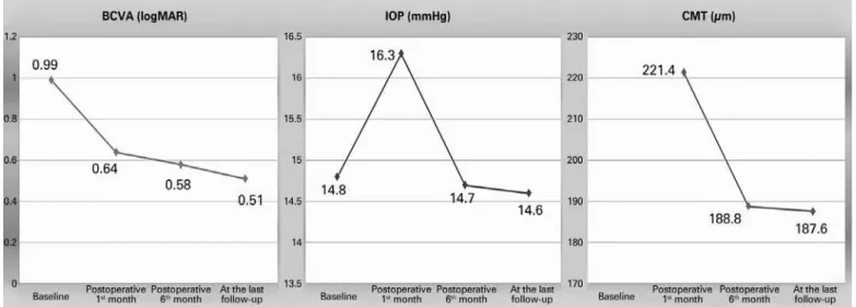
MHV was measured on the basis of preoperative SD-OCT, which captured the widest cross-sectional image of the hole. Because the outer plexiform layer (OPL) and outer nuclear layer (ONL) junctions are located just beneath the foveal contour, OPL was first marked in all preoperative macular scans for each study participant. For the purpose of this study, we pointed out the OPL level along both sides of the MH, and a border line connecting these points was drawn to estimate the possible location of the foveal contour using SD-OCT that captured the widest cross-sectional image of the hole. The space remaining under the imaginary border line of OPL was described as MHV (Figure 1). The aspect of such spaces had the shape of a conical frustum; thus, we calculated MHV for each study participant accor-ding to the formula used for calculating the volume of a truncated cone (Figure 2).
The data were stored in a computerized database and analyzed using Statistical Package for Scientific Studies for Windows v.16.0 (SPSS Inc., Chicago, IL, USA). Normal distribution analysis of the data was performed using the Kolmogorov-Smirnov test; moreover, the chi-square test, the paired t-test, Student’s t-test, and the Mann- Whitney U test were performed for statistical analyses. Pearson’s correlation coefficient test was also used to analyze the association among MHV, postoperative CMT, and pre- and postoperative BCVA scores. A p value of <0.05 was considered statistically significant.
RESULTS
Of the 30 patients with a mean age of 63.9 ± 11.0 (range, 15-76) years, 18 (60.0%) were females and 12 (40.0%) were males. Three par ticipants (10.0%) had diabetes mellitus and five (16.7%) had hy-pertension; however, none of the subjects demonstrated signs of
Figure 1. Retinal layers are shown in an SD-OCT image (A). An imaginary border line demonstrating the outer plexiform layer (OPL) was drawn to estimate the possible localization of the foveal contour on SD-OCT that captured the widest cross-sectional image of the hole (B).
A
re tinopathy. Thirteen eyes (39.4%) were pseudophakic and 20 (60.6%) were phakic. Mean preoperative BCVA, intraocular pressure (IOP), and MHV were identified as 0.99 ± 0.36 (range, 0.3-2.0) logMAR, 14.7 ± 2.1
(range, 11-20) mmHg, and 0.139 ± 0.076 (range, 0.004-0.318) mm3,
respectively. Surgery was performed within 6 months of the onset of clinical symptoms; thus, MH diagnosis was established soon after. Perifoveally located intraretinal cysts relevant to a newly developed
MH were found on preoperative macular SD-OCT of all participants. These scans revealed VMT in 16 eyes (48.5%) and a true retinal oper-culum in 10 eyes (30.3%) preoperatively. Although early anatomical success was achieved with MH surgery in all cases, MH recurred in four eyes (12.1%) after the mean follow-up of 16.3 ± 14.3 (range, 3-50) months. Demographic and ophthalmological findings are summa-rized in tables 1 and 2.
Figure 2. Calculation of the macular hole volume (MHV) according to the formula that gives the volume of a truncated cone.
Table 1. Demographic and initial ophthalmological indings
All study eyes (n=33) Eyes with c-MH (n=29) Eyes with r-MH (n=4) p value
Age (years) 63.900 ± 11.000 61.800 ± 14.400 70.200 ± 4.500 0.139
Preoperative BCVA (logMAR) 00.990 ± 00.360 00.930 ± 00.330 01.400 ± 0.350 0.013*
Preoperative IOP (mmHg) 14.700 ± 02.100 14.900 ± 02.200 13.700 ± 1.000 0.322
Macular hole volume (mm3) 00.139 ± 00.076 00.138 ± 00.076 00.149 ± 0.075 0.777
Gender 0.643
Female 60.0% 61.5% 50.0%
Male 40.0% 38.5% 50.0%
Concomitant disease
Diabetes mellitus 10.0% 11.5% - 0.500
Hypertension 16.7% 19.2% - 0.367
Preoperative VMT
VMT 16 (48.5%) 16 (55.2%) - 0.038*
A true retinal operculum 10 (30.3%) 10 (34.5%) - 0.159
Follow-up (months) 16.3 ± 14.3 16.3 ± 14.4 15.8 ± 15.2 0.939
c-MH= closed macular hole; r-MH= recurrent macular hole; BCVA= best-corrected visual acuity; IOP= intraocular pressure; VMT= vitromacular traction; *= Statistical significance.
Table 2. Postoperative ophthalmologic indings in eyes with and without successful surgery
All study eyes (n=33) Eyes with c-MH (n=29) Eyes with r-MH (n=4) p value
Follow-up (months) 16.30 ± 14.3 016.30 ± 14.40 15.80 ± 15.2 0.939*
Postoperative BCVA (logMAR) 00.64 ± 0.38 000.51 ± 00.31 01.60 ± 00.0 <0.001*
Postoperative IOP (mmHg) 14.60 ± 1.90 014.70 ± 01.90 14.00 ± 01.8 0.502*
Postoperative CMT (µm) 187.60 ± 67.80
Reestablishment of ELM 20 (60.6%) 20 (69.0%) - 0.008*
Reestablishment of EZ 13 (39.4%) 13 (44.8%) - 0.085*
Reestablishment of COST line 08 (24.2%) 08 (27.6%) - 0.227*
Reestablishment of RPE band 28 (84.8%) 25 (86.2%) 3 (75.0%) 0.558*
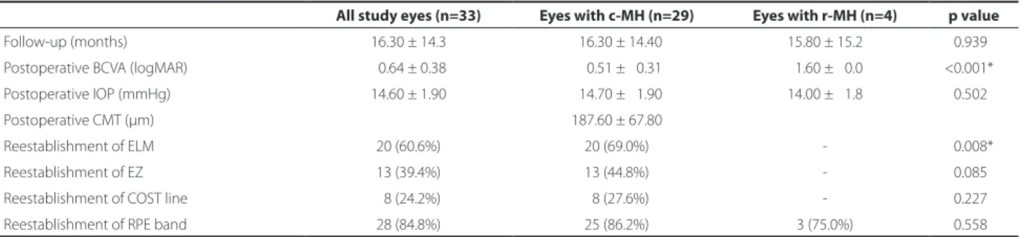
Figure 3. Changes in best-corrected visual acuity (BCVA), intraocular pressure (IOP), and central macular thickness (CMT).
Mean BCVA and IOP were found to be 0.64 ± 0.38 (range, 0.0-1.6) logMAR and 14.6 ± 1.9 (range, 11-20) mmHg (p<0.001 and p=0.723), respectively, at the last follow-up visit. When four eyes with recurrent disease were excluded, mean BCVA and CMT were found to be 0.51 ± 0.31 (range, 0.0-1.6) logMAR and 187.6 ± 67.8 (range, 60-247) µm, respectively, at the last follow-up in the remaining 29 eyes. Mean preo perative BCVA and MHV were 1.40 ± 0.35 logMAR and 0.149 ±
0.075 mm3, respectively, in eyes with recurrent disease, whereas these
scores were 0.93 ± 0.33 logMAR and 0.138 ± 0.076 mm3, respectively,
in successfully treated eyes (p=0.013 and p=0.777, respectively). VMT and a true retinal operculum were not found in any eyes with resistant MH; however, such vitreomacular interface abnormalities were present in 55.2% and 34.5% of eyes with closed MH (p=0.038 and p=0.159, respectively). Changes in BCVA, CMT, and IOP are shown in figure 3. While abnormalities were preoperatively present in the foveal ellipsoid zone (EZ) of all participants, SD-OCT revealed re-es tablishment of EZ in 13 eyre-es (39.4%). There was no correlation between preoperative MHV and postoperative EZ re-establishment (p=0.713). However, better postoperative BCVA scores were observed in patients with re-established EZ (p=0.002).
Macular SD-OCT scans that were performed at the last fol low-up visit also depicted the re-establishment of the external limiting membrane (ELM) in 20 eyes (60.6%), cone outer segment tip (COST) line in eight eyes (24.2%), and retinal pigment epithelium (RPE) band in 28 eyes (84.8%). Preoperative MHV was not found to be correlated with the re-establishment of ELM, COST line, or RPE band (p=0.268, p=0.283, and p=0.920, respectively). No statistical corre-lation was found between final BCVA and re-establishment of RPE band (p=0.411); however, reformed ELM and COST line provided a better visual prognosis in cases undergoing MH surgery (p=0.003 and p=0.005, respectively). Postoperative ocular features are summarized in table 2.
Although no statistical correlations were found between MHV and postoperative BCVA (p=0.588) and between MHV and disease recurrence (p=0.544), a negative correlation was present between MHV and CMT that was measured at the last follow-up (p=0.040). Furthermore, there was a strong correlation between pre- and pos-toperative BCVA scores (p<0.001). The results of Pearson’s correlation coefficient test for the parameters studied are given in table 3.
DISCUSSION
Recent advances in MH surgery techniques have contributed to more successful postoperative anatomical and associated
functio-nal prognoses(6-11). However, anatomical closure of a hole does not
always correlate with a good functional result(6,17,18). Although various
preoperative SD-OCT parameters have been examined to estimate postoperative visual prognosis, studies have reported the lack of any consistent volumetric or structural feature that is closely correlated
with anatomical and functional surgical successes(15-18). In this study,
there was no statistical difference in MHV measurements between eyes with and without disease recurrence; however, lower preopera-tive BCVA scores were found in eyes with relapsing MH. Furthermore, in our study population, preoperative SD-OCT revealed that neither VMT nor a true retinal operculum was found in any of the eyes with a recurrent hole.
Defects of foveal photoreceptors and abnormalities of external retinal microstructures that can be observed in SD-OCT images of surgically treated eyes with MH have been studied, with the goal of defining any association between such problems and the develop-ment of poorer vision. Furthermore, a significant correlation between the presence of such abnormalities and postoperative visual
progno-sis has been recently reported(16-20). In this study, MHV was not found
to be associated with the postoperative re-establishment of any of
Table 3. Pearson’s correlation coeicient test results for studied parameters
Studied parameters p value r value
Macular hole volume Preoperative BCVA (logMAR) 0.294* 0.188
Macular hole volume Final BCVA (logMAR) 0.588* 0.113
Preoperative BCVA (logMAR) Postoperative BCVA (logMAR) <0.001* 0.660 Macular hole volume Postoperative CMT in cases with c-MH (n=29) 0.040* -0.383
A C E G
B D F H
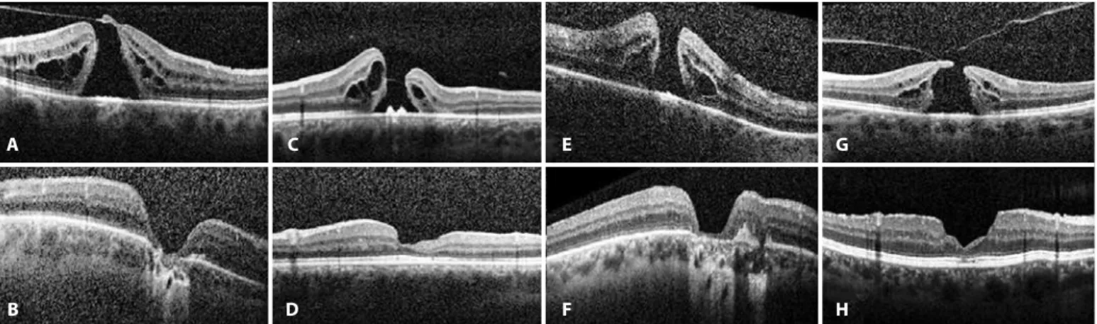
Figure 4. Preoperative (A, C, E, and G) and postoperative (B, D, F, and H) SD-OCT of patients with macular atrophy after 4-port 23-gauge PPV combined with ILM peeling and intrao-cular 20% SF6 tamponade.
the studied parameters of external retinal microstructure. Improved RPE band integrity on successful MH surgery was also not found to be associated with final BCVA scores; however, re-establishment of ELM, EZ, and COST line was associated with a better visual prognosis.
On the contrary, some published studies have reported a ne-gative correlation between preoperative MH dimensions measured
using OCT and postoperative visual recovery(21-26). The MH base
dia meter, which was measured at the RPE level, and the minimum diameter of the hole, which represented the shortest extent of MH, appear to constitute prognostic factors for postoperative anatomical
and functional outcomes. Freeman et al.(6),similarly found that MH
with smaller diameters were associated with a better visual progno-sis. Moreover, a hole form factor (HFF) was calculated from the base and minimum diameters to estimate the postoperative anatomical
success rate. Ullrich et al.(21) reported significantly larger base
dia-meters and greater minimum MH diadia-meters in cases with recurrent holes. Furthermore, postoperative visual prognosis was found to be negatively correlated with both the base and minimum diameters of the hole. The authors also stated that all cases with HFF of >0.9 were surgically treated with a single intervention; however, the anatomical success rate was 67% in patients with HFF of <0.5.
Ruiz-Moreno et al.(22) reported the sensitivity of various parame ters
used to estimate visual prognosis after MH surgery. They de monstrated that postoperative BCVA was statistically correlated with base and mi-nimum diameters of MH, the MH index, which is defined as the ratio of the hole height to the base diameter, and the tractional hole index, which is defined as the ratio of the maximum height to the minimum diameter of MH. Base area, base diameter, top area, top diameter, minimum diameter, MH height, and MHV were found to be signifi-cantly correlated with initial BCVA in a computer algorithm-supported
study by Xu et al(23). Moreover, they reported a significant correlation
between BCVA at postoperative sixth month and MHV, base area and diameter, as well as height-to-base diameter ratio; however, after per-forming multivariate analyses, only base area and MHV were found to be significant predictors of six-month postoperative BCVA.
Pilli et al.(26) recommended performing ILM peeling to achieve
a better visual outcome because a close correlation between inner macular volume and BCVA was observed. In our study, although no statistical correlations were found between MHV and postoperative BCVA or MHV and disease recurrence, a negative correlation was pre-sent between MHV and CMT measured at the last follow-up (p=0.04, r=-0.383). Because postoperative CMT scores were within the favora-ble normal range in the entire study population, except in cases with recurring MH, lower CMT scores were considered to be associated with a more severe macular atrophy (Figure 4). Visual acuity is directly
related to both the extent and centricity of the macular atrophy(27,28).
However, preoperative BCVA score was the only factor consistently affecting postoperative BCVA in our study population.
The major limitation of this study was the lack of the use of real-ti me three-dimensional volumetric analysis of MH. Although the shape of each MH varies in its microarchitecture, we used the widest two-dimensional cross-sectional SD-OCT image of all studied holes. Nevertheless, we believe that within a close range of deviation, every MHV, regardless of the varying shape of the hole, may simply be pre-dicted using the parameters of the formula that gives the volume of a truncated cone. The second limitation was that 20% of our study participants had concomitant diabetes mellitus, hypertension, or both. However, we deliberately selected the surgically treated cases with a newly developed idiopathic MH who did not have any other retinal abnormalities, including retinopathies and a previous history of retinal detachment. In addition, the coexistence of diabetes and/ or hypertension may result in some cellular and microarchitectural changes within the retinal tissue, even in cases with no signs of reti-nopathy. Finally, the retrospective nature of this study and the lack of a large study population reflect other limitations.
Visual acuity may not improve as expected in some patients even when anatomical closure of the hole is attained following MH sur-gery. This may be related with postoperative macular atrophy, which can be diagnosed on the basis of a reduced CMT score on SD-OCT. In this study, greater MHV was associated with lower postoperative CMT scores. To the best of our knowledge, this is the first study in-vestigating the association between preoperative MHV and macular atrophy. Long-term prospective trials with large cohorts are required to assess the correlation of macular volumetric features with posto-perative anatomical and functional prognoses.
REFERENCES
1. Bainbridge J, Herbert E, Gregor Z. Macular holes: vitreoretinal relationships and sur-gical approaches. Eye (Lond). 2008;22(10):1301-9.
2. Steel DH, Lotery AJ. Idiopathic vitreomacular traction and macular hole: a com-prehensive review of pathophysiology, diagnosis, and treatment. Eye (Lond). 2013; 27(Suppl 1):S1-S21.
3. Williamson TH, Lee E. Idiopathic macular hole: analysis of visual outcomes and the use of indocyanine green or brilliant blue for internal limiting membrane peel. Graefes Arch Clin Exp Ophthalmol. 2014;252(3):395-400.
4. Tirelli F, Sasso P, Scupola A. Idiopathic macular hole: post-operative morpho-func-tional assessment and prognostic factors for recovery of visual acuity. Ann 1st Super Sanita. 2013;49(3):313-6.
5. Kelly NE, Wendel RT. Vitreous surgery for idiopathic macular holes: results of a pilot study. Arch Ophthalmol. 1991;109(5):654-9.
7. Spiteri Cornish K, Lois N, Scott NW, Burr J, Cook J, Boachie C, et al. Vitrectomy with internal membrane peeling versus no peeling for idiopathic full-thickness macular hole. Ophthalmology. 2014;121(3):649-55.
8. Lois N, Burr J, Norrie J, Vale L, Cook J, McDonald A, Boachie C, Ternent L, McPherson G; Full-thickness Macular Hole and Internal Limiting Membrane Peeling Study (FILMS) Group. Internal limiting membrane peeling versus no peeling for idiopathic full-thi-ckness macular hole: a pragmatic randomized controlled trial. Invest Ophthalmol Vis Sci. 2011;52(3):1586-92.
9. Morizane Y, Shiraga F, Kimura S, Hosokawa M, Shiode Y, Kawata T, et al. Autologous transplantation of the internal limiting membrane for refractory macular holes. Am J Ophthalmol. 2014;157(4):861-9.
10. Mahalingam P, Sambhav K. Surgical outcomes of inverted internal limiting membra-ne flap technique for large macular hole. Indian J Ophthalmol. 2013;61(10):601-3. 11. Xirou T, Theodossiadis PG, Apostolopoulos M, Kabanarou SA, Feretis E, Ladas ID, et
al. Macular hole surgery with short-acting gas and short-duration face-down posi-tioning. Clin Ophthalmol. 2012;6:1107-12.
12. Margherio RR, Margherio AR, Williams GA, Chow DR, Banach MJ. Effect of perifoveal tissue dissection in the management of acute idiopathic full-thickness macular holes. Arch Ophthalmol. 2000;118(4):495-8.
13. Lai MM, Williams GA. Anatomical and visual outcomes of idiopathic macular hole sur-gery with internal limiting membrane removal using low-concentration indocyanine green. Retina. 2007;27(4):477-82.
14. Altaweel M, Ip M. Macular hole: improved understanding of pathogenesis, staging, and management based on optical coherence tomography. Semin Ophthalmol. 2003;18(2):58-66.
15. Villate N, Lee JE, Venkatraman A, Smiddy WE. Photoreceptor layer features in eyes with closed macular holes: optical coherence tomography findings and correlation with visual outcomes. Am J Ophthalmol. 2005;139(2):280-9.
16. Bonnabel A, Bron AM, Isaico R, Dugas B, Nicot F, Creuzot-Garcher C. Long-term anatomical and functional outcomes of idiopathic macular hole surgery. The yield of spectral-domain OCT combined with microperimetry. Graefes Arch Clin Exp Oph-thalmol. 2013;251(11):2505-11.
17. Chen WC, Wang Y, Li XX. Morphologic and functional evaluation before and after suc-cessful macular hole surgery using spectral-domain optical coherence tomography combined with microperimetry. Retina. 2012;32(9):1733-42.
18. Matsumiya W, Kusuhara S, Shimoyama T, Honda S, Tsukahara Y, Negi A. Predictive value of preoperative optical coherence tomography for visual outcome following macular hole surgery: effects of imaging alignment. Jpn J Ophthalmol. 2013; 57(3):308-15.
19. Oh J, Smiddy WE, Flynn HW Jr, Gregori G, Lujan B. Photoreceptor inner/outer segment defect imaging by spectral domain OCT and visual prognosis after macular hole surgery. Invest Ophthalmol Vis Sci. 2010;51(3):1651-8.
20. Kao TY, Yang CM, Yeh PT, Huang JY, Yang CH. The value of combining autofluorescen-ce and optical coherenautofluorescen-ce tomography in predicting the visual prognosis of sealed macular holes. Am J Ophthalmol. 2013;156(1):149-56.
21. Ullrich S, Haritoglou C, Gass C, Schaumberger M, Ulbig MW, Kampik A. Macular hole size as a prognostic factor in macular hole surgery. Br J Ophthalmol. 2002;86(4): 390-3.
22. Ruiz-Moreno JM, Staicu C, Pinero DP, Montero J, Lugo F, Amat P. Optical coherence tomography predictive factors for macular hole surgery outcome. Br J Ophthalmol. 2008;92(5):640-4.
23. Xu D, Yuan A, Kaiser PK, Srivastava SK, Singh RP, Sears JE, et al. A novel segmentation algorithm for volumetric analysis of macular hole boundaries identified with optical coherence tomography. Invest Ophthalmol Vis Sci. 2013;54(1):163-9.
24. Duker JS, Kaiser PK, Binder S, de Smet MD, Gaudric A, Reichel E, et al. The International Vitreomacular Traction Study Group classification of vitreomacular adhesion, traction, and macular hole. Ophthalmology. 2013;120(12):2611-9.
25. Haritoglou C, Neubauer AS, Reiniger IW, Priglinger SG, Gass CA, Kampik A. Long--term functional outcome of macular hole surgery correlated to optical coherence tomography measurements. Clin Exp Ophthalmol. 2007;35(3):208-13. Comment in: Clin Experiment Ophthalmol. 2007;35(3):203.
26. Pilli S, Zawadzki RJ, Werner JS, Park SS. Visual outcome correlates with inner macular volume in eyes with surgically closed macular hole. Retina. 2012;32(10):2085-95. 27. Brader HS, Ying GS, Martin ER, Maguire MG;Complications of age-related macular
degeneration prevention trial (CAPT) research group. Characteristics of incident geographic atrophy in the complications of age-related macular degeneration pre-vention trial. Ophthalmology. 2013;120(9):1871-9.
28. Holz FG, Strauss EC, Schmitz-Valckenberg S, van Lookeren Campagne M. Geographic atrophy: clinical features and potential therapeutic approaches. Ophthalmology. 2014;121(5):1079-91.