Sediment examination of urine without
physical-chemical alterations
Sedimentoscopia de parciais de urina sem alterações físico-químicas
Diego Z. Nascimento; Mayara D. Pickler; Gabriela M. Marques; Fabiana S. Trevisol; Ana Luisa O. Martins
Universidade do Sul de Santa Catarina (Unisul), Santa Catarina, Brazil.
First submission on 11/10/17; last submission on 04/20/18; accepted for publication on 05/14/18; published on 06/20/18
ABSTRACT
Introduction: It is from the urine sediment examination that many conclusions are drawn by the professional responsible for diagnosis. Because that is a test frequently run in laboratories, it is desirable to reduce costs and time to exam sediment in urine samples considered normal. Objectives: To evaluate the importance of sediment microscopy in urine without physical and chemical alterations. Material and method: This is an epidemiological study of a cross-sectional design developed at a college laboratory based on reports of urine tests
performed from January to July, 2017. For comparison between means, Student’s t-test, Pearson’s chi-square test and Spearman’s correlation were done. The level of significance was 5%. Results: We analyzed 7,734 urine reports, with 2,530 (32.7%) results without physical-chemical changes. Patients had a mean age of 39 (± 23) years, most of them were males (61.7%). Regarding leukocyte quantification, 2.3% of the patients had a number higher than the reference values, and in relation to red blood cells, 1.7% of the patients exceeded these values. It was observed that the amount of leukocytes found in the urine specimens without physical-chemical alterations (p < 0.007) was reduced by 0.4 with each year of age. Conclusion: From the data of the present study, one can conclude that the majority of patients without physical-chemical changes do not have any changes of clinical relevance in sediment analysis.
Key words: microscopy; urinalysis; diagnostic tests routine.
INTRODUCTION
Studies reveal, by means of drawings, that our ancestors already examined urine. Logically, those examinations did not have today’s specificity and technology, but some observations could be made when the evaluation consisted of the physical aspect of urine, such as its turbidity, viscosity, and color(1). The importance of urinalysis lies the range of analyses that can be performed facilitating diagnosis. It is a test that goes beyond provision of essential information about the functional state of kidneys(2), being possible to diagnose several diseases by observing urine color, density, aspect, presence of leukocytes, bacteria, blood, glucose, urobilinogen, bilirubin, nitrite, and urine sediments(3). Urinalysis is a commonly expeditious, reliable, accurate, safe, and low-cost procedure(4).
Urinalysis consists of three steps: physical examination, chemical test, and microscopic examination of urine sediment.
The last phase is the most complex, because it needs a trained professional, has higher cost and also demands time in laboratory
routine(5). Physical examination consists of assessment of urine color, aspect, density, and odor(6, 7). Chemical examination is conducted with reagent strips, aimed at determining urine elements in a quick and simple manner(8-10).
Urine sediment examination is a clinically important step of urinalysis, especially when the sample presents alterations in the physical and chemical phases(11). For this reason, an adequate evaluation of urine sediments is necessary, including all found components(12).
Sediment examination is a simple and low-cost procedure, but it does not receive, most of times, the necessary attention: the time employed in this activity and the cost of professionals qualified for this function are not considered, what makes the practice moderately complex(2, 13, 14). For analysis of urine sediment, the presence of a capable professional is necessary, and this process generates cost
and time demands. Considering the great diversity of urinalysis costs in Brazil, where most laboratories perform the sediment analysis in all samples, further studies are necessary to determine the current importance of urine sediment analysis in urinalysis in the presence of normal physical and chemical steps, protecting the exam result, with no consequences to patients. The objective of this work is to relate the absence of alterations in physical and chemical urine analyses to possible alterations in sediment analysis.
MATERIAL AND METHOD
This is an epidemiological study of cross-sectional design conducted at a college laboratory in the south of Santa Catarina, which carries out tests in the areas of biochemistry, hematology, immunology, parasitology and urinalysis. The laboratory, founded on March 1st, 1974, treats the academic community and the wider public, as private patients or under coverage of a health insurance, offering laboratory tests with agility, easy access, and advanced technology. Around 10,000 urinalyses are performed in that laboratory each year.
By means of this study, researchers assessed urinalysis reports from patients at Laboratório de Análises Clínicas da Universidade do Sul de Santa Catarina (LAC-Unisul) regarding tests performed from January to July, 2017. At LAC-Unisul, urinalyses are carried out the following way: after sample homogenization, aspect and color of urine are verified. Aspect can be classified into clear, slightly cloudy, or turbid. Color can vary among pale yellow, bright yellow, amber yellow, red and green.
Chemical analysis is conducted after immersion of reagent strips into 10 ml of the sample, and reading in the semiautomatic analyzer Urisys 1100 (Roche®). Urine sediment examination is carried out after centrifugation of the 10 ml of urine at 1500 rpm for five minutes; the supernatant is discarded and 1 ml saline solution is added to the sediment. After sediment homogenization with saline solution, 25 μl of the dilution are placed at a Kcell chamber.
Leukocyte and erythrocyte counting is done using a 40× objective; the elements are counted in nine reticles of the chamber and the obtained number is multiplied by 1,000. In case no element (leukocytes and erythrocytes) is found, result is released as “below 1,000”. In case of many elements, one reticle is counted and multiplied by 9,000. The reference value for leukocytes and erythrocytes is 10,000 per ml.
Cells and casts are observed in 10× objectives, and average count is conducted in 10 fields. Classification varies among no, rare, few, many, and numerous elements. Bacteriuria is classified
into discreet, moderate or intense. The presence of yeasts, crystals, mucus, protozoa and sperm (just in male patients) is reported.
Patients’ reports are digitized by the sector in charge, and next, checked and released by skilled professionals.
Internal control of reactive strips of the analyzer is done daily, using a control sample before beginning the routine. If there is any doubt about visualized structures on the microscope or on cell counting, the responsible professional must order analysis by another colleague. Once a month, a sample is selected to be analyzed by all skilled professionals, for result comparison. Equipment calibration is weekly done, or when there are problems regarding internal control, by means of a standard reagent strip provided by the manufacturer. The laboratory is associated with Sociedade Brasileira de Análises Clínicas (SBAC), which monthly sends samples to the different sectors for internal quality control, by means of Programa Nacional de Controle de Qualidade (PNCQ).
Researchers had access to the following information about each patient: results of physical-chemical analysis and urine sediment examination, age, sex, use of medications, presence of chronic diseases, and pregnancy. Access to this set of information was obtained by the laboratory electronic system and the printed records of the semiautomatic analyzer. The used inclusion criterion comprised all urine tests performed between January and July, 2017; the exclusion criterion, urine tests with physical and chemical alterations.
This study was carried out with approval by the Ethics Research Committee of Unisul. The collected data were digitized in the Microsoft Office Excel program, and statistical analysis was done in the SPSS version 21 (IBM, Armonk, New York, USA) software. Descriptive statistics was used for data presentation, with absolute and relative frequency for categorical variables, and mean and standard deviation (SD) for quantitative variables. For comparison between variables of interest, Pearson’s chi-squared test was used, to verify association between the presence of erythrocytes and leukocytes in urinalysis according to patients’ sex. Student’s t-test was used for comparison of mean ages between the presence or absence of erythrocytes and leukocytes in urine. Correlation of age and leukocytes and erythrocytes was also analyzed, using non-parametric statistics with the employment of Spearman’s correlation. The established significance level was 5%.
RESULTS
In this group of patients, the most frequent diseases were: diabetes, high blood pressure, hypothyroidism, anxiety, and depression. The main encountered medications were: contraceptives, antidepressants, benzodiazepines, antimicrobials, hypoglycemic drugs, anti-hypertensive agents, and synthetic hormones.
In the reports of urine tests with no physical-chemical alterations, adopting the minimum abnormal reference parameters, we found: leukocytes, erythrocytes, epithelial cells, mucus, and sperm (Table 2).
Among the urinalysis reports, the following crystals were found: calcium oxalate in 32 patients, amorphous phosphate in five, and uric acid in one. Twenty-one patients presented hyaline casts in urine sediment analysis. Bacteria were found in four reports, belonging to females.
Among reports, leukocytes were prevalent in females, 38 (3.9%) of the tests presented leukocytes in urine sediment analysis, considering p < 0.007.
We also observed that for each year of age, the quantity of leukocytes is reduced by 0.4 in urinalyses with no physical-chemical alterations, considering p < 0.05.
DISCUSSION
In the present study, 2,530 (32.71%) of the patients did not present physical-chemical alterations at urinalysis. Among those, only 32 (4%) presented alterations of clinical relevance in urine sediment examination. These data allow questioning the necessity of microscopy in urine samples without physical-chemical alterations.
Urine is extremely important for the diagnosis of different clinical conditions; it is the precursor sample of laboratory medicine. Its composition varies according to hour of the day, physical condition, diet, and individual’s health status. Water, urea, and sodium chloride are the three main components of urine, and alterations in its composition are a sign of possible diseases. Urinary volume is also variable, depending on age, water and liquid intake, metabolism, blood pressure, dietary intake, action of aldosterone and other hormones, besides a series of other factors(15).
Due to the several diagnoses obtained based on a urine sample, the search for urinalysis is evident, with a number of 7,734 tests performed in our laboratory for this study in a period of seven months, with a daily average of 36.8 tests. In other studied scientific articles, the number of urine samples evaluated was smaller than 1,200, such as that by Bottini and Garlipp (2006)(13), in which 1,140 samples were analyzed by common optical microscopy and flow cytometry; that by Feitosa et al. (2009)(6), which evaluated accuracy of simple urine test for diagnosis of urinary tract infections in 230 pregnant women; and that by
Heggendornn et al. (2014)(16), whose objective was to assess the importance of microscopic urine sediment analysis in 247 samples with no physical-chemical alterations. A study analyzing a larger number of samples, exactly 10,234, was conducted by
Costaval et al. (2001)(2), which evaluated the importance of sediment analysis in urine samples with normal physical-chemical characteristics.
Urinalysis is the most commonly used test for screening of bacteriuria and urinary infection(6). However, in the reports of patients with tests without physical-chemical alterations, sediment analysis was practically irrelevant, with just four reports presenting bacteriuria. All of them were females, with the presence of epithelial cells, considering, therefore, that these bacteria come from vaginal microbiota. The exact confirmation of bacteriuria TABLE 2 − Cells and elements found in urinalysis with
no physical chemical alterations
Variables n (%)
Leukocytes (thousand/ml)
0 808 (31.9)
0-10 1,665 (65.8)
11-15 48 (1.9)
> 15 9 (0.4)
Erythrocytes (thousand/ml)
0 674 (26.6)
0-10 1,813 (71.7)
11-15 38 (1.5)
> 15 5 (0.2)
Epithelial cells 1,580 (62.5)
Mucus 82 (3.2)
Sperm 16 (0.6)
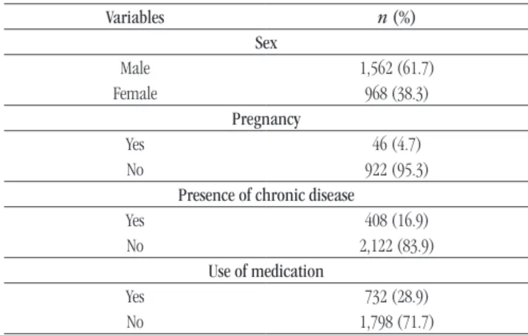
TABLE 1 − Characteristics of 2,530 patients with mean age of 39 (± 23) years, with no physical-chemical alterations in urinalysis
Variables n (%)
Sex
Male 1,562 (61.7)
Female 968 (38.3)
Pregnancy
Yes 46 (4.7)
No 922 (95.3)
Presence of chronic disease
Yes 408 (16.9)
No 2,122 (83.9)
Use of medication
Yes 732 (28.9)
is determined by culture of the mid-stream urine sample. In the study by Santos et al. (2017)(17), 29 (25.7%) positive results were analyzed of urine culture with antibiogram, in which there was prevalence of urinary tract infection in women positive for
Escherichia coli bacterium.
Other studies show that urine tests with no physical-chemical alterations have low presence of bacteria. At a study with 247 urine samples, just five (2%) presented increased bacterial flora(16), while in the study by de Costaval et al. (2001)(2) , among the 5,000 sampleswith no physical-chemical alterations there was presence of bacteria in seven.
In the results of the present study, 2,472 (97.7%) patients had results within the reference values (up to 10,000/ml) for leukocytes and 2,487 (98.3%) for erythrocytes. Based on the found numbers, we question to which point sediment analysis is relevant for erythrocyte and leukocyte visualization in samples with no physical-chemical alterations. In another study, the number of urine tests with no physical-chemical alterations with presence of leukocytes was also low, with just 26 (0.5%) among the 5,000 analyzed samples(2). The presence of erythrocytes in urine tests with no physical-chemical alterations was also low in another study, in which among the 247 observed samples, just 11 (4.5%) presented hematuria(16).
The association of reduced leukocytes in urine tests with no physical-chemical alterations for each year of age can be explained by the progressive alterations that occur with age. Both immunities, innate and adaptive, show age-related alterations, which seem to be crucial for healthy ageing and survival. Innate immune system is composed of, besides physical, mechanical, physiological and inflammatory barriers, cell barriers that phagocytose strange particles and microorganisms, eliminating them. Those cells defend the body against possible pathogens, and immune senescence of innate response is quite complex, seen that there is evidence of alterations resulting from ageing of cell components(18).
In the observed reports that presented no physical-chemical alterations, 21 showed the presence of hyaline casts. Hyaline casts present homogeneous colorless matrix and are the most commonly found in sediment analysis. Up to two hyaline casts per field is considered normal, they can result from extenuating exercise, dehydration, exposure to heat and emotional stress(19). No patients of this study presented a number considered elevated for hyaline casts.
In 1,580 (62.5%) urine tests with no physical-chemical alterations, epithelial cells were found, with 526 (33.3%) being from females. Most of those cells come from vaginal epithelium,
what means, therefore, contamination. The layer of epithelial cells from the vagina constitutes the initial contact point between microorganisms and the host’s genital tract(20). Moreover, in the loop of Henle, precisely in the ascending limb, there are thick walls, designated thick ascending limb; they have epithelial cells presenting high metabolic activity and are capable of active reabsorption of sodium, potassium, and chloride. They can be excreted in urine normally, what can explain the presence of epithelial cells present in the urines of male patients(21).
The presence of long, slender and wavy filaments or threads of mucus observed in 82 (3.2 %) samples does not have clinical significance, being from possible contamination during collection; they can even be increased in infectious or inflammatory processes in samples with physical-chemical alterations(10).
The color change that happens at a physical urine test, urine pH, glycosuria, proteinuria, and detection of ketone bodies are some examples of tests not comprised in sediment analysis, what makes urinalysis be possibly divided into two tests: physical-chemical and sediment analysis. They can be ordered together, to help clinical diagnosis, or in isolation, according to actual necessity, optimizing time and cost for patients and laboratories. The aim of reducing operating costs in urinalysis, stressing the sediment analysis step, is an issue dealt with in the latest congresses of Clinical Pathology, whose objective is wide economy due to the great demand of those tests.
Although sediment analysis is a simple technique and helps diagnosis of several diseases, including kidney diseases, allowing identify diverse elements with different diagnostic relevance(16), it has some obstacles that hinder its capacity to be considered an ideal marker of kidney diseases, because it requires extreme competence by the analyst(3). The cost of sediment analysis in total exam is presented in the literature. The largest world potency, the United States, for example, has a projection of cost reduction of approximately US$ 44 million in annual economy in the whole country, with the exclusion of sediment analysis from normal urine samples(2). The time for analysis of urine sediment is around two
CONCLUSION
The importance of performing an adequate sediment examination is attributed to the conclusions obtained by means of the exam, because based on the results, medical decisions are taken. Based on data from the present study, we conclude most patients with no physical-chemical alterations do not have
alterations of clinical relevance in sediment analysis, what would make microscopy irrelevant for the samples of these individuals. We suggest, based on these results, that accuracy studies for evaluation of chemical test be performed comparing also reports whose physical-chemical tests had alterations, ensuring thus, measures of economy to the laboratory without impairing the test quality.
REFERENCES
1. Strasinger SK, Lorenzo MS. Urinálise e fluídos biológicos. 3 ed. São Paulo: Editorial Premier; 2000.
2. Costaval JA, Massote AP, Cerqueira CMM, Costaval AP, Auler A, Martins GJ. Qual o valor da sedimentoscopia em urinas com características físico-químicas normais? J Bras Patol Med Lab [Internet]. 2001 Jul; 37(4): 261-5. Available at: http://www.scielo.br/pdf/jbpml/v37n4/a07v37n4.pdf. 3. Perazella MA. The urine sediment as a biomarker of kidney disease. AJKD [Internet]. 2015 Nov; 66(5): 748-55. Available at: http://www. sciencedirect.com/science/article/pii/S0272638615006071.
4. Rabinovitch A, Arzoumanian L, Curcio KM, Dougherty B, Halim AB. GP16-A3: urinalysis; approved guideline. 3ed. Wayne: Clinical & Laboratory Standards Institute; 2009. Available at: https://clsi.org/ media/1382/gp16a3_sample.pdf.
5. Simerville JA, Maxted WC, Pahira JJ. Urinalysis: a comprehensive review. AFP [Internet]. 2015 Mar; 71(6): 1153-62. Available at: http://www.aafp. org/afp/2005/0315/p1153.html.
6. Feitosa DCA, Silva MG, Parada CMGL. Acurácia do exame de urina simples para diagnóstico de infecções do trato urinário em gestantes de
RESUMO
Introdução: É a partir da sedimentoscopia do parcial de urina que muitas conclusões são tiradas pelo profissional responsável pelo diagnóstico. Por ser um exame frequentemente realizado nos laboratórios, é importante a redução dos custos e do tempo para a realização da sedimentoscopia em amostras de urina consideradas normais. Objetivo: Avaliar a importância da sedimentoscopia em urinas sem alterações físico-químicas. Material e método: Trata-se de um estudo epidemiológico de delineamento transversal desenvolvido em um laboratório-escola a partir de laudos referentes aos parciais de urina realizados de janeiro a julho de 2017. Para comparação entre médias, foi realizado teste t de Student, qui-quadrado de Pearson e teste de correlação de Spearman. O nível de significância estabelecido foi de 5%. Resultados: Foram analisados 7.734 laudos de parciais de urinas, com 2.530 (32,7%) resultados de parciais sem alterações físico-químicas. Os pacientes tinham idade média de 39 (± 23) anos, a maior parte do sexo masculino (61,7%). Com relação à quantificação de leucócitos, 2,3% dos pacientes apresentaram número superior aos valores de referência, e com relação às hemácias, 1,7% deles ultrapassaram esses valores. Observou-se que a cada um ano de idade a mais para o indivíduo, reduz-se em 0,4 a quantidade de leucócitos encontrados nos parciais de urina sem alterações físico-químicas (p < 0,007). Conclusão: A partir dos dados do presente estudo, conclui-se que a maior parte dos pacientes sem alterações físico-químicas não possui quaisquer alterações de relevância clínica na sedimentoscopia.
Unitermos: microscopia; urinálise; testes diagnósticos de rotina.
baixo risco. Rev Latinoam Enferm [Internet]. 2009 Ago; 17(4): 507-13. Available at: http://www.scielo.br/pdf/rlae/v17n4/pt_12.pdf.
7. Campana GA, Oplustil CP, Faro LB. Tendências em medicina laboratorial. J Bras Patol Med Lab [Internet]. 2011 Ago; 47(4): 399-408. Available at: http://www.scielo.br/scielo.php?script=sci_arttext&pid =S1676-24442011000400003.
8. Lima AO, Soares JB, Greco JB, Galizzi J, Cançado JR. Métodos de laboratório aplicados à clínica: técnica e interpretação. 8 ed. Rio de Janeiro: Guanabara Koogan; 2001.
9. Colombeli ASS, Falkenberg M. Comparação de bulas de duas marcas de tiras reagentes utilizadas no exame químico de urina. J Bras Patol Med Lab [Internet]. 2006 Abr; 42(2): 85-93. Available at: http://www.scielo.br/ pdf/jbpml/v42n2/a05v42n2.pdf.
10. Mundt LA, Shanahan K. Exame de urina e de fluidos corporais de Graff. 2 ed. Porto Alegre: Artmed; 2012.
degruyter.com/view/j/cclm.2015.53.issue-s2/cclm-2015-0479/cclm-2015-0479.xml.
13. Bottini PV, Garlipp CR. Urinálise: comparação entre microscopia óptica e citometria de fluxo. J Bras Patol Med Lab [Internet]. 2006 Jun; 42(3): 157-62. Available at: http://www.scielo.br/pdf/jbpml/v42n3/ a03v42n3.pdf.
14. Andersen H, Daae LNW, Wien TN. Urine microscopy – an important diagnostic tool. Tidsskr Nor Legeforen [Internet]. 2014 Sept; 18(134): 1765-7. Available at: http://tidsskriftet.no/en/2014/09/perspectives/urine-microscopy-important-diagnostic-tool.
15. Eaton DC, Pooler JP. Fisiologia renal de Vander. 8 ed. Artmed; 2016. 16. Heggendornn LH, Silva NA, Cunha GA. Urinálise: a importância da sedimentoscopia em exames físico-químicos normais. Rev Eletr de Bio [Internet]. 2014; 7(4): 431-43. Available at: https://revistas.pucsp.br/ index.php/reb/article/view/20177.
17. Santos AG, Silva DF, Moraes TI. Prevalência de positividade bacteriana em exames de urina de um laboratório particular em Itapevi. Rev Saud Foc [Internet]. 2017; 1(9): 300-7. Available at: http:// www.unifia.edu.br/revista_eletronica/revistas/saude_foco/artigos/ ano2017/036_artigo.pdf.
18. Agondi RC, Rizzo LV, Kalil J, Barros MT. Imunossenescência. Rev Bras Alerg Imunopatol [Internet]. 2012 Sept; 35(5): 169-76. Available at: http://www.sbai.org.br/revistas/vol355/Imunossenescencia.pdf. 19. Strasinger SF. Urinálise e fluidos biológicos. 5 ed. Editorial Premier Ltda; 2009.
20. Linhares IM, Giraldo PC, Baracat EC. Novos conhecimentos sobre a flora bacteriana vaginal. Rev Assoc Med Bras [Internet]. 2010; 56(3): 370-4. Available at: http://www.scielo.br/pdf/ramb/v56n3/v56n3a26.pdf. 21. Guyton AC, Hall JE. Tratado de fisiologia médica. 11 ed. Rio de Janeiro: Elsevier; 2006.
CORRESPONDING AUTHOR
Diego Zapelini do Nascimento
Av. José Acácio Moreira, 787; Dehon; CEP: 88704-900; Tubarão-SC, Brasil; Phone/Fax: +55 (48) 3621-3334/(48) 99156-1522; e-mail: diegozapnasc@gmail.com.