ABSTRACT
The objective of this systematic review was to select articles including chest X-ray or
chest CT findings in patients who developed pulmonary tuberculosis following solid
organ transplantation (lung, kidney, or liver). The following search terms were used:
“tuberculosis”; “transplants”; “transplantation”; “mycobacterium”; and “lung”. The
databases used in this review were PubMed and the Brazilian Biblioteca Virtual em
Saúde
(Virtual Health Library). We selected articles in English, Portuguese, or Spanish,
regardless of the year of publication, that met the selection criteria in their title, abstract,
or body of text. Articles with no data on chest CT or chest X-ray findings were excluded,
as were those not related to solid organ transplantation or pulmonary tuberculosis. We
selected 29 articles involving a collective total of 219 patients. The largest samples were
in studies conducted in Brazil and South Korea (78 and 35 patients, respectively). The
imaging findings were subdivided into five common patterns. The imaging findings
varied depending on the transplanted organ in these patients. In liver and lung transplant
recipients, the most common pattern was the classic one for pulmonary tuberculosis
(cavitation and “tree-in-bud” nodules), which is similar to the findings for pulmonary
tuberculosis in the general population. The proportion of cases showing a miliary pattern
and lymph node enlargement, which is most similar to the pattern seen in patients
coinfected with tuberculosis and HIV, was highest among the kidney transplant recipients.
Further studies evaluating clinical data, such as immunosuppression regimens, are
needed in order to improve understanding of the distribution of these imaging patterns
in this population.
Keywords:
Tomography, X-ray computed; Radiography; Tuberculosis, pulmonary; Lung/
transplantation; Kidney/transplantation; Liver/transplantation.
Chest X-ray and chest CT findings in
patients diagnosed with pulmonary
tuberculosis following solid organ
transplantation: a systematic review
Irai Luis Giacomelli
1,a, Roberto Schuhmacher Neto
1,b, Edson Marchiori
2,c,
Marisa Pereira
1,d, Bruno Hochhegger
1,eCorrespondence to:
Irai Luis Giacomelli. Irmandade da Santa Casa de Misericórdia de Porto Alegre, Rua Professor Annes Dias, 295, Centro Histórico, CEP 90020-090, Porto Alegre, RS, Brasil.
Tel.: 55 51 8190-9256. E-mail: iraigiacomelli@gmail.com Financial support: None.
INTRODUCTION
Pulmonary tuberculosis is an infection that is spread by
airborne transmission and has a major impact on morbidity
and mortality in several countries. In the year 2014, the
global incidence of tuberculosis was approximately 133
cases/100,000 population, underdeveloped countries
accounting for the majority of those cases, being 281
cases/100,000 population in Africa and approximately
33 cases/100,000 population in Brazil.
(1,2)Pulmonary tuberculosis occurs in two principal forms:
primary, responsible for only 5% of cases, in which the
inhaled tuberculosis bacillus infects the airway and is
not immediately contained by the host immunity; and
post-primary, responsible for 95% of cases, in which
the principal focus of pulmonary infection is contained
by the host immunity, with subsequent reactivation of
the disease.
The incidence of pulmonary tuberculosis can be up
to 20 times higher among recipients of solid organ
transplants than among immunocompetent individuals
in areas where tuberculosis is not endemic.
(3,4)The clinical manifestations of pulmonary tuberculosis
in immunosuppressed patients, including solid organ
transplant recipients, can often be attenuated, the typical
signs and symptoms, including fever, productive cough,
and night sweats, often being absent, which hinders and
delays the correct diagnosis.
For immunosuppressed patients with acute or subacute
respiratory symptoms, CT is the imaging modality of
choice, often strongly suggesting the diagnostic hypothesis
of pulmonary tuberculosis. Many radiological findings
have been described in this disease, including the miliary
pattern, consolidations, ground-glass attenuation opacities,
cavitation with centrilobular “tree-in-bud” nodules, diffuse
pulmonary infiltrates, mediastinal or hilar lymph node
enlargement, and pleural effusion.
(4-6)There have been few studies reporting the tomographic
findings of pulmonary tuberculosis in patients undergoing
1. Irmandade da Santa Casa deMisericórdia de Porto Alegre, Porto Alegre (RS) Brasil.
2. Universidade Federal do Rio de Janeiro, Rio de Janeiro (RJ) Brasil.
a. http://orcid.org/0000-0003-0166-5082 b. http://orcid.org/0000-0002-3758-5001 c. http://orcid.org/0000-0001-8797-7380 d. http://orcid.org/0000-0002-8432-2247 e. http://orcid.org/0000-0003-1984-4636
Submitted: 11 January 2018. Accepted: 2 March 2018.
Study carried out at the Irmandade da
solid organ transplantation. The objective of the
present study was to conduct a systematic review of
the literature in order to identify the main radiological
patterns of tuberculosis in this population.
METHODS
Search strategies
For this systematic review, we followed the precepts
of the Cochrane Handbook for Systematic Reviews of
Interventions,
(7)which involve formulating the research
question; locating and selecting scientific articles; and
critically evaluating the articles selected. The research
question used was as follows: What are the presentations
of pulmonary tuberculosis on chest X-ray and chest CT
in solid organ transplant recipients? The research was
carried out by five researchers, four of whom carried
out the searches for articles in an independent and
blinded fashion, whereas the fifth was the reviewer,
being consulted in cases of uncertainty in order to
reach a consensus. The following search terms were
used: “tuberculosis”; “transplants”; “transplantation”;
“mycobacterium”; and “lung”. Those search terms were
selected from the list of descriptors available from
the U.S. National Library of Medicine Medical Subject
Headings and the Brazilian
Descritores em Ciências da
Saúde da Biblioteca Virtual em Saúde
(Virtual Health
Library Descriptors in the Health Sciences). For the
research, the following online databases were used:
PubMed, which includes Medline and the Cochrane
Library; and the Virtual Health Library, which includes
LILACS, the Spanish Bibliographic Index of the Health
Sciences, and SciELO. The searches were conducted
between January and October of 2016.
Selection criteria
We selected articles in English, Portuguese, or Spanish,
published between January of 1980 and October of
2017, involving human subjects, in which the title,
abstract, or body of the text had some relationship with
the study objective. Duplicate articles were excluded,
as were those for which abstracts were not available,
those that did not contain information on chest X-ray
or chest CT findings, and those that were not related to
solid organ transplantation or pulmonary tuberculosis.
No search filters were applied. The article selection
process is depicted as a flow chart in Figure 1, according
to the recommendations of the preferred reporting
items for systematic reviews and meta-analyses.
(8)Data analysis
On the basis of the reading of the abstracts of the
studies identified, the full texts of the selected articles
were retrieved. After the full texts of the articles had
been read, the following data were extracted: author
names, year of publication, the country where the
research was conducted, sample size, patient age,
patient genders, time from transplantation to diagnosis
of tuberculosis, transplanted organ, chest CT findings,
and chest X-ray findings. The selected articles were
divided, by their study design, into case series and
case reports.
The results obtained from the evaluation of the
selected articles served as the basis for the evaluation
of the demographic data related to the patients in the
sample and chest imaging data. The chest imaging data
were divided into five presentation groups, according
to the predominant finding: miliary nodules; cavitation
and centrilobular “tree-in-bud” nodular pattern;
consolidation and ground-glass attenuation; mediastinal
lymph node enlargement; and pleural effusion. This
classification followed the criteria established by the
Fleischner Society
.
(9)For articles that discriminated the presentation of
tuberculosis as pulmonary only, without additional
details, the chest imaging data were classified as the
Figure 1. Selection of the articles analyzed in the present review. Abstracts selected
(n = 37)
Abstracts excluded (n = 8)
Other concomitant diseases, in 3; lack of individualization for lung transplantation in 4;
and other pulmonary infections, in 1
Full texts selected for inclusion in the study (n = 29)
Articles identified through database searches (n = 262)
Articles identified from other sources (n = 0)
typical presentation of tuberculosis and were inserted
into the cavitation and centrilobular in “tree-in-bud”
nodular pattern group, as were those for articles
that described small pleural effusion, because the
classification was based on the predominant pattern.
Three abstracts were excluded because the pulmonary
tuberculosis patients evaluated also had pulmonary
Kaposi’s sarcoma or pulmonary infection.
(10-12)Four
cases series were excluded for generalizing imaging
findings to recipients of more than one solid organ
transplant,
(13-16)as was one case series for generalizing
imaging findings to patients with tuberculosis or
other respiratory infections.
(17)The demographic data
presented in two articles were censored, because they
combined groups of interest (thoracic pathologies) with
other groups (nonthoracic pathologies).
(5,18)RESULTS
From among the articles involving solid organ
transplant recipients with pulmonary tuberculosis, we
selected 16 case series
(5,18-32)and 13 case reports
(33-45)in which chest imaging findings were available, with
a collective total of 219 patients. The data had been
obtained in countries on all continents. Among the
selected studies, the largest patient samples (78 and
35 patients, respectively) were in a study conducted
in Brazil and a study conducted in South Korea, as
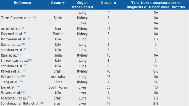
can be seen in Table 1.
(26,29,31,32)Pulmonary tuberculosis occurs most commonly in
men, who accounted for 65% and 72% of the sample,
respectively, in the two most representative studies.
(26,29)The majority of patients with pulmonary tuberculosis
were between the fourth and sixth decades of life. The
diagnosis of pulmonary tuberculosis was made 3-12
months after transplantation (Table 1).
The incidence of tuberculosis cases in relation to
the number of transplants of a given organ at each
institution ranged from 0.09% to 4.7% of the cases,
with a mean incidence of 1.12%. We identified 53
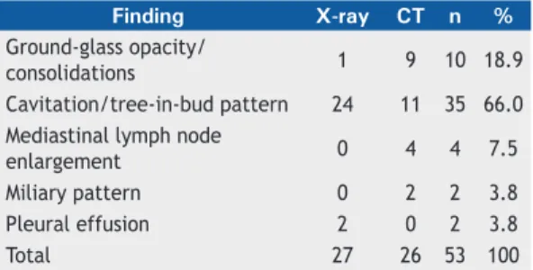
cases of pulmonary tuberculosis among lung transplant
recipients, with a predominance of the cavitation/
tree-in-bud pattern in 35 (66%) of the cases (Table 2).
The largest patient sample was composed of kidney
transplant recipients (96 patients); the most common
pattern was cavitation and centrilobular nodules in a
tree-in-bud pattern, which was seen in approximately
one third of those patients, followed by the “lymph
node enlargement” and “pleural effusion” categories,
which together also accounted for a third of the cases
(Table 3). We identified 51 liver transplant recipients,
62% of whom had cavitation and centrilobular nodules
in a bud-tree pattern (Table 4).
DISCUSSION
To our knowledge, this is the first systematic review
of chest imaging findings in solid organ transplant
recipients diagnosed with pulmonary tuberculosis. A
collective total of 219 cases were analyzed. Among the
219 cases analyzed, 96, 70, and 53 were in kidney,
liver, and lung transplant recipients, respectively.
This proportional distribution of cases of pulmonary
tuberculosis probably reflects the similar proportional
distribution of transplantations by organ.
In the articles selected (i.e., those that contained
imaging findings), the prevalence of tuberculosis
among transplant recipients ranged from 0.09% to
4.7%, thus not representing the total number of cases,
which was not the purpose of the present study. These
proportions allow us to infer only an approximate value
for the true prevalence.
Table 1. Data obtained from the case series selected in the present systematic review.
Reference
Country
Organ
transplanted
Cases, n
Time from transplantation to
diagnosis of tuberculosis, months
Torre-Cisneros et al.
(5)Spain
Lung
4
NA
Kidney
6
NA
Liver
7
NA
Aslani et al.
(18)Iran
Kidney
16
NA
Kaaroud et al.
(19)Tunisia
Kidney
6
NA
Mortensen et al.
(20)USA
Lung
3
3.7
Kesten et al.
(21)USA
Lung
2
3
Schulma et al.
(22)USA
Lung
2
3
Ram et al.
(23)India
Kidney
16
NA
Shreeniwas et al.
(24)USA
Lung
1
3
Schulma et al.
(25)USA
Lung
2
11
Pereira et al.
(26)Brazil
Kidney
40
8.6
Malouf et al.
(27)Australia
Lung
12
NA
Jiang et al.
(28)China
Kidney
7
12
Lyu et al.
(29)South Korea
Liver
35
10
Meyers et al.
(30)USA
Liver
9
NA
Giacomelli et al.
(31)Brazil
Lung
19
3.2
Schuhmacher Neto et al.
(32)Brazil
Liver
19
2.6
The studies with the highest numbers of patients
included were conducted in Brazil, South Korea,
India, and Iran. It is necessary to emphasize that
tuberculosis is endemic only in certain countries,
unlike most other opportunistic diseases, which are
ubiquitous. The incidence of pulmonary tuberculosis
among transplant recipients will always be related to
the incidence of tuberculosis in the region in which
the patient and donor reside.
Among the patient samples evaluated, there was a
predominance of men, with an approximate male:female
ratio of 2:1. However, it should be borne in mind that
approximately half of the articles did not provide
demographic data or extrapolated them to diseases
other than tuberculosis and were therefore not included.
The majority of the patients were between the fourth
and sixth decades of life. A complete evaluation of
demographic findings might be more appropriate if
all articles on solid organ transplant recipients with
tuberculosis were evaluated, rather than only those
including chest imaging findings.
Data on the time from transplantation to the diagnosis
of tuberculosis were present in approximately half of
the studies of lung or kidney transplant recipients and
most of those of liver transplant recipientsThe time from
transplantation to the diagnosis of tuberculosis ranged
from 3 to 11 months for lung transplant recipients,
with medians between 3 and 4 months, compared
with 8-12 months for kidney transplant recipients and
2.6-12 months for liver transplant recipients.
Approximately 66% of the lung transplant recipients
with tuberculosis showed a typical pattern of pulmonary
involvement (cavitations and the tree-in-bud pattern),
atypical patterns occurring in approximately one third
of cases. Unlike the lung transplant recipients, only 34
of the 96 kidney transplant recipients—approximately
one third—showed the classic presentation of pulmonary
tuberculosis, whereas approximately one quarter showed
a miliary presentation and approximately one third
showed a predominance of lymph node enlargement
or pleural effusion.
Most (76%) of the data obtained for kidney transplant
recipients were from CT findings, as were all (100%)
of the data obtained for liver transplant recipients. In
the latter case, the majority (67.2%) had the typical
presentation of pulmonary tuberculosis. None of the
liver transplant recipients showed a predominance of
pleural effusion.
In patients with tuberculosis/HIV coinfection,
pulmonary tuberculosis was found to be most often
accompanied by lymph node enlargement and
miliary disease.
(46)Hilar and mediastinal lymph node
enlargement occurred in 60% of such patients.
(47,48)On the basis of our results, we can infer that the
presentation of pulmonary tuberculosis in kidney
transplant recipients tend to be most similar to that
of patients with tuberculosis/HIV coinfection (i.e., a
greater proportion of cases of lymph node enlargement
and miliary involvement); the same was not true
for lung and liver transplant recipients, in whom the
presentation tended to be more similar to that seen
in the general population.
In the articles selected for this review, there were
other presentations of pulmonary tuberculosis not
defined in the classification described in the methods
section. Boedefeld et al.
(35)reported a case of pulmonary
tuberculosis accompanied by pericardial involvement.
There were also two reported cases of transplant
recipients presenting with pulmonary tuberculosis in
the form of masses.
(39,45)There were reports of pulmonary tuberculosis in solid
organ transplant recipients with normal chest X-rays
findings, as well as in healthy patients, with incidence
rates that vary widely across studies. Lyu et al.
(29)also
identified patients with normal chest CT findings who
developed pulmonary tuberculosis. Therefore, normal
chest X-rays do not exclude a diagnosis of pulmonary
tuberculosis in solid organ transplant recipients. The
incidence of that combination might be better evaluated
in clinical studies of tuberculosis in this population. In a
case report, Carlsen et al.
(42)stated that the presence
of calcified mediastinal lymph nodes can raise the
suspicion of a diagnosis of tuberculosis.
Table 2. Chest X-ray and chest CT findings in lung transplant recipients.
Finding
X-ray
CT
n
%
Ground-glass opacity/
consolidations
1
9
10 18.9
Cavitation/tree-in-bud pattern
24
11
35 66.0
Mediastinal lymph node
enlargement
0
4
4
7.5
Miliary pattern
0
2
2
3.8
Pleural effusion
2
0
2
3.8
Total
27
26
53
100
Table 3. Chest X-ray and chest CT findings in kidney transplant recipients.
Finding
X-ray
CT
n
%
Ground-glass opacity/
consolidations
0
9
9
9.38
Cavitation/tree-in-bud pattern
5
29
34 35.4
Mediastinal lymph node
enlargement
6
8
14 14.6
Miliary pattern
1
22
23
24
Pleural effusion
11
5
16 16.7
Total
23
73
96
100
Table 4. Chest X-ray and chest CT findings in liver transplant recipients.
Finding
X-ray
CT
n
%
Ground-glass opacity/
consolidations
0
1
1
1.4
Cavitation/tree-in-bud pattern
0
47
47 67.2
Mediastinal lymph node
enlargement
0
10
10 14.3
Miliary pattern
0
12
12 17.1
Total
0
70
70
100
In summary, the majority of lung and liver transplant
recipients with pulmonary tuberculosis show the classic
cavitation and tree-in-bud nodular presentation (66.0
and 67.2%, respectively). However, that presentation
is seen in only one third of kidney transplant recipients
with pulmonary tuberculosis, in whom the presentation is
similar to that seen in patients coinfected with tuberculosis
and HIV. Studies evaluating sociodemographic differences
and, in particular, the immunosuppressive regimen could
help identify new hypotheses for the predominance of
the atypical presentation of pulmonary tuberculosis in
kidney transplant recipients.
REFERENCES
1. World Health Organization [homepage on the Internet]. Geneva: World Health Organization; C2016 [cited 2016 Aug 7]. Media centre: Tuberculosis; [about 8 screens]. Available from: http://www.who.int/ mediacentre/factsheets/fs104/en/
2. Brasil. Ministério da Saúde. Secretaria de Vigilância em Saúde. [homepage on the Internet]. Brasília: Ministério da Saúde; [cited 2016 Jun 10]. Tuberculose - 2015: Detectar, tratar e curar: desafios e estratégias brasileiras frente à tuberculose. Boletim Epidemiológico. 2015;46(09). [Adobe Acrobat document, 19p.]. Available from: http:// portalarquivos2.saude.gov.br/images/pdf/2015/marco/27/2015-007---BE-Tuberculose---para-substitui----o-no-site.pdf
3. Subramanian A, Dorman S; AST Infectious Diseases Community of Practice. Mycobacterium tuberculosis in solid organ transplant recipients. Am J Transplant. 2009;9 Suppl 4:S57-62. https://doi. org/10.1111/j.1600-6143.2009.02894.x
4. Singh N, Paterson DL. Mycobacterium tuberculosis infection in solid-organ transplant recipients: impact and implications for management. Clin Infect Dis. 1998;27(5):1266-77. https://doi.org/10.1086/514993
5. Torre-Cisneros J, Doblas A, Aguado JM, San Juan R, Blanes M, Montejo M, et al. Tuberculosis after solid-organ transplant: incidence, risk factors, and clinical characteristics in the RESITRA (Spanish Network of Infection in Transplantation) cohort. Clin Infect Dis. 2009;48(12):1657-65. https://doi.org/10.1086/599035
6. Kiyono K, Sone S, Sakai F, Imai Y, Watanabe T, Izuno I, et al. The number and size of normal mediastinal lymph nodes: a postmortem study. AJR Am J Roentgenol. 1988;150(4):771-6. https://doi. org/10.2214/ajr.150.4.771
7. Higgins JPT, Green S, editors. Cochrane Handbook for Systematic Reviews of Interventions Version 5.1.0 [updated March 2011]. The Cochrane Collaboration; 2011.
8. Moher D, Liberati A, Tetzlaff J, Altman DG; PRISMA Group. Preferred reporting items for systematic reviews and meta-analyses: the PRISMA statement. PLoS Med. 2009;6(7):e1000097. https://doi. org/10.1371/journal.pmed.1000097
9. Hansell DM, Bankier AA, MacMahon H, McLoud TC, Müller NL, Remy J. Fleischner Society: glossary of terms for thoracic imaging. Radiology. 2008;246(3):697-722. https://doi.org/10.1148/ radiol.2462070712
10. Kalra V, Agarwal SK, Khilnani GC, Kapil A, Dar L, Singh UB, et al. Spectrum of pulmonary infections in renal transplant recipients in the tropics: a single center study. Int Urol Nephrol. 2005;37(3):551-9. https://doi.org/10.1007/s11255-005-4012-9
11. Rathi M, Gundlapalli S, Ramachandran R, Mohindra S, Kaur H, Kumar V, et al. A rare case of Cytomegalovirus, Scedosporium apiospermum and Mycobacterium tuberculosis in a renal transplant recipient. BMC Infect Dis. 2014;14:259. https://doi.org/10.1186/1471-2334-14-259
12. Krayem AB, Abdullah LS, Raweuily EA, Wali SO, Rawas MM, Samman YS, et al. The diagnostic challenge of pulmonary Kaposi’s sarcoma with pulmonary tuberculosis in a renal transplant recipient: a case report. Transplantation. 2001;71(10):1488-91. https://doi. org/10.1097/00007890-200105270-00024
13. Tabarsi P. Farshidpour M, Marjani M, Baghaei P, Yoisefzadeh A, Najafizadeh K, et al. Mycobacterial infection and the impact of rifabutin treatment in organ transplant recipients: a single-center study. Saudi J Kidney Dis Transpl. 2015;26(1):6-11. https://doi. org/10.4103/1319-2442.148710
14. Schultz V, Marroni CA, Amorim CS, Baethgen LF, Pasqualotto AC. Risk factors for hepatotoxicity in solid organ transplants recipients being treated for tuberculosis. Transplant Proc. 2014;46(10):3606-10. https://doi.org/10.1016/j.transproceed.2014.09.148
15. Singh N, Patterson DL. Mycobacterium tuberculosis infection in solid-organ transplant recipients: impact and implications for management. Clin Infect Dis. 1998;27(5):1266-77. https://doi.org/10.1086/514993
16. Lopez de Castilla D, Schluger NW. Tuberculosis following solid organ transplantation. Transpl Infect Dis. 2010;12(2):106-12. https://doi.
org/10.1111/j.1399-3062.2009.00475.x
17. Eyüboğlu FÖ, Küpeli E, Bozbaş SS, Ozen ZE, Akkurt ES, Aydoğan C, et al. Evaluation of pulmonary infections in solid organ transplant recipients: 12 years of experience. Transplant Proc. 2013;45(10):3458-61. https://doi.org/10.1016/j.transproceed.2013.09.024
18. Aslani J, Einollahi B. Prevalence of tuberculosis after renal transplantation in Iran. Transplant Proc. 2001;33(5):2804-5. https:// doi.org/10.1016/S0041-1345(01)02197-2
19. Kaaroud H, Beji S, Boubaker K, Abderrahim E, Ben Hamida F, Ben Abdallah TB, et al. Tuberculosis after renal transplantation. Transplant Proc. 2007;39(4):1012-3. https://doi.org/10.1016/j. transproceed.2007.02.032
20. Mortensen E, Hellinger W, Keller C, Cowan LS, Shaw T, Hwang S, et al. Three cases of donor-derived pulmonary tuberculosis in lung transplant recipients and review of 12 previously reported cases: opportunities for early diagnosis and prevention. Transpl Infect Dis. 2014;16(1):67-75. https://doi.org/10.1111/tid.12171
21. Kesten S, Chaparro C. Mycobacterial infections in lung transplant recipients. Chest. 1999;115(3):741-5. https://doi.org/10.1378/ chest.115.3.741
22. Schulma LL, Htun T, Staniloae C, McGregor CC, Austin JH. Pulmonary nodules and masses after lung and heart-lung transplantation. J Thorac Imaging. 2000;15(3):173-9. https://doi.org/10.1097/00005382-200007000-00004
23. Ram R, Swarnalatha G, Prasad N, Dakshinamurty KV. Tuberculosis in renal transplant recipients. Transpl Infect Dis. 2007;9(2):97-101. https://doi.org/10.1111/j.1399-3062.2006.00182.x
24. Shreeniwas R, Schulman LL, Berkmen YA, McGregor CC, Austin JH. Opportunistic bronchopulmonary infections after lung transplantation: clinical and radiographic findings. Radiology. 1996;200(2):349-56. https://doi.org/10.1148/radiology.200.2.8685324
25. Schulma LL, Scully B, McGregor CC, Austin JH. Pulmonary tuberculosis after lung transplantation. Chest. 1997;111(5):1459-62. https://doi.org/10.1378/chest.111.5.1459
26. Pereira M, Gazzoni FF, Marchiori E, Irion K, Moreira J, Giacomelli IL, et al. High-resolution CT findings of pulmonary Mycobacterium tuberculosis infection in renal transplant recipients. Br J Radiol. 2016;89(1058):20150686. https://doi.org/10.1259/bjr.20150686
27. Malouf MA, Glanville AL. The spectrum of mycobacterial infection after lung transplantation. Am J Respir Crit Care Med. 1999;160(5 Pt 1):1611-6. https://doi.org/10.1164/ajrccm.160.5.9808113
28. Jiang T, Xue F, Zheng X, Yu H, Tao X, Xiao X, et al. Clinical data and CT findings of pulmonary infection caused by different pathogens after kidney transplantation. Eur J Radiol. 2012;81(6):1347-52. https://doi. org/10.1016/j.ejrad.2011.03.070
29. Lyu J, Lee SG, Hwang S, Lee SO, Cho OH, Chae EJ, et al. Chest computed tomography is more likely to show latent tuberculosis foci than simple chest radiography in liver transplant candidates. Liver Transpl. 2011;17(8):963-8. https://doi.org/10.1002/lt.22319
30. Meyers BR, Papanicolaou GA, Sheiner P, Emre S, Miller C. Tuberculosis in orthotopic liver transplant patients: increased toxicity
of recommended agents; cure of disseminated infection with
nonconventional regimens. Transplantation. 2000;69(1):64-9. https:// doi.org/10.1097/00007890-200001150-00013
31. Giacomelli IL, Schuhmacher Neto R, Nin CS, Cassano PS, Pereira M1, Moreira JDS, et al. High-resolution computed tomography findings of pulmonary tuberculosis in lung transplant recipients. J Bras Pneumol. 2017;43(4):270-273. https://doi.org/10.1590/s1806-37562016000000306
32. Schuhmacher Neto R, Giacomelli IL, Schuller Nin C, da Silva Moreira J, Comaru Pasqualotto A, Marchiori E, et al. High-resolution CT findings of pulmonary tuberculosis in liver transplant patients. Clin Radiol. 2017;72(10):899.e9-899.e14. https://doi.org/10.1016/j. crad.2017.05.006
33. Winthrop KL, Kubak BM, Pegues DA, Hufana C, Costamagna P,
Desmond E, et al. Transmission of mycobacterium tuberculosis via lung transplantation. Am J Transplant. 2004;4(9):1529-33. https://doi. org/10.1111/j.1600-6143.2004.00536.x
34. Ardalan MR, Shoja MM, Ghabili K. Concomitant pulmonary tuberculosis and tuberculous appendicitis in a recipient of a renal transplant: a case report. J Med Case Rep. 2011;5:191. https://doi. org/10.1186/1752-1947-5-191
35. Boedefeld RL, Eby J, Boedefeld WM 2nd, Stanley D, Lau LC, Kern JA, et al. Fatal Mycobacterium tuberculosis infection in a lung transplant recipient. J Heart Lung Transplant. 2008;27(10):1176-8. https://doi.org/10.1016/j.healun.2008.07.009
36. Shitrit D, Bendayan D, Saute M, Kramer MR. Multidrug resistant tuberculosis following lung transplantation: treatment with pulmonary resection. Thorax. 2004;59(1):79-80.
37. Miller RA, Lanza LA, Kline JN, Geist LJ. Mycobacterium tuberculosis in lung transplant recipients. Am J Respir Crit Care Med. 1995;152(1):374-6. https://doi.org/10.1164/ajrccm.152.1.7599848
38. Place S, Knoop C, Remmelinsk M, Baldassarre S, Van Vooren JP, Jacobs F, et al. Paradoxical worsening of tuberculosis in a heart-lung transplant recipient. Transpl Infect Dis. 2007;9(3):219-24. https://doi. org/10.1111/j.1399-3062.2006.00194.x
39. Lee J. Yew WW, Wong CF, Wong PC, Chiu CS. Multidrug-resistant tuberculosis in a lung transplant recipient. J Heart Lung Transplant. 2003;22(10):1168-73. https://doi.org/10.1016/S1053-2498(02)01189-0
40. Kumar D, Budev M, Koval C, Hellinger WC, Gordon SM, Tomford JW. Donor-derived tuberculosis (TB) infection in lung transplant despite following recommended algorithm. Am J Transplant.
2013;13(8):2225-6. https://doi.org/10.1111/ajt.12344
41. Kukrej N, Cook GJ, Pattison JM. Positron-emission tomography used to diagnose tuberculosis in a renal transplant patient. Am J Transplant. 2002;2(1):105-7. https://doi.org/10.1034/j.1600-6143.2002.020117.x
42. Carlsen SE, Bergin CJ. Reactivation of tuberculosis in a donor lung after transplantation. AJR Am J Roentgenol. 1990;154(3):495-7. https://doi.org/10.2214/ajr.154.3.2106211
43. Wong KK, Lim ST, Yeung CK, Ng WL, Ong GB. Disseminated tuberculosis in a renal transplant recipient. Aust N Z J Surg. 1983;53(2):173-5. https://doi.org/10.1111/j.1445-2197.1983.tb02422.x
44. Duggal R, Rajwanshi A, Gupta N, Lal A, Singhal M. Polymicrobial lung infection in postrenal transplant recipient diagnosed by fine-needle aspiration cytology. Diagn Cytopathol. 2010;38(4):294-6.
45. Tan BH, Cheah FK, Chew S, Ahmed Q. A renal transplant recipient with pulmonary nodules. Transpl Infect Dis. 2005;7(1):18-25. https:// doi.org/10.1111/j.1399-3062.2005.00080.x
46. Saurborn DP, Fishman JE, Boiselle PM. The imaging spectrum of pulmonary tuberculosis in AIDS. J Thorac Imaging. 2002;17(1):28-33. https://doi.org/10.1097/00005382-200201000-00003
47. Castañer E, Gallardo X, Mata JM, Esteba L. Radiologic approach to the diagnosis of infectious pulmonary diseases in patients infected with the human immunodeficiency virus. Eur J Radiol. 2004;51(2):114-29. https://doi.org/10.1016/j.ejrad.2004.03.008