Dieulafoy’s disease of the bronchial tree: a case report
Doença de Dieulafoy da árvore brônquica: relato de caso
Massoud Baghai Wadji
I, Athena Farahzadi
IIFiruzgar Hospital, Iran University of Medical Sciences, Tehran, Iran
ABSTRACT
CONTEXT: Dieulafoy’s disease of the bronchial tree is a very rare condition. Few cases have been reported in the literature. It can be asymptomatic or manifest with massive hemoptysis. This disease should be considered among heavy smokers when recurrent massive hemoptysis is present amid otherwise normal indings. The treatment can be arterial embolization or surgical intervention.
CASE REPORT: A 16-year-old girl was admitted to the emergency department due to hemoptysis with an unknown lesion in the bronchi. She had sufered massive hemoptysis and respiratory failure one week be-fore admission. Fiberoptic bronchoscopy revealed a lesion in the bronchus of the right lower lobe, which was suspected to be a Dieulafoy lesion. Segmentectomy of the right lower lobe and excision of the lesion was carried out. The outcome for this patient was excellent.
CONCLUSION: Dieulafoy’s disease is a rare vascular anomaly and it is extremely rare in the bronchial tree. In bronchial Dieulafoy’s disease, selective embolization has been suggested as a method for cessation of bleeding. Nevertheless, standard anatomical lung resection is a safe and curative alternative.
RESUMO
CONTEXTO: A doença de Dieulafoy da árvore brônquica é uma condição muito rara, poucos casos foram descritos na literatura. Pode ser assintomática ou manifestar-se com hemoptise maciça. Esta doença deve ser considerada em fumadores pesados quando eles têm recorrentes hemoptises maciças sem outros achados anormais. O tratamento pode ser tanto embolização arterial como intervenção cirúrgica.
RELATO DE CASO: Uma menina de 16 anos foi admitida no Serviço de Urgências devido a hemoptise com uma lesão nos brônquios de origem desconhecida. Havia sofrido hemoptise maciça e insuiciência respiratória uma semana antes da admissão. A broncoscopia de ibra óptica relevou lesão no brônquio do lobo inferior direito, com suspeita de ser lesão de Dieulafoy. Foi realizada uma segmentectomia do lobo inferior direito com excisão da lesão. O resultado da paciente foi excelente.
CONCLUSÃO: A doença de Dieulafoy é uma anomalia vascular rara, sendo extremamente rara na árvore brônquica. Na doença de Dieulafoy bronquial, embolização seletiva tem sido sugerida como método para cessação do sangramento; no entanto, a habitual resseção anatômica do pulmão é uma alternativa segura e curativa.
IMD. Associate Professor of Surgery, Firuzgar Hospital, Iran University of Medical Sciences, Tehran, Iran.
IIMD. Resident of General Surgery, Iran University of Medical Sciences, Rasool Akram Hospital, Shahrara, Tehran, Iran.
KEY WORDS: Dieulafoy disease. Bronchi. Hemoptysis. Pulmonary artery. Lung lobectomy.
INTRODUCTION
Dieulafoy’s disease of the bronchial tree is a very rare disease. Few cases have been reported in the literature. It can be asymp-tomatic or can manifest with massive hemoptysis. his disease should be considered among heavy smokers with recurrent mas-sive hemoptysis.1
he diagnosis can be conirmed by means of bronchoscopy, which shows aberrant arterial bleeding in the bronchial tree. Imaging, consisting of either normal chest X-ray or chest com-puted tomography (CT) scan, can be helpful in making the diag-nosis, through ruling out other causes of hemoptysis.
he treatment usually comprises arterial embolization. If this method is unavailable or unsuccessful, surgery can be another option for achieving a deinitive cure.
Here, we report a case of Dieulafoy’s disease in a girl who pre-sented with massive hemoptysis, which was diagnosed by means of bronchoscopy and treated through segmentectomy.
CASE REPORT
A 16-year-old nonsmoking girl was referred to our hospital because of an episode of massive hemoptysis. She had been admit-ted to a local hospital one week earlier because of this symptom and had developed respiratory failure, requiring mechanical ven-tilation for two days. Ater extubation and cessation of bleeding, she was referred to our hospital for further evaluation.
On admission to the thoracic surgery department, she was conscious and extubated, without respiratory distress, but mildly anxious. Her vital signs were stable and she was afebrile. Physical examination on the head and neck, chest, abdomen and extremities showed that these were normal. Oxygen saturation in the ambient air was 98%. here was no longer any hemoptysis.
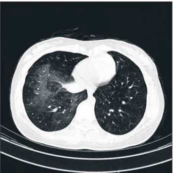
Laboratory data including white blood cell (WBC) and plate-let counts, hemoglobin and hematocrit, prothrombin time, par-tial thromboplastin time (PTT) and international normalized ratio (INR) were within normal limits. A chest X-ray was normal, while chest CT scans showed some patchy haziness in the right lower lobe and a very small lesion in the distal bronchus interme-dius (Figures 1 and 2). he imaging did not show any atelecta-sis, honeycomb appearance, cavitation, consolidation or tumoral lesion. Common causes of massive bleeding like bronchiectasis, carcinoid tumor, tuberculosis, arteriovenous (AV) malforma-tions and other condimalforma-tions were less likely to be the reason for the bleeding in this girl.
On the next day, iberoptic bronchoscopy was performed and this showed a lesion at the beginning of the bronchus of the basal segments of the right lower lobe, without evidence of active bleeding. he lesion originated from the mucosal surface, with a small clot over it. he mucosa surrounding the lesion was abso-lutely normal (Figure 3). No biopsy was taken, because of the
suspicion of Dieulafoy’s disease and the risk of bleeding. Given the lack of expertise in bronchial angiography and embolization at our center, we preferred surgical treatment. herefore, within an elective setting and ater hemorrhaging had ceased, basal seg-mentectomy of the right lower lobe was carried out in a planned manner, by means of right lateral thoracotomy. he superior seg-ment of the right lower lobe remained intact (Figure 4). here was
Figure 1. Chest X-ray showing nearly normal lung ield.
Figure 2. Computed tomography scan of the chest
no intraoperative inding except for consolidation of the paren-chyma of the diseased lobe, most probably due to hemorrhage. he operation was performed without any diiculty because of normal anatomical integrity.
An intraoperative frozen section study was negative for any malignant condition.
he patient had a very smooth and uneventful postoperative course, in which she only presented pain, which could be con-trolled with ordinary analgesics. She was discharged on the sixth postoperative day. At an outpatient visit one week later, she did not have any serious complaint. Moreover, in the third, sixth and eighteenth months of follow-up, she was still asymptomatic with-out recurrence of any kind of hemoptysis.
Although the diagnosis of this disease was clinical, further pathological studies showed few dilated vessels in the submu-cosa. his was compatible with a diagnosis of Dieulafoy’s disease (Figure 5).
DISCUSSION
Dieulafoy’s disease is a rare vascular anomaly consisting of a dys-plastic artery in the submucosa. It is mostly seen in the gastroin-testinal tract and is extremely rare in the bronchial tree. To the best of our knowledge, there are only a few reports of Dieulafoy’s disease of the bronchial tree in the English-language literature (Tables 1 and 21-8). Accordingly, the natural history of this disease
and the preferred treatment are not known well. On the other hand, the mortality rate in the absence of any treatment rises to more than 50%.1
he pathogenesis of this disease is also unclear, but most reports state that it occurs in heavy smokers and presents with massive and recurrent hemoptysis. Dieulafoy’s disease of the bronchus may have a congenital origin, arising from either the systemic or the pulmo-nary circulation.2 Spontaneous bleeding has been described in these
cases, but bleeding in such cases oten occurs ater a biopsy on a lesion that has not been diagnosed as a vascular anomaly. Age and tobacco use have an inluence on occurrences of this disease.1,3
Dieulafoy’s disease can be suspected when there is severe or massive hemoptysis in the absence of any signiicant abnormality
Figure 4. Right thoracic cavity after basal segmentectomy on the right lower lobe. The arrow shows upper segment of right lower lobe.
Figure 5. Histological section through bronchial Dieulafoy lesion (arrow: dilated hypertrophic submucosal artery; 2.5 X magniication, hematoxylin and eosin staining).
on either chest X-ray or chest CT scan and in the absence of any medical or surgical history, as in our patient’s case. Bronchoscopy, preferably using a iberoptic when the bleeding is not severe, may be diagnostic. It will usually make it possible to ind both the source and the cause of the bleeding.3 he characteristics of the lesion
are nonspeciic, but it can be suspected when a small (usually less than 1 cm) sessile non-pulsatile nodular lesion with a white cap and apparently normal mucosa is seen.2
It has been suggested that, ater the diagnosis has been made, angiography and embolization can be the preferred treatment5,6
and that surgical resection would only be needed in a few cases.4
However, the failure rate of embolization is not negligible, whereas surgery alone or ater failure of embolization has had a success rate of nearly 100% in all reports.7 Nevertheless, angioembolization is
less invasive than surgery, and both physicians and patients pre-fer it as the irst attempt to halt the bleeding. In the event of sur-gical intervention, since the lesion is usually located in a lobar or segmental bronchus, the surgery should be carried out as an ana-tomical segmentectomy or lobectomy. Alternatively, bronchoplas-tic procedures can be performed if the lesion is located in a major bronchus. here is a lack of long-term follow-up in the reports on patients who have undergone embolization alone.7
Although the bleeding recurrence rate in patients whose hem-orrhaging has stopped spontaneously is not known, physicians cannot take the risk of not initiating any interventions. If selective embolization is unavailable or if it fails, surgery can be lifesaving.
Even in patients whose bleeding stops spontaneously, surgery can have a role in prevention of life-threatening hemoptysis.
CONCLUSION
Dieulafoy’s disease is a rare vascular anomaly and is extremely rare in the bronchial tree. It should be considered as a diagnosis when there is severe or massive hemoptysis in an otherwise nor-mal patient who has nearly nornor-mal chest imaging. Bronchoscopy is diagnostic. In bronchial Dieulafoy’s disease, selective emboli-zation has been suggested as a method for cessation of bleeding. When angioembolization fails or is unavailable, surgical resec-tion consisting of either segmentectomy or lobectomy can be life-saving for these patients.
REFERENCES
1. Barisione EE, Ferretti GG, Ravera SS, Salio MM. Dieulafoy’s disease of the
bronchus: a possible mistake. Multidiscip Respir Med. 2012;7(1):40.
2. Fang Y, Wu Q, Wang B. Dieulafoy’s disease of the bronchus: report of a
case and review of the literature. J Cardiothorac Surg. 2014;9:191.
3. Smith B, Hart D, Alam N. Dieulafoy’s disease of the bronchus: a rare
cause of massive hemoptysis. Respirol Case Rep. 2014;2(2):55-6.
4. Savale L, Parrot A, Khalil A, et al. Cryptogenetic hemoptysis: from a
benign to a life-threatening pathological vascular condition. Am J
Respir Crit Care Med. 2007;175(11):1181-5.
5. Bhatia P, Hendy MS, Li-Kam-Wa E, Bowyer PK. Recurrent embolotherapy
in Dieulafoy’s disease of the bronchus. Can Respir J. 2003;10(6):331-3.
Table 1. Articles relating to Dieulafoy’s disease that were found through searching the medical literature databases (November 22, 2016)
Database Search strategies Papers found Related papers
MEDLINE (via PubMed
((Dieulafoy’s disease) OR Dieulafoys OR Dieulafoy)) AND
((“Bronchial Diseases”[Mesh]) OR (Bronchial Disease) OR (Bronchial Disease) OR (Disease, Bronchial) OR (Diseases, Bronchial) OR (Bronchial tree) OR Bronchus)
Filters: Case Reports
17 8
LILACS (via BVS) Dieulafoy 31 0
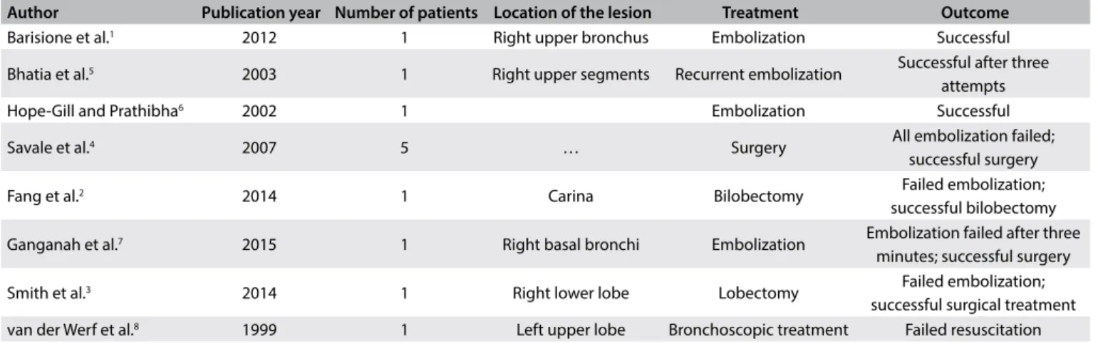
Table 2. A review of Dieulafoy’s disease reported in the medical literature
Author Publication year Number of patients Location of the lesion Treatment Outcome
Barisione et al.1 2012 1 Right upper bronchus Embolization Successful
Bhatia et al.5 2003 1 Right upper segments Recurrent embolization Successful after three attempts
Hope-Gill and Prathibha6 2002 1 Embolization Successful
Savale et al.4 2007 5 … Surgery All embolization failed;
successful surgery
Fang et al.2 2014 1 Carina Bilobectomy Failed embolization;
successful bilobectomy
Ganganah et al.7 2015 1 Right basal bronchi Embolization Embolization failed after three minutes; successful surgery
Smith et al.3 2014 1 Right lower lobe Lobectomy Failed embolization;
6. Hope-Gill B, Prathibha BV. Bronchoscopic and angiographic indings
in Dieulafoy’s disease of the bronchus. Hosp Med. 2002;63(3):178-9.
7. Ganganah O, Guo S, Chiniah M, Sah SK, Wu J. Endobronchial ultrasound
and bronchial artery embolization for Dieulafoy’s disease of the bronchus
in a teenager: A case report. Respir Med Case Rep. 2015;16:20-3.
8. van der Werf TS, Timmer A, Zijlstra JG. Fatal haemorrhage from Dieulafoy’s
disease of the bronchus. Thorax. 1999;54(2):184-5.
Acknowledgement: We would like to thank Dr. Shakira Ghafoor for
editing the manuscript and Dr. Francisco Ferreira e Silva for translating
the title, abstract and keywords into Portuguese
Conlict of interest: None
Sources of funding: None
Date of irst submission: September 30, 2016
Last received: November 11, 2016
Accepted: November 19, 2016
Address for correspondence:
Massoud Baghai-Wadji
Iran University of Medical Sciences, Firuzgar Hospital
Karimkhan St. Beh Afarin Str. Tehran, Iran
Tel. 989131415262