Received June 14, 2010 and accepted July 11, 2011. Corresponding author: lutakada@gmail.com
Ovarian response of Suffolk ewes to estrous synchronization using
short-term protocol
Luciana Takada1, Sony Dimas Bicudo1, Carlos Frederico de Carvalho Rodrigues2, Lia de Alencar Coelho3, Luiz Claudio Nogueira Mendes4, Sílvia Helena Venturoli Perri4
1UNESP/FMVZ – Faculdade de Medicina Veterinária e Zootecnia, Departamento de Reprodução Animal, Distrito de Rubião Júnior
s/n, 18618-970 – Botucatu, SP, Brazil.
2Unidade de Pesquisa e Desenvolvimento, Agência Paulista de Tecnologia dos Agronegócios, Rodovia Gladys Bernades Minhoto, Km 62,
18200-970 – Itapetininga, SP, Brazil.
3USP/FZEA – Departamento de Zootecnia, Av. Duque de Caxias Norte, 225 – 13635-900 – Pirassununga, SP, Brazil.
4UNESP/FMVA – Faculdade de Medicina Veterinária de Araçatuba, R. Clóvis Pestana, 793 – 16050-680 – Araçatuba, SP, Brazil
ABSTRACT - The efficacy of estrus synchronization using short-term protocol was evaluated by ultrasound exams in Suffolk ewes during the pre-breeding season. The control Group (n = 12) was synchronized by treatment for 12 days with vaginal sponges impregnated with medroxyprogesterone acetate, and 400 IU eCG at sponge withdrawal. Experimental groups I, II and III kept the sponge in place for 4 days, and 100 µg of PGF2αwas administered at sponge withdrawal. Additionally, Group I (n = 12) had 0.1 mg of estradiol benzoate (EB) administered during sponge placement and 50 µg of GnRH 48 hours after sponge removal. Group II (n = 6) had 35 mg of progesterone (P4) injected, and 0.1 mg of EB administered during sponge placement, 400 IU eCG at withdrawal and 48 hours after, 50 µg GnRH were administrated. Group III (n = 12) had 35 mg of P4 and 0.2 mg of EB administered at sponge placement, 400 IU eCG at withdrawal, and 50 µg of GnRH was administrated after 56 hours. Ovaries were monitored through ultrasound scanning. Concerning the first wave, no difference was detected between the control group and the experimental groups. However, the characteristics of ovulatory wave were significantly different between the groups. The duration of the follicular wave was shorter for Group III than for Group II. The follicle in Group I reached its maximum diameter before the Group II. The diameter of the follicle at the sponge withdrawal in the control group was larger than in Group I. After sponge withdrawal, the follicular growth rate was smaller in the control group than in Group III. The maximum diameter of the follicle in Group II was larger than in the other groups.The short-term protocol in which estrogen was used did not synchronize the emergence of the wave of follicular development.
Key Words: eCG, estradiol benzoate, follicular dynamics, progesterone www.sbz.org.br
Introduction
The induction of estrus in ewes during seasonal anoestrous was accomplished with intravaginal sponges impregnated with progestagens, medroxyprogesterone acetate (MAP) (Evans et al., 2004), inserted from 12 to 14 days. Subsequently, equine chorionic gonadotropin (eCG) was administered (Zeleke et al., 2005). Nevertheless, the fertility of anestrous ewes treated with progestagen ranged from 22 to 70% (Evans et al., 2001).
Long term progestagen estrus synchronization protocols can affect follicular dynamics and fertility of ewes (Viñoles et al., 2001; Diskin et al., 2002). Initially, a supraluteal effect is expected, which means that an increase in follicular renewal may occur. In the end, however, a subluteal effect may happen and decrease the speed of follicular renewal (Hamra et al., 1986). The use of progestagen during a short
time would be an alternative solution because it could improve estrus synchronization in ewes (Husein et al., 2007).
A combination of progestagen with eCG was used in ewes for 6 days and resulted in good fecundity. It was enough to induce and synchronize estrus in approximately 90% of the animals (Ataman et al., 2006).
et al., 2006). The interval between the estrogen injection and the recruitment of a new wave depends on the dose administered (Martínez et al., 2005).
In order to develop methods for the estrous cycle control, the aim of the present study is to evaluate the effects of exogenous hormones during the estrous cycle in ewes. Short-term protocols were used and estradiol benzoate was associated with progestagen, prostaglandin analogue, eCG and GnRH. Ewes were monitored by ultrasound scanning.
Material and Methods
The experiment was performed in accordance with the ethical standards and approved by the Comitê de Ética em Pesquisa (Research Ethics Committee) in Botucatu FMVZ-UNESP. The experiment was carried out during the pre-breeding season (from October to December 2002) in the Laboratório de Biotecnologia Aplicada à Reprodução de Ovinos e Caprinos in Botucatu FMVZ-UNESP in Brazil: latitude 22ºS.
Forty-two 2-7-year old female adult Suffolk sheep (ewes) of 40-80-kg body weight and a vasectomized male sheep (ram) were used. The sternum of the ram was impregnated with powder that had been previously mixed with vegetable oil. Ram was introduced to ewes after sponge withdrawal. Ewes which were receptive and stood for aspects of their estrus and ovarian activity at mounting by the ram were considered in estrus. These ewes were also marked with ink.
After adaptation period, the animals were kept in 3 × 3-m stalls under natural conditions of light receiving water and mineral salt ad libitum, roughage such as alfalfa, and salt
formulated for sheep (Nutrumin®).
The animals were randomly assigned to groups according to protocol type. The experimental groups were divided into four groups: Control Group, Group I, Group II and Group III. Intravaginal sponges impregnated with 60 mg of Medroxyprogesterone acetate (Evigest®- Agribands) were inserted into the animals from Groups I, II and III and removed after four days. 100 µg PGF2α(D-cloprostenol sodium, Preloban®, Intervet) were administered i.m. at
sponge removal. Additionally:
Control Group (n = 12): Intravaginal sponges were administered for 12 days. 400 IU of equine chorionic gonadotropin (eCG, Folligon®, Intervet) were administered
i.m. on the day of sponge withdrawal.
Experimental Group I (n = 12): 0.1 mg estradiol benzoate (EB, Estrogin®, Farmavet) was administered i.m. on the day
of sponge insertion. 50 µg GnRH (Gonadorelin, Fertagil®,
Intervet) was given i.m. 48 h after sponge removal.
Experimental Group II (n = 6): Ewes were given 35 mg progesterone and 0.1 mg EB i.m. on the day of sponge insertion. 400 IU eCG were administered i.m. at sponge withdrawal. 50 µg GnRH were given i.m. 48 h after sponge removal.
Experimental Group III (n = 12): Animals were given 35 mg progesterone and 0.2 mg EB i.m. on the day of sponge insertion. 400 IU eCG were administered i.m. at sponge withdrawal. 50 µg GnRH were administered i.m. 56 h after sponge removal.
Ultrasound examinations commenced two days before induction of estrus. Follicular dynamics were monitored every 24 h by using a B-mode scanner (SSD-500, Aloka, Japan) equippedwith a7.5 MHz linear-array transrectal transducer (Model UST-660-7.5, Aloka, Japan). It was designed for transrectual prostrate examination in humans; however, it has also been validated for application in sheep (Schrick et al., 1993; Ravindra et al., 1994). The transducer was inserted into the rectum and detected the urinary bladder and uterus. The ovarian was located by rotating the transducer laterally and, when necessary, the image was frozen. Follicular development was monitored until ovulation had occurred or after 48 h of administration of GnRH.
Two-dimensional (2 D) images from the ovaries were obtained, afterwards data was tabulated so that ovarian response could be identified and monitored during the following days. Measurements of each follicle were carried out and the mean follicular diameter was based upon measurements in two dimensions.
Estrus induction protocols were used and the characteristic waves of follicular growth were observed and identified as ‘first wave’, ‘second wave’ and ‘ovulatory wave’. A follicular wave growth was defined as one or more ovarian follicles emerged, which grew in size from 2 ≥ 4.5 mm in diameter, within interval of 48 h (Bartlewski et al., 1998). If more than one follicle reached the same maximum size, the follicle that attained the same maximum diameter and/or remained at its maximum size for the longest period of time was regarded as the largest follicle of the wave (Bartlewski et al., 1998).
follicle, interwave interval, number of small, medium and large follicles in each interwave interval, the number of follicles larger than 4.5 mm and the number of ovulatory follicles. Follicles were classified as small (2 to3 mm in diameter), medium (3.0 to 4.5 mm in diameter) and large (greater than 4.5 mm in diameter). This was determined by the method described by Meikle et al. (2001).
Data were examined for normality and homogeneity of variances. They were analyzed in accordance with the analysis of variance and the means were compared using the test of Tukey (5% significance level).
Parametric statistics were used and the number of small, medium and large (>4.5 mm) ovulatory follicles was transformed into log (X + 1). Data were subjected to analysis of variance and the means were compared using the test of Tukey (5% significance level). The non-parametric Kruskal-Wallis test was used to analyze the data, day of wave emergence after treatment and wave emergence before sponge removal. The t test of Student for paired samples was used to compare the first and second wave in the control group. Statistical analysis was carried out using the SAS (Statistical Analysis System, version 6.12, 1997; version 8, 1999).
Results and Discussion
Ultrasound imaging demonstrated that the follicular growth occurred in a wave-like pattern of follicular development in ewes. Futhermore, follicles emerged from a pool of 2–5-mm diameter follicles and grew before regression. Similar results were observed in Western White-Faced ewes (Bartlewski et al., 1999).
Atresia did not occur in small follicles where estrogen was administered at doses of 0.1 mg to 0.2 mg at sponge insertion. Moreover, the follicle continued to grow, which means that it did not interfere with the growth or the diameter of the largest follicle. Since follicular wave emergence occurred after treatment(Table 1), the emergence of a new follicular wave was not synchronous; this can be verified by a new wave start after treatment. This differs from observations in anestrous ewes in which follicle wave emergence was suppressed after administration of E2 (estradiol 17β E2) (Meikle et al., 2001) and after MAP/E2 treatment with a synchronized emergence of a new follicular wave (Barrett et al., 2008).
It has been hypothesized that estrogen administration in the beginning of the treatment would suppress growth of large follicles, induce atresia in small follicles and synchronize follicular wave emergence. Bó et al. (2000) compared different doses of E2 (estradiol 17β E2) in cows and found that administration of 0.1 mg E2 increased the levels of E2 for only 10 h. This was insufficient to induce suppression of follicular growth. Moreover, the group did not differ from the control group that had received sesame oil. The present experiment had similar results; i.e., doses of estrogen were insufficient to produce long term effects and, consequently, induce emergence of a new follicular wave. The administration of 5 mg E2 in heifers indicated higher estradiol levels in the blood stream for 42 h and led to follicular regression and emergence of a new follicular wave regardless of the stage of development of the dominant follicle from the first follicular wave (Bó et al., 2000).
The sponges remained in place for 12 days. Follicular growth occurred in 33% (4/12) of the animals in the control
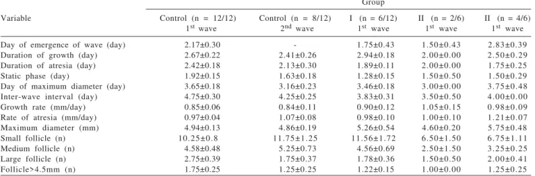
SEM - standard error of mean. P - wave variables between groups (P>0.05); mm - millimeter; n - number. Control groupMAP for 12 days and eCG at sponge withdrawal Group I 0.1 mg EB and MAP for 4 days, PGF2αat sponge withdrawal, and GnRH48 h after sponge removal. Group II 0.1 mg EB, 35 mg injectable progesterone and MAP for 4 days, PGF2α and eCG at sponge removal and GnRH 48 hours after sponge withdrawal. Group III 0.2 mg EB, 35 mg injectable progesterone and MAP for 4 days, PGF2α and eCG at sponge removal and GnRH 56 hours after sponge withdrawal. The day of the emergence of the second wave was not available in the control group after initiation of treatment.
Group
Variable Control (n = 12/12) Control (n = 8/12) I (n = 6/12) II (n = 2/6) II (n = 4/6) 1st wave 2nd wave 1st wave 1st wave 1st wave
Day of emergence of wave (day) 2.17±0.30 - 1.75±0.43 1.50±0.43 2.83±0.39
Duration of growth (day) 2.67±0.22 2.41±0.26 2.94±0.18 2.00±0.00 2.50±0.29
Duration of atresia (day) 2.42±0.18 2.13±0.30 1.89±0.11 2.00±0.00 1.75±0.25
Static phase (day) 1.92±0.15 1.63±0.18 1.28±0.15 1.50±0.50 1.50±0.29
Day of maximum diameter (day) 3.65±0.18 3.16±0.23 3.46±0.18 3.00±0.00 3.75±0.48
Inter-wave interval (day) 4.75±0.30 4.25±0.25 3.83±0.31 3.50±0.50 4.00±0.00
Growth rate (mm/day) 0.85±0.06 0.84±0.11 0.90±0.12 1.05±0.15 0.98±0.09
Rate of atresia (mm/day) 0.97±0.04 1.07±0.08 0.98±0.10 1.00±0.10 1.21±0.07
Maximum diameter (mm) 4.94±0.13 4.86±0.19 5.26±0.54 4.60±0.20 5.75±0.48
Small follicle (n) 10.25±0.8 11.75±1.25 11.56±1.72 6.50±1.50 6.75±1.11
Medium follicle (n) 4.58±0.48 5.25±0.73 4.56±0.69 2.50±1.50 3.25±0.25
Large follicle (n) 2.75±0.39 1.75±0.37 1.78±0.36 1.50±0.50 2.00±0.41
Follicle>4.5mm (n) 1.75±0.25 1.25±0.25 1.22±0.15 1.00±0.00 1.25±0.25
Group (long term protocol MAP sponges for 12 days and eCG) and they showed one-wave follicular growth (first wave) whereas 66% (8/12) of the animals showed two follicular waves (second wave). No significant differences (P>0.05) were seen between the variables. The characteristics of the first and second wave in control ewes were not statistically different from the other groups (Table 1).
Sponges were left in place for 4 days in all experimental groups. Thus, ewes had the first follicular wave and it was shown in 50% (6/12) of the animals in Group I (short-term protocol MAP sponges for 4 days, 0.1 mg EB and GnRH), 33% (2/6) of the animals in Group II (short-term protocol MAP for 4 days, 0.1 mg EB, eCG and GnRH), and 33% (4/12) of the animals in Group III (short-term protocol MAP for 4 days, 0.2 mg EB, eCG and GnRH). There were no significant differences between the control and experimental groups (Table 1).
The control and experimental groups had interwave intervals (Table 1) that were similar to Leyva et al. (1998) and Meikle et al. (2001).
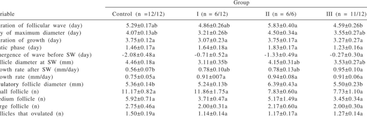
Follicular waves of the ovulatory follicle were observed in 100% (12/12) of the animals in the long term protocol and eCG and 100% of the animals in short-term protocol, 0.1 mg EB, eCG and GnRH, 50% (6/12) of the animals in short-term protocol, 0.1 mg EB and GnRH and 91% (11/12) of the animals in short-term protocol, 0.2 mg EB, eCG and GnRH (Table 2).
The duration of the follicular wave of ovulatory follicle was shorter (P<0.05) in short-term protocol, 0.2 mg EB, eCG and GnRH than in short-term protocol, 0.1 mg EB, eCG and GnRH (Table 2). Follicular wave of the ovulatory follicle originated from the second wave in 4 animals and third wave
in 8 animals after sponge insertion. Similar results were observed in anestrous ewes following the same protocol. Barrett et al. (2004) reported that the ovulatory follicles originated from different waves before sponge withdrawal. The development of antral follicles larger than 2 mm was observed in Suffolk ewes. There were no significant differences in the number of small, medium and large follicles in the control and experimental groups during treatment. No differences were observed in the first wave of follicular growth and the ovulatory follicular wave at the beginning of breeding season. These results (Table 1 and 2) are similar to those obtained in previous studies in which the number of small (2 to 3.5 mm), medium (4 to 5 mm) and large (greater than 5 mm) follicles were compared at the beginning, middle and end of the breeding season (Bister et al., 1999). These authors concluded that the number of small, medium and large follicles did not differ across the periods of time. They were also similar during the first and second wave and during the ovulatory wave. Therefore, similar results were obtained in the present study.
The duration of the follicular wave of ovulatory follicle was shorter (P<0.05) in short-term protocol, 0.2 mg EB, eCG and GnRH than in short-term protocol, 0.1 mg EB, eCG and GnRH. The duration ranged from 4.59±0.26 days in short-term protocol, 0.2 mg EB, eCG and GnRH to 5.83±0.40 days in short-term protocol, 0.1 mg EB, eCG and GnRH (Table 2). The day of maximum follicular diameter occurred earlier (P<0.05) in short-term protocol, 0.1 mg EB and GnRH than in short-term protocol, 0.1 mg EB, eCG and GnRH after the emergence of the follicular wave of the ovulatory follicle. The duration ranged from 3.21±0.26 days in short-term protocol, 0.1 mg EB and GnRH to 4.50±0.34 days in
short-Means followed by the same letter in the row do not differ significantly at P>0.05; SEM = standard error of mean; mm= millimeter; SW= sponge withdrawal; n= number. Control group MAP for 12 days and eCG at sponge withdrawal. Group I 0.1 mg EB and MAP for 4 days, PGF2αat sponge withdrawal, and GnRH 48 h after sponge removal.
Group II 0.1 mg EB, 35 mg injectable progesterone and MAP for 4 days, PGF2α and eCG at sponge removal and GnRH 48 hours after sponge withdrawal.
Group III 0.2 mg EB, 35 mg injectable progesterone and MAP for 4 days, PGF2α and eCG at sponge removal and GnRH 56 hours after sponge withdrawal.
Group
Variable Control (n =12/12) I (n = 6/12) II (n = 6/6) III (n = 11/12)
Duration of follicular wave (day) 5.29±0.17ab 4.86±0.26ab 5.83±0.40a 4.59±0.26b
Day of maximum diameter (day) 4.07±0.13ab 3.21±0.26b 4.50±0.34a 3.55±0.27ab
Duration of growth (day) 3.75±0.12a 3.07±0.23a 3.75±0.17a 3.27±0.27a
Static phase (day) 1.46±0.17a 1.64±0.18a 1.83±0.17a 1.23±0.16a
Emergence of wave before SW (day) -2.08±0.48a -0.71±0.52a -1.33±0.49a -0.27±0.30a
Follicle diameter at SW (mm) 4.46±0.18a 3.11±0.35b 4.15±0.31ab 3.53±0.27ab
Growth rate after SW (mm/day) 0.56±0.07b 0.78±0.10ab 0.78±0.13ab 0.95±0.10a
Growth rate (mm/day) 0.75±0.05a 0.91±007a 0.94±0.08a 0.91±0.06a
Ovulatory follicle diameter (mm) 5.36±0.14b 5.24±0.13b 6.39±0.43a 5.50±0.23b
Small follicle (n) 11.17±0.82a 11.86±1.75a 7.83±0.60a 7.73±1.10a
Medium follicle (n) 5.92±0.71a 3.71±0.47a 5.17±1.49a 3.45±0.34a
Large follicle (n) 2.75±0.46a 2.00±0.31a 2.17±0.60a 2.00±0.30a
Follicles that ovulated (n) 1.50±0.19a 1.14±0.14a 1.17±0.17a 1.27±0.14a
term protocol, 0.1 mg EB, eCG and GnRH (Table 2). There were no differences (P>0.05) in the length of growth, growth rate, static phase, wave emergence before sponge withdrawal, number of small, medium and large follicles, and number of ovulatory follicles in the control and experimental groups (Table 2).
The emergence of a follicular wave occurred between –2.08±0.48 days in long term protocol and eCG, –0.27±0.30 days in short-term protocol, 0.2 mg EB, eCG and GnRH and –0.71±0.52 days in short-term protocol, 0.1 mg EB and GnRH before sponge removal (Table 2). Similar results were found by Viñoles et al. (2001) (–2.2±0.8 days, 0.5±0.5 days and 0.4±1.1 days, respectively). Furthermore, Leyva et al. (1998) used the same long term protocol and reported that the wave began on days –3.4±0.4. The results of that study and the current one differ from one day.
The follicle size was statistically larger (P<0.05) in the long term protocol and eCG than in short-term protocol, 0.1 mg EB, eCG and GnRH at sponge removal. Nevertheless, follicle growth rate was slower (P<0.05) in long term protocol and eCG than in short-term protocol, 0.2 mg EB, eCG and GnRH. A possible explanation for this reduction in follicular growth is that the control ewes had ovulatory follicles emerging two days earlier. Therefore, the animals in the long term protocol and eCG) had a slower growth rate at sponge withdrawal (Table 2).
In all groups, control and experimental, the maximum diameter of the ovulatory follicles was similar to those described by Ravindra et al. (1994), Leyva et al. (1998), Bister et al. (1999) and Gonzalez de Bulnes et al. (2001) (Table 2). In previous studies, Barrett et al. (2004) reported that ovulatory follicles were smaller during the anestrous season (6.1±0.1 mm) than those observed during the breeding season (7.5±0.5 mm). Similar results were also obtained in the present study. In mares, low levels of circulating LH appear to be partially responsible for the reduction in follicular diameter during anestrous season (Donadeu & Watson, 2007).
The preovulatory follicle was the largest growing follicle present in both ovaries when luteolysis was induced (Gonzalez de Bulnes et al., 2001). Moreover, the preovulatory follicle emerged from the pool of small antral follicles during regression of the largest follicle. Similar findings were reported by McNatty et al. (1982). All follicles larger than 2 mm present at luteolysis are able to ovulate (Tsonis et al., 1984).
The number of follicles per ovulatory wave (1.14±0.14 to 1.50±0.19) indicates that Suffolk ewes are low fecundity sheep breeds as its ovulation rate is 1.2±0.4. This is in accordance with Bister et al. (1999). Similar results were also
obtained in other ewes breeds. The mean number of follicles per wave was 1.3±0.2 in Bartlewski et al. (1998) and 1.4± 0.3 in Letelier et al. (2009). The dominance exercised by large follicles was not clearly evident among ewes of this study, based on the definition of dominance for cattle (Ginther et al., 1989). In the present study, there were no differences between the two largest follicles in the anovulatory wave. Notwithstanding, the preovulatory follicle of the ovulatory wave appears to suppress the number of follicles and the growth of other follicles before ovulation. Similar findings were reported by Ravindra et al. (1994). Furthermore, Lopez -Sebastian et al. (1997) demonstrated that the ovulation rate in monovular ewes would be limited by high levels of follicular atresia rather than by low levels of recruitment of a group of growing follicles.
Conclusions
The effects of short-term (4 days) medroxyprogesterone acetate administration with 0.1 mg and 0.2 mg of estradiol benzoate were insufficient to synchronize the emergence of a new follicular wave or improve synchronization of estrus or ovulation. These results were compared to traditional long term protocol in Suffolk ewes in the non-breeding season.
References
ATAMAN, M.B.; AKÖZ, M.; AKMAN, O. Induction of synchronized oestrus in Akkaraman cross-bred ewes during breeding and anestrus seasons: the use of short-term and long-term progesterone treatments. Revista de Medicina Veterinária, v.157, n.5, p.257-260, 2006.
BARRETT, D.M.W.; BARTLEWSKI, P.M.; BATISTA-ARTEAGA, M. et al. Ultrasound and endocrine evaluation of the ovarian response to a single dose of 500 IU of eCG following a 12-day treatment with progestagen-releasing intravaginal sponges in the breeding and nonbreeding seasons in ewes. Theriogenology, v.61, n.2-3, p.311-327, 2004.
BARRETT, D.M.W.; BARTLEWSKI P.M.; DUGGAVATHI, R. et al. Synchronization of follicular wave emergence in the seasonally anestrous ewe: 2The effects of estradiol with or without medroxyprogesterone acetate. Theriogenology, v.69, n.7, p.827-836, 2008.
BARTLEWSKI, P.M. Ovarian follicular dynamics during anoestrus in ewes. Journal of Reproduction and Fertility, v.113, n.12, p.275-285, 1998.
BARTLEWSKI, P.M.; BEARD, A.P.; RAWLINGS, N.C. Ultrassonographic study of luteal function in breeds of sheep with different ovulation rates. Theriogenology, v.2, n.1, p.115-130, 1999.
BINELLI, M.; IBIAPINA, B.T.; BISINOTTO, R.S. Bases fisiológicas, farmacológicas e endócrinas dos tratamentos de sincronização do crescimento folicular e da ovulação. Acta Scientiae Veterinariae, v.34 (Supl 1), p.1-7, 2006. BISTER, J.L.; NOEL, B.; PERRAD, B. et al. Control of ovarian
BÓ, G.A.; ADAMS, G.P.; PIERSON, R.A. et al. Follicular waves dinamics after estradiol-17b treatment of heifers with or without a progestagen implant. Theriogenology, v.41, n.8, p.1555-1569, 1994. BÓ, G.A.; ADAMS, G.P.; PIERSON, R.A. et al. Exogenous control
of follicular wave emergence in cattle. Theriogenology, v.43, n.1, p.31-40, 1995.
BÓ, G.A.; BERGFELT, D.R.; BROGLIATTI, G.T. et al. Local versus systemic effects of exogenous estradiol-17b on ovarian follicular dynamics in heifers with progestagen implants. Animal Reproduction Science, v.59, n.3-4, p.141-157, 2000. CAVALIER, J.; KINDER, J.E.; DEATH, G. et al. Effect of 48 h
treatment with 17b oestradiol or progesterone on follicular wave emergence and syncrony of ovulation in Bos indicus cows when administred at the end of a period of progesterone treatment. Animal Reproduction Science, v.46, n.3-4, p.187-201, 1997. DISKIN, M.G.; AUSTIN, E.J.; ROCHE, J.F. Exogenous hormonal manipulation of ovarian activity in cattle. Domestic Animal Endocrinology, v.23, n.1-2, p.211-228, 2002.
DONADEU, F.X.; WATSON, E.D. Seasonal changes in ovarian activity: lessons learnt from the horse. Animal Reproduction Science, v.100, n.3-4, p.225-242, 2007.
EVANS, A.C.O.; FLYNN, J.D.; QUINN, K.M. et al. Ovulation of aged follicles does not affect embryo quality or fertility after a 14 day progestagen estrus synchronization protocol in ewes. Theriogenology, v.56, n.5, p.923-936, 2001.
EVANS, A.C.O.; DUFFY, P.; CROSBY, A.T.F. et al. Effect of ram exposure at the end of progestagen treatment on estrus synchronization and fertility during the breeding season in ewes. Animal Reproduction Science, v.84, n.3-4, p.349-358, 2004. GINTHER, O.J.; KASTELIC, J.P.; KNOPF, L. Intraovarian relationship among dominante and subordinate follicles and the corpus luteum in heifers. Theriogenology, v.32, n.5, p.787-795, 1989. GONZALEZ DE BULNES, A.A.; MORENO, J.S.; GARCIA, L.M.
et al. Origin of the preovulatory follicle in Mouflon sheep (Ovis gmelini musimon) and effect on growth of remaining follicles during the follicular phase of oestrous cycle. Animal Reproduction Science, v.65, n.3-4, p.265-272, 2001. HAMRA, A.H.; MASSRI, Y.G.; MARCEK, J.M. et al. Plasma
progesterone levels in ewes treated with progesterone-controlled internal drug-release dispensers, implants and sponges. Animal Reproduction Science, v.11, p.187-194, 1986.
HUSEIN, M.Q.; ABABNEH, M.M.; ABU-RUMAN, D.S. The effects of short or long term FGA treatment with or without eCG on reproductive performance of ewes bred out-of-season. American Journal of Animal and Veterinary Science, v.2, n.1, p.23-28, 2007.
LETELIER C.A.; CONTRERAS-SOLIS, I.; GARCIA-FERNANDEZ, R.A. et al. Ovarian follicular dynamics and plasma steroid concentrations are not significantly different in ewes given intravaginal sponges containing either 20 or 40 mg of fluorogestone acetate. Theriogenology, v.71, n.4, p.676-682, 2009. LEYVA, A.; BUCKRELL, B.C.; WALTON, J.S. Follicular activity
and ovulation regulated by exogenous progestagen and PMSG in anestrous ewes. Theriogenology, v.50, n.3, p.377-393, 1998.
LOPEZ-SEBASTIAN, A.; GONZALEZ-BULNES, A.; SANTIAGO-MORENO, J. et al. Patterns of follicular development during the estrous cycle in monovular Merino del Pais ewes. Animal Reproduction Science, v.48, n.2-4, p.279-291, 1997. MARTÍNEZ, M.F.; KASTELIC, J.P.; BÓ, G.A. et al. Effects of
oestradiol and some of its esters on gonadotrophin release and ovarian follicular dynamics in CIDR-treated beef cattle. Animal Reproduction Science, v.86, n.1-2, p.37-52, 2005. McNATTY, K.P.; BALL, K.; GIBB, M. et al. Induction of cyclic ovarian activity in seasonally anoestrous ewes with exogenous GnRH. Journal of Reproduction and Fertility, v.64, n.1, p.93-96, 1982.
MEIKLE, A.; FORSBERG, E.G.; GARÓFALO, E.G. et al. Circulating gonadotrophins and follicular dynamics in anestrous ewes after treatment with estradiol-17. Animal Reproduction Science, v.67, n.1-2, p.79-90, 2001.
RAVINDRA, J.P.; RAWLINGS, N.C.; EVANS, AC.O. et al. Ultrasonic study of ovarian follicular dynamics in ewes during the estrous cycle. Journal of Reproduction and Fertility, v.101, n.2, p.501-509, 1994.
SCHRICK, F.N.; SURFACE, R.A.; PRITCHARD, J.Y. et al. Ovarian structures during the estrous cycle and early pregnancy in ewes. Biological of Reproduction, v.49, n.6, p.1133-1140, 1 9 9 3 .
TSONIS, C.G.; CAHILL, L.P.; CARSON, R.S. et al. Identification at the onset of luteolysis of follicles capable of ovulation in the ewe. Journal of Reproduction and Fertility, v.70, n.2, p.609-614, 1984.
VIÑOLES, C.; FORSBERG, M.; BANCHERO, G. et al. Effect of long-term and short-term progestagen treatment on follicular development and pregnancy rate in cyclic ewes. Theriogenology, v.55, n.4, p.993-1004, 2001.