Article
Printed in Brazil - ©2018 Sociedade Brasileira de Química*e-mail: claudia.rezendeufrj@gmail.com
Antimicrobial Diterpene from the Brazilian Termite
Nasutitermes macrocephalus
(Isoptera: Termitidae: Natutitermitinae)
Márcia N. S. de la Cruz,a Helvécio M. dos Santos Júnior,a Denilson F. Oliveirab and Claudia M. Rezende*,a
aInstituto de Química, Universidade Federal do Rio de Janeiro, 21941-909 Rio de Janeiro-RJ, Brazil
bDepartamento de Química, Universidade Federal de Lavras, 37200-000 Lavras-MG, Brazil
Termites are insects with a complex social organization on castes among which soldiers are the only responsible for the colony defense. The soldiers of the Nasutitermes genus use
chemical defense, which comprises a mixture of mono, sesqui and mainly diterpenes. The new diterpene 2β,3α-dihydroxy-trinervita-1(15),11-diene, found in Nasutitermes macrocephalus,
was isolated using preparative high performance liquid chromatography (HPLC) and identified by gas chromatography-mass spectrometry (GC-MS) and nuclear magnetic resonance (NMR) techniques. The absolute stereochemistry determined was supported by ab initio calculations
and by comparison of the experimental circular dichroism data with the theoretical results. The isolated diterpene was evaluated against the bacteria Staphylococcus aureus (Gram-positive) and Escherichia coli (Gram-negative), and the fungi Candida albicans and Aspergillus brasiliensis. The
isolated diterpene presented minimal inhibitory concentrations of 62.5 and 62.5 µg mL-1 against
S. aureus and C. albicans, respectively.
Keywords:Nasutitermes macrocephalus, biological activities, HPLC
Introduction
Termites are insects present all over the world, with about 2750 species living in tropical and subtropical regions.1 They are social insects with castes responsible for
distinct functions: workers are tasked to build the mound and to feed the others castes; king and queen respond for reproduction, resulting in eggs that become larvae that may become workers, soldiers or winged (secondary breeding).2,3 Soldiers are responsible for the defense of
the mound and, depending on the species they will present mechanical and/or chemical defense.1,2,4,5 The Nasutitermes
genus (Isoptera: Termitidae: Nasutitermitinae) is the most evolved among the termites since their soldiers use only
chemical defense, avoiding contact with the enemies.5
They produce and storage a mixture of mono, sesqui and diterpenes in the frontal gland, resulting in a sticky secretion that is ejected against the enemies.6-10 The great
success of the Nasutitermes evolution has been attributed
to this defense method.2 The monoterpenes are the same
compounds found in plants, like α-pinene, β-pinene,
limonene and terpinolene, and they are associated with
the protection of the colony from fungal infections.11
Sesquiterpenes are in minor quantity, as found in
N. gracilirostris, N. torresi, N. triodiae, N. octopilis
and N. macrocephalus.9,12,13 The skeletons of the
diterpenes found in termites (trinervitanes, kempanes and rippertanes) are broadly described in all species,14 and are
related to both species communication, as they are blind,
and to defense.15 Although there are several studies on
the identification of these diterpenes, just a few of them focused on the biological activities of those substances. For example, one study presented five compounds with antibacterial activity against Bacillus subtilis and Escherichia coli.16 Therefore, several molecules with
interesting biological activities produced by species in the Nasutitermes genus, remains to be studied.
As a consequence, our research group recently studied the biological activities and chemical composition
of the crude extract from N. macrocephalus soldiers,
which inhibited the growth of methicillin resistant
Staphylococcus aureus and Candida albicans.13 To continue
spectrometric methods and underwent antibacterial and antifungal assays to evaluate its biological activities.
Experimental
General experimental procedures
Optical rotation was measured in a PerkinElmer 341 LC polarimeter. Electronic circular dichroism (ECD) spectrum was obtained in a JASCO, J-715, Circular Dichroism (CD) Spectropolarimeter, using acetonitrile as solvent. 1D and 2D nuclear magnetic resonance (NMR) spectra were recorded in a Bruker Ultrafield Avance 600 MHz
or 400 MHz spectrometer, with CDCl3 as solvent.
EIMS analysis were performed in an HP GC-EIMS (gas chromatography with electron ionization mass spectrometry) (mass selective detector model 5937 and GC model G1530A) (70 eV) spectrometer with a HP-5 column (30 m × 250 µm × 0.25 µm). The HRESIMS (high-resolution electrospray ionization mass spectrometry) spectra in a positive mode were recorded in a Waters QTOF Micro mass spectrometer with direct infusion on the ionization source. Fractionation and isolation were performed in a preparative high performance liquid chromatograph (HPLC) Shimadzu Prominence 20A system with a Phenomenex Gemini C-18 preparative column (21.2 × 250 mm, 5 µm).
Termite material
The mound was collected at Fiocruz campus
(22°52’53.24’’S, 43°14’40.73’’W, 8 m above sea level
(ASL), Rio de Janeiro, RJ, Brazil) from a Leucaena sp.
Benth. tree and identified as Nasutitermes macrocephalus
(Isoptera: Termitidae: Nasutitermitinae) by Dsc Maurício Rocha from University of São Paulo (USP) Museum, where a specimen has been deposited (MZUSP 15803).
Extraction, GC-EIMS analysis, and isolation
Soldiers (21.5 g) of N. macrocephalus were separated
from the other castes and crushed in CH2Cl2 at room
temperature for 5 min. The resulting mixture was filtered and dried under a nitrogen flow to yield 824.2 mg of a
crude extract. An aliquot of this material (1 mg mL-1 in
CH2Cl2) was analyzed by GC-EIMS, with the injector set
at 280 °C, detector at 290 °C, injection volume of 1 µL, and
nitrogen as gas carrier at 1.2 mL min-1. The temperature
was initially maintained at 45 °C for 5 min, and then raised to 130 °C at a rate of 12 °C min-1. From 130 to 230 °C and
from 230 to 290 °C, the temperature was raised at rates of 33 and 1.5 °C min-1, respectively. The total time of analysis
amounted to 60.0 min. Part of the CH2Cl2 extract (602.7 mg)
was dissolved in methanol (12 mL, 50.2 mg mL-1) and
fractionated (volume of each injection: 800 µL) on an
HPLC system using CH3CN:H2O (75:25) at flow ratio of
20 mL min-1 as mobile phase. The process was monitored at λ 210 nm for 55.0 min to select the fractions to be collected, which were dried under reduced pressure. The fraction with the isolated diterpene (14.5 min) yielded 10.0 mg.
Antimicrobial assays
Both the CH2Cl2 extract and the isolated substance
underwent broth microdilution assays to determine their MIC (minimal inhibitory concentration) values.17 Briefly,
after two-fold serial dilution of test samples, the wells were inoculated with 10 µL of a bacterial suspension of E. coli
ATCC 8739 or methicilin-resistant S. aureus BMB 9393
(MRSA) in Müeller-Hinton, or inoculated with 100 µL
of RPMI-MOPS pH 7.2 containing C. albicans ATCC
10231 or A. brasiliensis ATCC 16404. The microplates
were incubated overnight at 37 °C for MRSA and for 48 h at room temperature (28-30 °C) for fungi. The negative control was pure medium, and positive controls comprised inoculated growth medium. The results were based on visual growth of microorganisms. The lowest concentrations of the samples that inhibited the growth of microorganisms were considered their MIC values. The visual observations were confirmed after aseptically addition of resazurin (30 µL) to the microplate wells, followed by incubation at 37 °C for 1 h.
Computational methods
Initially, the isolated substance underwent a c o n f o r m a t i o n a l s e a r c h u s i n g t h e s o f t wa r e
Open3Dalign 2.103.18 One thousand molecular dynamics
simulations were carried out for the substance at 1,000 K, with a 1 fs time step for 1 ps using the MMFF94 force field, and considering the solvent (water) implicitly using the GBSA model. The most stable conformation in each simulation was optimized with the MMFF94S force field and ranked according to the final energy. The most stable conformation and those up to 10 kcal mol-1 from the most
stable underwent optimization with the Hamiltonian
PM719 using the software MOPAC 2012.20,21 In this step
the solvent (acetonitrile, dielectric constant = 37.5) was implicitly considered using the conductor-like screening model (COSMO). The most stable conformation and those up to 10 kJ mol-1 distant from the most stable were
optimized in a density functional theory (DFT) calculation
at the B3LYP/Def2-TZVP-RIJCOSX22-27 level of theory
in this calculation to consider the solvent (acetonitrile) implicitly. As no conformation could be found up to 20 kJ mol-1 from the most stable, only one was used in the
next step that comprised time-dependent density functional theory (TD-DFT) calculation at the same level of theory. A total of 20 excited states were calculated, and only singlet excited states were considered. The DFT optimization of the conformations and TD-DFT calculation were also performed at the B3LYP/6-311G (2d,2p)29 level of theory.
Rotational and oscillator strengths were respectively converted into CD and UV (ultraviolet) spectra using the
software SPECDIS 1.51.30 These conversions were carried
out employing Gaussian curves with half the bandwidth of the CD band at 1/e peak height (σ) equal to 0.29 eV.
2β,3α-Dihydroxytrinervita-1(15),11-diene
White resin (10.0 mg); [α]D20 –5.0 (c 0.046 CHCl3); CD
(0.33 mM, CH3CN) λmax / nm (∆ε): 200 (14.3), 220 (–15.8),
240 (–0.3); EIMS (70 eV) m/z: 304 [M+·] (1), 286 (13), 271
(19), 255 (6), 215 (4), 207 (13), 201 (5), 190 (8), 175 (19), 159 (11), 147 (12), 135 (40), 119 (27), 107 (29), 91 (30), 79 (21), 67 (22), 55 (26), 44 (100). HRESIMS, ESI (+):
m/z [M + H]+ 305.2472 (calcd. for C20H33O2+ 305.2475).
Results and Discussion
According to Everaerts et al.9 and Braekman et al.,31
there is no difference in the final results of diterpene composition when crushing just the heads or the entire bodies of the termites. Therefore, the entire bodies of
N. macrocephalus soldiers were employed in the present
study to obtain larger mass of extracted substances.
Fractionation of the CH2Cl2 extract yielded 10 mg of a
white resin whose molecular formula (C20H32O2) was
determined through HRESIMS by the protonated molecule at m/z 305.2472 [M + H]+.
The structure elucidation (relative stereochemistry) of the isolated compound was carried out using 1D (1H, 13C)
and 2D (HSQC (heteronuclear single quantum correlation) (1J
HC), HMBC (heteronuclear multiple bond correlation)
(2,3J
HC), COSY (correlation spectroscopy) and NOESY
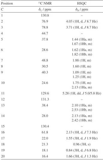
(nuclear Overhauser effect spectroscopy)) NMR techniques (Table 1). Chemical shifts are given in parts per million
(ppm). On the basis of 13C NMR data, 20 resonances were
assigned to four methyl groups (d 22.0, 21.3, 18.1 and 16.4), six methylenes (d 37.8, 28.6, 40.3, 24.6, 38.4 and 28.0), six methines (d 76.9, 78.8, 48.8, 30.5, 129.6 and 61.8), and four quaternary carbons (d 130.8, 44.7, 131.3 and 130.4).
The 1H NMR spectrum showed signals consistent with the
presence of four methyls at dH 1.55 (d, J 1.9 Hz, CH3-17),
0.96 (CH3-18), 0.84 (d, J 6.8 Hz, CH3-19), and 1.66 (d,
J 1.3 Hz, CH3-20), the first one and the last being allylic
methyls, the second is bonded to a quaternary carbon, and the third bonded to a tertiary carbon.
The coupling of a methyl group (C-18, dH 0.96) bonded
to a tertiary carbon bearing a proton with a signal at dH 1.60
(H-8), with which the methyl protons were coupled with constant of 6.8 Hz, was confirmed in the COSY experiment (3J
HH). The signal associated with the methine double bond
proton at dH 5.28 (dd, J 5.0/5.8 Hz H-11), connected to
C-11 (dc 129.6) according to the HSQC experiment, was
coupling to C-9 (dC 40.3, 3JCH), C-10 (dC 24.6, 2JCH), C-13
(dC 38.4, 3JCH), and C-20 (dC 16.4, 3JCH), according to the
HMBC experiment. This result was important to confirm the 1H and 13C chemical shifts attributed to these groups.
Although the methine proton at dH 1.88 (H-7) showed a
multiplet, the methine proton at dH 2.13 (H-16) exhibited
a doublet with a constant of 7.3 Hz. The coupling of
these hydrogens was also confirmed in the COSY (3J
HH)
spectrum. The carbinolic protons presented a coupling
Table 1. 1H and 13C NMR data for 2β,3α -dihydroxytrinervita-1(15),11-diene and their correlation by HSQC (1J
HC)
Position 13C NMR HSQC
C dC / ppm dH / ppm
1 130.8 –
2 76.9 4.03 (1H, d, J 8.7 Hz)
3 78.8 3.71 (1H, d, J 8.7 Hz)
4 44.7 –
5 37.8 1.44 (1Ha, m)
1.67 (1Hb, m)
6 28.6 1.62 (1Ha, m)
1.82 (1Hb, m)
7 48.8 1.88 (1H, m)
8 30.5 1.60 (1H, m)
9 40.3 1.09 (1H, m)
1.25 (1H, m)
10 24.6 1.75 (1H, m)
2.13 (1Ha, m)
11 129.6 5.28 (1H, dd, J 5.0/5.8 Hz)
12 131.3 –
13 38.4 2.10 (1Ha, m)
2.53 (1Hb, m)
14 28.0 2.13 (1Ha, m)
2.42 (1Hb, m)
15 130.4 –
16 61.8 2.13 (1H, d, J 7.3 Hz)
17 22.0 1.55 (3H, d, J 1.9 Hz)
18 21.3 0.96 (3H, s)
19 18.1 0.84 (3H, d, J 6.8 Hz)
constant of 8.7 Hz, that is characteristic of Haxial–Haxial
coupling in a saturated six members ring. This corroborates the trans relationship between the hydroxyl groups. NOE
correlations observed through the NOESY spectrum show interactions of H-3 with H-6a and H-14a (both up), confirming the relative configurations of both C-3 and C-8 (both hydroxyl and methyl groups in axial position). H-2 correlations with H-16 and H-18 in the NOESY spectrum also confirmed the relative configurations of C-2, C-16 and C-18 (Figure 1). NOESY spectrum also shows interaction of H-8 with H-16, also confirming the configurations of C-8 and C-16. In conclusion, interpretation of the NMR data leads to the structure 2β,3α -dihydroxy-trinervita-1(15),11-diene, which is a new diterpene (Figure 1).
There was no doubt about the relative configuration of the isolated substance. Therefore, aiming to discover its absolute configuration by comparing its experimental and theoretical ECD spectra (Figure 2), a conformational search was performed using molecular mechanics methods, which afforded eight conformations. The energy difference between the most and the less stable
was 8.8046 kcal mol-1. When all these conformations
underwent optimization through semiempirical calculations only four of them presented total energies corresponding to above 1% of the Boltzmann distribution. Therefore, only these four conformations were further optimized through DFT calculations, according to which one had energy corresponding to more than 99% of the Boltzmann distribution at both levels of theory employed.
The root-mean-square deviation between atomic positions of superimposed conformations resulting from both DFT calculations, accounting to more than 99% of the Boltzmann distribution, was only 0.074 Å. Thus, these conformations are practically the same and are in agreement with the NMR data. For example, in the conformation obtained at the B3LYP/Def2-TZVP/RIJCOSX level of theory, the distance between H-3 with H-6a is 1.942 Å,
while between H-3 and H8 is 2.761 Å. Regarding H-2, its distances from H-16 and H-18 are 3.917 and 2.274 Å, respectively (Figure 3). All these values are in accordance with the observed NOE.
Both theoretical ECD spectra, obtained through TD-DFT calculations at both levels of theory for the isolated substance, were in accordance with the experimental one, suggesting that the attributed configuration was correct. Based on the data described, the configuration of the substance was established as (2R,3R,4S,7S,8R,11E,16S
)-2,3-dihydroxytrinervita-1(15),11-diene.
Figure 1. (a) Key 1H-13C couplings at two and three bonds distant (HMBC, 2,3J
HC) of 2β,3α-dihydroxy-trinervita-1(15),11-diene and (b) key NOE correlations (NOESY).
Figure 2. Experimental (a) and theoretical electronic circular
dichroism spectra for 2β,3α-dihydroxy-trinervita-1(15),11-diene. The theoretical spectra were obtained through time-dependent density functional theory at two levels: B3LYP/Def2-TZVP-RIJCOSX (b) and B3LYP/6-311G-2d2p (c).
Figure 3. Three-dimensional structure of the most stable conformation of
The isolated compound was evaluated against a series of fungal and bacterial strains. The bacteria
methicillin-resistant Staphylococcus aureus BMB 9393 (clinical
isolate) (MRSA) and Escherichia coli ATCC 8739 were
chosen for antibacterial assay due to the association with hospital acquired infections worldwide and gastrointestinal infections, respectively.32Candida albicans ATCC 10231
and Aspergillus brasiliensis ATCC 16404 were used in the
antifungal assay, since they are responsible for candidiasis and lung infections, respectively.33 The isolated diterpene
showed activity against both MRSA (MIC = 62.5 µg mL-1)
and C. albicans (MIC = 62.5 µg mL-1), and no activity
against the Gram-negative bacterium E. coli was
observed. It was also inactive against the filamentous fungus A. brasiliensis. The positive controls ciprofloxacin
and amphotericin B presented MIC values of 0.5 and 0.125 µg mL-1, for antibacterial and antifungal activity,
respectively. This result contributes to know the role of diterpenes produced by the termites, and help to understand the chemical ecology of termites.
Conclusions
In this study a diterpene trinervitene was isolated
from termite soldiers of Nasutitermes microcephalus and
presented antimicrobial activities against S. aureus and
C. albicans.
This work contributes to the knowledge of termite chemical composition, which is fundamental to understand the success of termites development and adaptation to many different environments.
Supplementary Information
Supplementary data (chromatogram, mass spectrum, 1H
and 13C NMR spectra, COSY, HSQC, HMBC correlation
maps, and results through molecular mechanics) are available free of charge at http://jbcs.sbq.org.br as PDF file.
Acknowledgments
The authors would like to thank the DIRAC/Fiocruz, Dr Mauricio Rocha of the University of São Paulo Museum, Dr Antonio Gilberto Ferreira of the University of São Carlos, Dr Daniela S. Alviano of Federal University of Rio de Janeiro, and Centro Nacional de Supercomputação (CESUP) from Federal University of Rio Grande do Sul (UFRGS) where part of the computational work was carried out. We also thank CNPq (Conselho Nacional de Desenvolvimento Científico e Tecnológico), CAPES (Coordenação de Aperfeiçoamento de Pessoal de Nível
Superior), FAPERJ (Fundação Carlos Chagas Filho de Amparo à Pesquisa do Estado do Rio de Janeiro) and FAPEMIG (Fundação de Amparo à Pesquisa do Estado de Minas Gerais), for financial support and fellowships.
References
1. Constantino, R.; Pap. Avulsos Zool. 1999, 40, 387.
2. Prestwich, G.; Sci. Am. 1983, 249, 68.
3. Laurent, P.; Daloze, D.; Pasteels, J. M.; Braekman, J. C.; J. Nat. Prod. 2005, 68, 532.
4. Prestwich, G.; Biochem. Syst. Ecol. 1979, 7, 211.
5. Prestwich, G.; Annu. Rev. Entomol. 1984, 29, 201. 6. Baker, R.; Walmsley, S.; Tetrahedron 1982, 38, 1899.
7. Prestwich, G.; Solheim, B.; Clardy, J.; Pilkiewicz, F.; Miura, I.; Tanis, S.; Nakanishi, K.; J. Am. Chem. Soc. 1977, 99, 8082.
8. Prestwich, G.; J. Chem. Ecol. 1979, 5, 459.
9. Everaerts, C.; Roisin, Y.; Le Quéré, J.-L.; Bonnard, O.; Pasteels, J.; J. Chem. Ecol. 1993, 19, 2865.
10. Prestwich, G.; Tetrahedron 1982, 38, 1911.
11. Rosengaus, R.; Lefebvre, M.; Traniello, J.; J. Chem. Ecol. 2000, 26, 21.
12. Prestwich, G.; Collins, M.; Biochem. Syst. Ecol. 1981, 9, 83. 13. Cruz, M. N. S.; Júnior, H. M. S.; Oliveira, D. F.; Costa-Lotufo,
L. V.; Ferreira, A. G.; Alviano, D. S.; Rezende, C. M.; Nat. Prod. Commun. 2013, 8, 69.
14. Cruz, M. N. S.; Júnior, H. M. S.; Rezende, C. M.; Alves, R. J. V.; Cancello, E. M.; Rocha, M. M.; Quim. Nova 2014, 37, 95.
15. Budesinsky, M.; Valterova, I.; Sémon, E.; Cancello, E.; Bordereau, C.; Tetrahedron 2005, 61, 10699.
16. Zhao, C.; Rickards, R.; Trowel, S.; Tetrahedron 2004, 60, 10753.
17. Clinical and Laboratory Standards Institute (CLSI); M27-A3, Reference Method for Broth Dilution Antifungal Susceptibility Testing of Yeasts; Approved Standard, 3rd ed.; CLSI: Wayne, PA,
USA, 2008; Clinical and Laboratory Standards Institute (CLSI); M38-A2, Reference Method for Broth Dilution Antifungal
Susceptibility Testing of Filamentous Fungi; Approved Standard, 2nd ed.; CLSI: Wayne, PA, USA, 2008; Clinical and
Laboratory Standards Institute (CLSI); M7-A4, Methods for Dilution Antimicrobial Susceptibility Tests for Bacteria that Grow Aerobically, 4th ed.; CLSI: Wayne, PA, USA, 2008.
18. Tosco, P.; Balle, T.; Shiri, F.; J. Comput.-Aided Mol. Des. 2011, 25, 777.
19. Stewart, J. J. P.; J. Mol. Model. 2013, 32, 19.
20. Stewart, J. J. P.; MOPAC2012, 13.284L Stewart Computational Chemistry: Colorado, Springs, CO, USA, 2012.
21. Maia, J. D. C.; Carvalho, G. A. U.; Júnior, C. P. M.; Santana, S. R.; Cabral, L. A. F.; Rocha, G. B.; J. Chem. Theory Comput.
2012, 8, 3072.
23. Lee, C.; Yang, W.; Parr, R. G.; Phys. Rev. B 1988, 37, 785. 24. Neese, F.; Wennmohs, F.; Hansen, A.; Becker, U.; Chem. Phys.
2009, 356, 98.
25. Weigend, F.; Ahlrichs, R.; Phys. Chem. Chem. Phys. 2005, 7,
3297.
26. Eichkorn, K.; Weigend, F.; Treutler, O.; Ahlrichs, R.; Theor. Chem. Acc. 1997, 97, 119.
27. Eichkorn, K.; Treutler, O.; Öhm, H.; Häser, M.; Ahlrichs, R.; Chem. Phys. Lett. 1995, 240, 283.
28. Neese, F.; Wiley Interdiscip. Rev.: Comput. Mol. Sci. 2012, 2,
73.
29. Krishnan, R.; Binkley, J. S.; Seeger, R.; Pople, J. A.; J. Chem. Phys. 1980, 72, 650.
30. Bruhn, T.; Schaumlöffel, A.; Hemberger, Y.; Bringmann, G.; Chirality 2013, 25, 243.
31. Braekman, J. C.; Daloze, D.; Dupont, A.; Pasteels, J. M.; Lefeuve, P.; Bordereau, C.; Declercq, J. P.; Van Meerssche, M.; Tetrahedron 1983, 39, 4237.
32. Ribeiro, A.; Dias, C.; Silva-Carvalho, M. C.; Berquó, L.; Ferreira, F. A.; Santos, R. N. S.; Ferreira-Carvalho, B. T.; Figueiredo, A. M.; J. Clin. Microbiol. 2005, 43, 1985.
33. Silva, A. C. R.; Lopes, P. M.; Azevedo, M. M. B.; Costa, D. C. M.; Alviano, C. S.; Alviano, D. S.; Molecules 2012, 17, 6305.
Submitted: April 10, 2017
Published online: September 14, 2017