Universidade de Lisboa
Faculdade de Medicina Dentária
Ethanol wet bonding: an in vitro new approach
Sara Micaela Nogueira Ribeiro
Dissertação
Mestrado Integrado em Medicina Dentária
Faculdade de Medicina Dentária
Ethanol wet bonding: an in vitro new approach
Sara Micaela Nogueira Ribeiro
Dissertação orientada pela Mestre Ana Pequeno
Mestrado Integrado em Medicina Dentária
Ethanol wet bonding: an in vitro new approach
“Habe nun, ach! Philosophie, Juristerei und Medizin, Und leider auch Theologie Durchaus studiert, mit heißem Bemühn. Da steh ich nun, ich armer Tor!”
Agradecimentos:
À minha orientadora, Mestre Ana Pequeno, por todo o apoio prestado durante a elaboração deste trabalho. Sempre disponível, prestável e interessada, de uma inteligência sagaz, consegue perceber aquilo que quero fazer. Sem a sua persistência, este trabalho não existiria nos nestes moldes. Obrigada por não ter desistido e ter conseguido que este trabalho fosse realizado no laboratório. Foi um prazer ter como mentora uma pessoa tão curiosa, inteligente e trabalhadora, sempre querendo fazer mais. Uma Mestre na verdadeira aceção da palavra.
Ao Professor Doutor Jaime Portugal, por nos ter aberto as portas do UICOB e por toda a ajuda prestada, especialmente com a análise estatística.
À Professora Doutora Sofia Arantes e Oliveira, pela sua disponibilidade e por tão bem nos ter recebido no UICOB. A forma como zela pelo bom funcionamento daquele espaço é notável, tornando o laboratório um local de um rigor exemplar.
À Mestre Filipa Chasqueira, por ser incansável e pela infindável ajuda que nos prestou. Sempre simpática, disponível e generosa na partilha dos seus conhecimentos, consegue aliar o melhor dos dois mundos: empatia e inteligência.
Ao Rui, meu colega neste desafio. Pelas horas de trabalho árduo no laboratório e o subsequente desespero quando as coisas não corriam como o esperado. Pela alegria, boa disposição, empenho e interesse, foi sem dúvida muito mais fácil realizar este trabalho graças a ti. E como já sabes, “O “R” é de Repeat!”
À minha professora da primária, Fernanda Magalhães, por ter contribuído em larga escala para a minha formação académica e pessoal. O seu rigor, seriedade e perfeccionismo foram determinantes para a minha formação. A professora foi, a nível académico, a pessoal responsável por me fornecer as bases para que hoje pudesse redigir este trabalho. Foi consigo que realizei o grande sonho que tinha enquanto criança:
Ethanol wet bonding: an in vitro new approach
aprender a ler. Obrigada por me ter incentivado sempre a querer fazer mais e melhor e pelo voto de confiança que sempre depositou em mim enquanto fui sua aluna.
À Andreia, minha colega e dupla do 5.º ano. Num ano tão exigente como este, precisamos de saber aprender a simplificar, algo que fazes com bastante destreza. Obrigada por teres ajudado a navegar neste mar, por vezes inóspito, que é o 5.º ano.
À minha amiga Filipa Silva por ser generosa e das pessoas com mais paciência que conheço. Obrigada pelo teu incentivo e amizades.
Aos meus avós, que partiram cedo demais.
Às minhas sete princesas, Miúda, Pica, Estrela, Boneca, D. Inércia de Jesus, Micki e Mel por me conseguirem sempre arrancar um sorriso. Pelas infindáveis horas de brincadeiras e beijinhos e por me todos os dias me ensinarem algo mais.
Aos meus pais, Maria Helena e José, pelo amor incondicional e por permitirem que lute pelos meus objetivos. Por serem das pessoas mais generosas que conheço e me incentivarem a continuar o meu caminho. Durante estes anos, têm sido o tronco que suporta e nutre a árvore e permite que os ramos brotem e as flores floresçam, e mais tarde, venham a dar frutos. Sem vocês, nada disto teria sido possível.
À minha irmã Catarina, outro dos ramos da árvore. Obrigada pelo apoio e por tudo auilo que me ensinas todos os dias.
Por fim, ao André. As árvores também dão livros. Livros que nos fazem pensar e questionar se o existencialismo será um humanismo. Obrigada por esteres sempre a meu lado e pela infindável ajuda que me prestas. És o meu Personal Jesus, someone who hears my prayers, someone who cares and someone who’s there. E somos mais fixes que o Sartre e a Simone.
Index:
Resumo ...
Abstract ...
Introduction ... 1
Objectives ... 7
Materials and Methods ... 8
Results ... 16
Discussion ... 21
Conclusion ... 27
Appendices ... I
Ethanol wet bonding: an in vitro new approach
List of tables and figures
Table 1 – Testing groups ... 9
Table 2 –Test for Equality of Variances of the µTBS in MPa ... 15
Table 3 – Descriptive statistics of the µTBS in MPa for the three experimental groups tested. Mean values that are not significantly different from one another share the same upper case letter at p<0,05. ... 16
Table 4 – One-way ANOVA test ... 17
Table 5 – Tukey HSD Post Hoc Test (α=0,05) ... 18
Table 6 – Tukey HSD test. Mean µTBS values in MPa for groups in homogeneous subsets are displayed. ... 18
Table 7 - Number of specimens according to the failure mode and premature failures of the three different experimental groups tested. ... 19
Table 8 – Number of specimens according to the failure mode and percentages of all experimental groups tested ... 20
Table 9 – Materials, Manufacturers, Components and Batch Numbers ... II Table 10 – Number of sticks obtained in each tooth ... III Figure 1 – Tooth attached to an acrylic holder with sticky wax. Root cutting 1-2 mm below the CEJ. ... 8
Figure 2 – Removal of the pulp tissues ... 8
Figure 3 – Removal of the occlusal enamel and superficial dentin ... 9
Figure 4 – Creating the smear layer on a mechanical grinder. ... 9
Figure 5 – Etching the dentin surface. ... 10
Figure 6 – Vigorous ethanol application ... 10
Figure 7 – Experimental hydrophobic primer being applied. ... 11
Figure 9 – Resin application build up, after being painted with waterproof ink. ... 12
Figure 10 - Specimen being cut in the “x” direction. ... 13
Figure 11 – Specimen being cut in the “y” direction. ... 13
Figure 12 – Sticks obtained after the final cut ... 13
Figure 13 – The specimen attached to a Geraldelli’s jig. ... 14
Figure 14 – Instron 4502, universal testing machine ... 14
Figure 15 – Box-whisker plots of the µTBS in MPa for the two different experimental groups tested. The median µTBS is represented by the central line. The box represents the interquartile range. The mean µTBS is represented by the diamond mark (♦). ... 16
Figure 16 – Mean µTBS value for each group. Mean values that are not significantly different from one another share the same upper case letter at p<0,05. ... 17
Figure 17 – Distribution of the specimens according to the failure mode of the three different experimental groups tested. ... 19
Figure 18 – Distribution of the specimens according to the failure mode of all experimental groups tested. ... 20
Ethanol wet bonding: an in vitro new approach
Abreviates
Bis-GMA Bisphenol A diglycyl methacrylaye EWB w/ P Ethanol wet bonding with primer EWB w/o P Ethanol wet bonding without primer
HEMA Hydroxyethylmethacrylate
MMPs Matrix metalloproteinases
PF Premature failures
WWB Water wet bonding
Ethanol wet bonding: an in vitro new approach
Resumo:
De acordo com o estado da arte actual, o grande desafio da adesão moderna prende-se com a elevada hidrofilia dos sistemas adesivos contemporâneos. Ao longo da evolução dos sistemas adesivos, a incorporação de monómeros hidrofílicos foi imperativa. No entanto, a incorporação desses monómeros tem efeitos nefastos a longo prazo, conduzindo à degradação hidrolítica e consequente compromisso da durabilidade da adesão dentária. Este problema é mais premente na dentina que no esmalte, pois tem um maior teor orgânico, estabelecendo forças de adesão menos estáveis e previsíveis.
Ao longo dos anos, várias abordagens foram propostas de forma a colmatar os problemas existentes. Durante os anos 90 do século passado, existiu uma grande revolução na abordagem utilizada: a adesão passou a ser feita sobre um substrato húmido, o que implicou uma transição da filosofia de dry bonding para wet bonding. No entanto, a adesão à dentina continua a não reproduzir os resultados desejados.
No início deste século, ocorreu uma nova mudança no paradigma da adesão e surgiu a filosofia de ethanol wet bonding. Esta abordagem ambiciona resolver os problemas relacionados com a elevada hidrofilia dos sistemas adesivos actuais. Desta forma, uma vez que os monómeros hidrofóbicos que compõem os adesivos são solúveis em etanol, foi proposta a aplicação de etanol sobre a dentina recém-condicionada e previamente à aplicação dos restantes componentes do sistema adesivo. Propõe-se que o etanol desidrate a superfície dentária desmineralizada, removendo a água residual e facilitando a infiltração dos monómeros. Na teoria, a as forças adesivas obtidas serão mais estáveis e a durabilidade da adesão será maior.
A literatura apresenta dois protocolos de aplicação do etanol. No primeiro, denominado de técnica progressiva, várias concentrações crescentes de etanol são aplicadas de forma sequencial. Este método apresenta resultados superiores e mais consistentes, no entanto, tem pouca aplicação na prática clínica pois requer muito tempo. O segundo protocolo, denominado de simplificado, preconiza a utilização de etanol na concentração de 100% durante um minuto. Este protocolo apresenta resultados mais variáveis, no entanto, devido à sua simplicidade, parece ser uma opção mais aliciante.
Objectivos: A abordagem ideal desta técnica deve englobar as vantagens dos dois
protocolos. O actual estudo preconiza a utilização de um protocolo híbrido, no qual a aplicação de duas concentrações crescentes de etanol é feita durante 60 segundos. Pretende-se obter uma técnica simples, através da utilização de etanol que pode ser
facilmente obtido até no supermercado. Assim, o objectivo deste estudo laboratorial é comparar as forças de adesão à dentina entre o protocolo proposto de ethanol wet bonding, variando a aplicação de primer, com a técnica convencional de water-wet bonding, através de testes de microtração (µTBS).
As hipóteses nulas deste estudo são: (1) o protocolo de adesão utilizado não influencia as forças de adesão obtidas; (2) a aplicação de primer não influencia as forças de adesão obtidas; (3) não existem diferenças na distribuição de fracturas entre os grupos estudados.
Materiais e métodos: Uma amostra conveniente composta por quinze molares
humanos recentemente extraídos, intactos e sem evidência macroscópica de cárie ou restaurações (n=15), foi distribuída aleatoriamente em três grupos, segundo a estratégia de adesão: Grupo WWB (controlo) – utilização do primer e adesivo do sistema etch-and-rinse de 3 passos Adper Scotchbond MultiPurpose (3M ESPE); o Grupo EWB w/ P – aplicação de concentrações crescentes de etanol (70% e 96%) durante 60 segundos, e utilização de um primer experimental hidrofóbico, obtido pela diluição do adesivo do sistema adesivo acima descrito em álcool a 96%; e o Grupo EWB w/o P – aplicação de concentrações crescentes de etanol (70% e 96%) durante 60 segundos, sem a utilização de qualquer primer. Todos os grupos utilizaram o mesmo adesivo Adper Scotchbond MultiPurpose Adhesive (3M ESPE).
Após a preparação dos dentes e, com o objetivo de formar uma smear layer semelhante à que é obtida em situações clínicas, a superfície dos dentes foi polida com papel abrasivo de carbureto de silício (SiC) de grão 600, durante 60 segundos sob refrigeração com água, numa máquina polidora (DAP-U, Struers, Denmark).
Procedeu-se de seguida à aplicação dos protocolos adesivos de acordo com a distribuição nos respetivos grupos experimentais. Em todas as amostras o adesivo foi fotopolimerizado durante 20 segundos com o fotopolimerizador Bluephase® 20i (G2), (Ivoclar Vivadent, Austria) com intensidade de 600 mW/cm2, controlado periodicamente por um radiómetro (Bluephase® meter, Ivoclar Vivadent, Austria). De seguida foi aplicada a resina composta Herculite™ XRV Ultra™ Dentine, na cor A2, (Kerr Italia S.r.I., Scafati (SA), Italy) em camadas de aproximadamente 2 mm fotopolimerizadas entre si durante 40 segundos. As faces vestibular, lingual, mesial e distal foram polimerizadas adicionalmente por mais 40 segundos cada. A face exterior da resina composta de todos os espécimes foi pintada com tinta à prova de água, com o objetivo de excluir do estudo os palitos nos quais a adesão foi feita em esmalte. Os dentes foram
Ethanol wet bonding: an in vitro new approach
armazenados em água destilada numa estufa de incubação durante 24h a 37ºC e registado o dia e a hora da reconstrução.
Posteriormente, foram efetuados cortes no eixo do “x” e do “y” com um disco de diamante, a baixa rotação e sob refrigeração com água, num micrótomo de tecidos duros (IsoMetTM Diamond Wafering Blade -10,2cmx0,3mm - Series 15HC, Buehler Ltd., Lake Bluff, IL, USA), com o objetivo de obter palitos com uma área de, aproximadamente, 0,7 mm2. Cada palito foi colado individualmente num suporte de Geraldelli´s, com cola de cianoacrilato, e testados um a um. Foram sujeitos a forças de tração numa máquina de Teste Universal, a uma velocidade de 1 mm/minuto até ocorrer fratura. Mediu-se a secção de cada espécimen fraturado com uma craveira digital e determinou-se a área em milímetros quadrados (mm2). A área de superfície de cada palito e a sua resistência à fratura, medida em KiloNewtons (KN), foram registadas e, a partir delas, calculadas as forças de adesão em MegaPascais (MPa). Cada fratura foi observada ao estereomicroscópio (Nikon, Japan) com uma ampliação de 10x, para se caracterizar o tipo de fratura ocorrida (coesiva, adesiva ou mista). Quando a fratura ocorreu exclusivamente na dentina foi denominada como coesiva de dentina (CD) e quando ocorreu exclusivamente na resina composta foi classificada como coesiva de compósito (CC). Quando a fratura ocorreu na interface dentina-adesivo, denominou-se de adesiva (A) e, quando atingiu tanto a dentina como a resina composta, foi denominada como mista (M). A análise estatística dos resultados foi realizada através do teste paramétrico ANOVA, após se ter verificado que a amostra seguia uma distribuição normal (os testes de Kolmogorov-Smirnov e Shapiro-Wilk foram usados para avaliar se os resultados seguiam uma distribuição normal; o teste de Levene foi usado para determinar a igualdade de variâncias). O intervalo de confiança definido foi de 95%. O número de palitos fraturados durante a sua preparação (palitos descolados) foi registado e os valores foram considerados para a análise estatística
Resultados: Foram testados 239 palitos, 111 correspondentes ao grupo WWB,
125 correspondentes ao grupo EWB w/P e 3 correspondentes ao grupo EWB w/o P. As forças de adesão quando a técnica de EWB w/P foi aplicada (27,1868 ± 11,91210 MPa) foram superiores às forças de adesão quando do grupo de WWB (25,6570 ± 5,36309 MPa). Ambos os grupos obtiveram forças de adesão superiores às obtidas pelo protocolo de EWB w/o P (2,4998 ± 0,34510 MPa). A análise estatística com ANOVA determinou a existência de diferenças estatisticamente significativas entre estes grupos (p=0,000). Análise estatística com o teste de Tukey permitiu apurar que existem diferenças
estatisticamente significativas entre quando se comprara o grupo de EWB w/o P com os outros dois grupos (p=0,001) e que não existem diferenças estatisticamente significativas entre os grupos de WWB e EWB w/ P (p=0,945). Após a observação do tipo de fratura ocorrida, verificou-se que, no total dos três grupos, 87,6% foram fraturas adesivas, 4,4% mistas, 6,2% coesivas de compósito e 2,7% coesivas de dentina.
Conclusões: Tendo em conta as limitações deste estudo laboratorial, pode-se
concluir que a técnica de EWB w/P apresenta resultados semelhantes aos obtidos pela técnica de WWB. No entanto, a técnica de EWB w/o P apresenta resultados inferiores aos outros dois grupos. Conclui-se também que não existem diferenças relativamente à distribuição de fracturas em cada um dos grupos Estudos futuros poderão avaliar o efeito a longo prazo do armazenamento em água no desempenho deste protocolo. Além disso, estudos em diferentes substratos, como por exemplo, em dentina terciária, após remoção da lesão de cárie. Bem como estudos de nanoinfiltração, em associação com estudos in vivo são necessários para avaliar o desempenho clínico desta nova classe de adesivos, para que possam ser utilizados futuramente com maior conhecimento.
Ethanol wet bonding: an in vitro new approach
Abstract:
Objetives: The purpose of the present study is to evaluate and compare the
immediate resin-dentin bond strength produced by WWB and by an experimental approach of the EWB technique, using microtensile bond strength (μTBS); and to clarify the influence of an experimental primer on the in vitro performance of EWB approach proposed in the present study. The null hypotheses tested were: (1) bonding protocol had no effect on the bond strength; (2) primer application had no effect on the bond strength; (3) there are no differences in the distribution of fractures across all tested groups.
Methods: Fifteen recently extracted human molars, intact and without
macroscopic evidence of caries or restorations, were assigned to three groups according to the etching strategy: Group WWB (control) – Adper Scotchbond Multipurpose Primer and Adhesive (3M ESPE) applied as a 3-step etch-and-rinse adhesive on moist dentin; Group EWB w/P – experimental series of increasing ethanol concentrations (70% and 96% during 60 seconds)applied, followed by an experimental hydrophobic primer, formulated by diluting 50 wt% Adper Scotchbond Multipurpose Adhesive (3M ESPE) in 96% ethanol; and EWB w/o P – experimental series of increasing ethanol concentrations (70% and 96% during 60 seconds) applied without any primer. The same adhesive was applied in all groups: Adper Scotchbond MultiPurpose (3M ESPE).After resin composite build-ups were performed, the teeth were stored in distilled water in an incubator for 24 hours at 37°C. Specimens were sectioned with a slow-speed diamond saw under water irrigation to obtain bonded sticks that were tested to failure in a universal testing machine at a crosshead speed of 1mm/minute. The statistical analysis of the results was performed with SPSS. A one-way ANOVA test was performed when the assumption of normality was valid.
Results: The mean µTBS to dentin of EWB w/ P was statistically similar to WWB
(p=0,001). The mean µTBS to dentin of EWB w/o P was statistically lower than both EWB w/P and WWB (p=0,945). Most fractures were adhesives (87,6%)
Conclusions: Within the limitations of the present laboratory study EWB w/ P
showed similar bonding effectiveness to WWB, after 24 hours. EWB w/o P showed lower bonding effectiveness when compared to the other two groups. There are no differences in the distribution of fractures across all tested groups.
Introduction:
Throughout the years, the revolution in modern dentistry has provided the means to accomplish a more conservative approach, leading to increasingly aesthetic treatments. The remarkable advances of dentin-bonding technology made possible the use of composites, a tooth-colored resin-bonded restorative material widely used in clinical practice. The clinical outcome of bonded restorations is intrinsically dependent of bonding to dentin, making improvements in this field a subject of continuing interest (Huang et al., 2011; Liu et al., 2011; Khoroushi et al, 2014).
Despite all improvements in adhesive systems, the longevity of the restorations is still a problematic issue. Dentin-bonding represents a challenge due to the high content of water and organic components in dentin, making it less stable and predictable than enamel-bonding (Breschi et al., 2008; Perdigão et al., 2013; Ayar et al., 2014; Khoroushi et al, 2014; Yesilyurt et al. 2015; Ayar, 2016).
Even though the immediate bond strength values of current adhesives have been shown to be quite high, aging leads to a significant decrease in resin-dentin bond strength. The hybrid layer at the adhesive interface degrades over time, weakening adhesion and ultimately leading to the loss of the bonded restoration. Hence, efforts have been made to extend the clinical lifetime of bonded restorations, focusing on enhancing the stability of the bond to tooth issue (Tay et al., 2007; Breschi et al., 2008; Liu et al., 2011).
With the aim of increasing the lifespan of the resin-dentin bond, the water wet bonding (WWB) technique was introduced in the early 1990’s. This technique seemed a promising way to prevent the collapse of the demineralized dentin collagen fibrils after acid etching, since the etched dentin is kept moist, increasing the penetration of the resin into the etched tooth surface. (Liu et al., 2011; Spencer et al., 2011; Mortazavi et al., 2012; Perdigão et al., 2013).
The presence of water in the etched dentin imposed a new bonding strategy and manufacturers had to develop new adhesive systems, since the main component of these systems, the bisphenol A diglycyl methacrylaye (Bis-GMA) monomer, has limited water solubility. To avoid this problem, manufacturers incorporated hydrophilic monomers, such as hydroxyethylmethacrylate (HEMA), into dentin adhesives. HEMA acted as a solvent for non-water-compatible resin monomers, enhancing wettability and reducing
the phase separation, making these resins more compatible with moisty acid-etch dentin (Van Landuyt et al.; 2007; Liu et al.; 2011; Perdigão et al., 2013).
Even though water plays a key role in enhancing the early stage of resin infiltration, it also promotes degradation of the resin interface. Improving the hydrophilic nature of these systems has several drawbacks, including the increased water adsorption after polymerization, which leads to plasticization of the adhesives and lowers their mechanical properties (Ito et al., 2005; Van Landuyt et al., 2007; Kostoryz et al., 2009; Liu et al., 2011; Spencer et al.; 2014; Tjäderhane, 2015).
Besides hydrolysis caused by water sorption, the durability of resin-dentin bond is affected by the residual water. Despite all advances, contemporary adhesives cannot replace free and unbound water from the interfibrillar spaces, creating water-filled channels within hybrid layers and leading to insufficient penetration of resin into the collagen fibrils. The exposed collagen fibrils, along with collagen fibrils poorly enveloped by resin, are vulnerable to slowly degradation by collagenolytic enzymes such as matrix metalloproteinases (MMPs) (De Munck et al., 2009; Kostoryz et al., 2009; Osorio et al., 2010; Kim et al., 2010; Liu et al., 2011; Pashley et al., 2011; Bertassoni et al., 2012).
Thus, all these factors contribute to a decrease of the resin-dentin bond strength over time, accelerating degradation of the resin-adhesive interface and leading to loss of restoration (De Munck et al., 2003; De Munck et al., 2004; Mazzoni et al., 2007; Vaidyanathan & Vaidyanathan, 2009; Pashley et al., 2011; Grégoire et al., 2013; Tjäderhane et al., 2013).
To overcome this issue, it has been proposed that future dentin adhesives should be rendered less hydrophilic and efforts have been made to find a technique that optimize the infiltration of hydrophobic monomers into the wet demineralized dentin and solve the problems associated with contemporary adhesive systems (Bertassoni et al., 2012; Mortazavi et al., 2012; Pei et al., 2012; Araújo et al., 2013; Souza Júnior, 2015).
In recent years, a paradigm shift led to the development of a new bonding philosophy known as ethanol-wet bonding (EWB). Firstly introduced by Pashley et al., 2007 as an experimental strategy, it relies on the idea that water replacement from interfibrillar and intrafibrillar spaces by ethanol through a dehydration/saturation process, provides a fairly hydrophobic, ethanol-suspended demineralized collagen matrix for
infiltration by resin monomers (Nishitani et al., 2006; Pashley et al., 2007; Osorio et al. 2010; Sadek et al.; 2010a). EWB embodies a major impact in adhesive dentistry, since the philosophy behind it reveals the critical barrier to progress in dentin bonding with etch-and-rinse and self-etch adhesives (Liu et al., 2011; Tjäderhane et al. 2013).
The concept of EWB may be explained in terms of solubility parameter theory. The rationale behind the use of ethanol is that miscibility of both hydrophobic and hydrophilic monomers in the ethanol-saturated dentin is better than those in the water-saturated dentin. These monomers are components in most of the dental adhesives currently available (Sadek et al., 2008; Hosaka et al., 2009; Sadek et al., 2010a; Ayar, 2016).
According to EWB concept, ethanol is applied prior to primer and adhesive, representing an extra step in bonding. Furthermore, this technique creates hybrid layers containing collagen fibrils with reduced fibrillar diameter and wider interfibrillar spaces, allowing better infiltration of hydrophobic resin and collagen encapsulation. Ethanol wet bonding also creates bonded interfaces with reduced micropermeability and forms a more hydrophobic hybrid layer. The obtained hybrid layer shows decreased water sorption and resin plasticization and increased resistance to cleavage of collagen, avoiding phase separation. Thereby, this prevents hybrid layer degradation, extending the longevity of resin-dentin bonds (Hosaka et al., 2009; Liu et al., 2011; Sauro et al., 2011; Tjäderhane, 2015).
Consequently, several laboratory studies have demonstrated that ethanol wet bonding technique results in bond strength values equal or higher than those produced by conventional adhesive techniques (Nishitani et al., 2006; Sadek et al., 2008; Hosaka et al., 2009; Huang et al., 2011; Araújo et al., 2013; Ayar, 2016).
Being a relatively new concept, there seems to be some disparity regarding to how ethanol application should be performed. The literature shows a myriad of protocols with great variability in terms of applied concentrations, number and time of applications. However, there are primarily two main protocols. (Sadek et al., 2008; Ayar, 2014).
The first one, known as full-dehydration protocol, comprises the application of series of increasing ethanol concentrations (50%, 70%, 80%, 95% and 100% ethanol three times for 30 seconds each). This progressive technique provides a gradual water replacement and avoids collapse of the interfibrillar spaces within the collagen matrix. It
should be stated that these ethanol concentrations are not easily available, which represents an evident disadvantage. Despite showing higher consistency of results, this approach is complex and time-consuming, becoming clinically impracticable (Pashley et al., 2007; Sadek et al., 2008; Ayar, 2014; Yesilyurt et al., 2015; Ayar 2016).
A second protocol, known as simplified technique, advocates the application of 100% ethanol concentration only once, for 60 seconds. Even though this user-friendly technique shows the potential for use in clinical practice, it is extremely technique sensitive. Special care should be taken to prevent the collapse of the collagen matrix caused by water evaporation during the transition from the water to the ethanol phase, as it can result in stiffening and stabilization of the matrix in its collapsed state. It has been shown that simplified technique is not able to adequately replace water within dentin, yielding lower bond strengths (Sadek et al., 2010b; Sadek et al., 2010c; Sauro et al., 2011; Guimarães et al., 2012; Li et al., 2012; Ayar, 2014).
For both techniques, it is imperative that ethanol application is meticulously performed. When water-saturated collagen is exposure to air, the surface tension present along the collagen interface can collapse the collagen matrix, inhibiting optimal infiltration of the adhesive monomers. Thus, after ethanol dehydration, adhesives should be readily applied in the ethanol-wet dentine to avoid the collapse of the collagen network (Sadek et al., 2010b; Sadek et al., 2010c; Huang et al., 2011; Liu et al. 2011).
It can be concluded that an ideal approach should embrace the advantages of both protocols. The present study advocates a hybrid protocol, which comprises the use of ascending ethanol concentrations (70% and 96% only once, for 30 seconds each) during 60 seconds. These two ethanol concentrations are easily available at a supermarket, which fulfils the principle of user-friendly dentistry.
Microtensile bond strength test
As previously stated, despite the rapid evolution of dental adhesive technology, the durability of the adhesive interface remains the Achilles heel of an adhesive restoration. The bedrock to avoid structural failure is that the stress applied must not
exceed the strength of the material. For dentin adhesion, this implies that the bonding failure can be avoided if the bond strength of resin to dentin is superior to the stress applied to a restoration (Van Meerbeek et al., 2010; Spencer et al., 2011).
In order to predict the performance of the bonding interface, diverse methodologies can be used. Clinical trials (in vivo studies) remain the ultimate method to assemble scientific evidence on the clinical effectiveness of a restorative procedure. Nevertheless, clinical trials are highly complex and their outcome depends upon diverse factors, such as patient compliance and the number of patients required (Perdigão & Lopes, 1999; Van Meerbeek et al., 2003).
Therefore, laboratory tests (in vitro studies) are used to predict the eventual clinical outcome. These tests can gather data on a specific parameter and evaluate the effect of a single variable, while keeping all other variables constant, using relatively unsophisticated and inexpensive test protocols and instruments (Swift et al., 1995).
Several methodologies can be used to measure the bonding effectiveness of adhesives to enamel and dentin. Bond strength tests are the most frequently used. The rationale behind this testing method is that the stronger the adhesion between tooth and biomaterial, the better it will resist stress imposed by resin polymerization and oral function. The bond strength can be measured statically (macro-shear, macro-tensile, micro-shear, micro-tensile) or dynamically (fatigue test) (Van Meerbeek et al., 2010).
In 1994, Sano et al. introduced the microtensile bond strength (µTBS) test. This methodology has been recognized as a versatile and reliable in vitro static test to quantify the bonding effectiveness and stability of adhesive biomaterials bonded to tooth structure. It appears to be able to discriminate adhesives better on their bonding performance than a traditional shear bond strength approach, being the most employed test in current scientific papers reporting on bond strengths (Pashley et al., 1995; Pashley et al., 1999; Van Meerbeek et al., 2010).
A long list of advantages is attributed to µTBS. It is an economic test (a single tooth origins multiple micro-specimens), with the ability to measure regional bond strengths (e.g. peripheral versus central dentin) and allows testing of both small areas and bonds to irregular surfaces. Pashley et al. (1995) stated that this test has more adhesive failures and fewer cohesive failures and allows the measure of higher interfacial bond. It also enables the calculation of means and variances for single teeth and examination of
the failed bonds by scanning electron microscopy, since the surface area is approximately 1 mm2.
Yet, some disadvantages of µTBS test have been reported. It is a laborious and technically demanding test and requires special equipment. Bond strengths lower than 5 MPa are not easily measured and this test requires samples so small that they dehydrate and damaged easily (Pashley et al., 1995; Pashley et al., 1999).
There is little information in the literature about the performance of EWB (Li et al,. 2012). While some in vitro studies have shown that EWB did not affect the bond strength, other studies demonstrated that bond strengths of both of hydrophilic and hydrophobic adhesive systems have been improved when this technique was used (Osorio et al. 2010).
Objectives:
Experimental in vitro study with the aim to evaluate and compare the immediate resin-dentin bond strength produced by WWB and by an experimental approach of the EWB technique, using microtensile bond strength (μTBS); and to clarify the influence of an experimental primer on the in vitro performance of EWB approach proposed in the present study, according to the following null hypothesis.
• Bonding protocol had no effect on the bond strength. • Primer application had no effect on the bond strength.
• There are no differences in the distribution of fractures across all tested groups.
Figure 1 – Tooth attached to an acrylic holder with
sticky wax. Root cutting 1-2 mm below the CEJ.
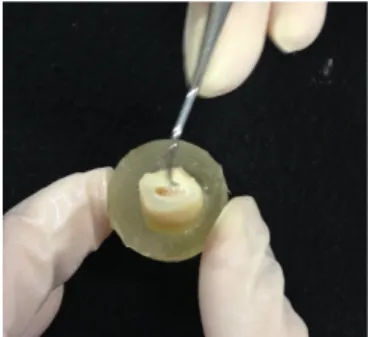
Figure 2 – Removal of the pulp
tissues.
Materials and methods:
1. Design of the study
A convenient sample of fifteen recently extracted human molars, intact and without macroscopic evidence of caries or restorations, was used on this study. Before their preparation, the teeth were randomly selected from a group of teeth firstly stored in 0,5% Chloramine T (Sigma Chemical Co., St Louis, MO, USA) at 4ºC for one week, according to the ISO/TS 11405 standard (ISO/TS 11405:2003) and then, left in distilled water at 4º C according to the ISO/TS 11405 standard (ISO/TS 11405:2003), no more than 6 months.
All teeth were cleaned under running water and all adherent tissues were removed using a periodontal scaler.
2. Teeth preparation
The teeth crowns were attached to an acrylic holder with sticky wax, perpendicular to the long axis of the tooth. Under constant distilled water irrigation and using a precision diamond disk at low speed (IsoMetTM Diamond Wafering Blade -10,2cmx0,3mm - Series 15HC, Buehler Ltd., Lake Bluff, IL, USA) on a hard tissue microtome (IsoMet® 1000 Precision Saw, Buehler Ltd. Ltd., Lake Buff, IL, USA), two cuts were made. The first cut was made parallel to the occlusal surface, 1-2 mm below the cementoenamel junction, to remove the roots and expose the pulp chamber (Figure 1). Then, the crowns were detached from the acrylic holders and the pulp tissues were removed from the pulp chamber with a dentin curette (Figure 2). The pulp chamber was then filled with cyanoacrylate glue (Loctite Super Cola 3 Precisão, Henkel, Germany) and the crowns were fixed with the same glue to the acrylic holders, by the sectioning surface.
Figure 3 – Removal of the occlusal enamel and
superficial dentin.
Figure 4 – Creating the smear layer on a
mechanical grinder.
A second cut was made parallel to the first one, in order to obtain mid-coronal dentin surfaces. This second cut removed both the occlusal enamel and superficial dentin of the molar crowns (Figure 3).
In order to create a uniform smear layer obtained in similar conditions of those occurring in clinic situations, the dentin surface was polished with 600-grit silica-carbide (SiC) sandpaper (CarbiMetTM SiC Abrasive Disk 600 [P1200] – 20,0mm – Buehler Ltd., Lake Bluff, IL, USA), for 60 seconds under water irrigation, on a mechanical grinder (DAP-U, Struers, Denmark), according to the ISO/TS 11405 standard (ISO/TS 11405:2003) (Figure 4).
3. Distribution and treatment of the crown segments
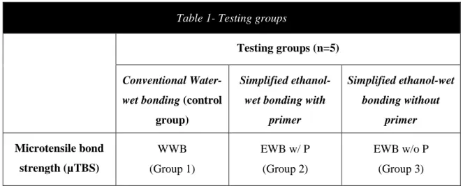
The fifteen crown segments were randomly assigned to one of the three groups (n=5), according to the different dentin conditioning methods (Table 1). The order in which the crown segments were treated was random, to avoid possible bias due to any particular sequence of treatment. All the treatment procedures were performed by the same operator.
Table 1- Testing groups
Testing groups (n=5) Conventional
Water-wet bonding (control group)
Simplified ethanol-wet bonding with
primer Simplified ethanol-wet bonding without primer Microtensile bond strength (µTBS) WWB (Group 1) EWB w/ P (Group 2) EWB w/o P (Group 3)
Figure 5 – Etching the dentin surface. Figure 6 – Vigorous ethanol application. In all groups, the dentin surfaces of the crown segments were etched for 15 seconds with a 37,5% phosphoric acid gel (Kerr Italia S.r.I., Scafati (SA), Italy). After acid etching, the surface was rinsed with water for 15 seconds (Figure 5). The excess of water was removed from the dentin surface using a moist cotton pellet so that the surface remained shiny and visibly moist, to prevent collapse of the collagen matrix.
In Group A, the control group, the water-wet bonding technique was performed using Adper Scotchbond Multi-Purpose Primer and Adhesive (3M ESPE, St. Paul, MN, USA). The primer was applied for 30 seconds to tooth surface with a disposable microbrush. The surface was then gently air-dried for 5 seconds, until it ceases to show any movement and the solvent was evaporated completely, forming a homogenous and slightly shiny film. If the dentin surface was overdried and didn’t remain visibly moist, a second coat of primer was applied.
For Groups B and C, an experimental simplified ethanol-wet bonding protocol was used. In both groups, after acid-etching, the dentin surface was treated with an experimental series of increasing ethanol concentrations: 70% and 96% (Continente, Portugal) ethanol applications, following a chemical dehydration protocol (Figure 6). Each concentration was applied by gently scrubbing for 30 seconds, using a disposable microbrush, giving a total application time of 60 seconds. Special attention was taken to ensure that the dentin surface was always visibly moist prior to the application of the subsequent higher ethanol concentration. After the ethanol application, excess ethanol was removed by gentle blotting with filter paper, leaving the dentin surface visibly moist.
Figure 7 – Experimental hydrophobic primer being
applied.
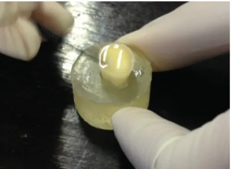
Figure 8 – Adhesive being
applied.
In Group B, after the chemical dehydration with ethanol that was previously described, an experimental primer was used. This experimental hydrophobic primer, formulated by combining 50 wt% resin monomers mixtures with 50 wt% ethanol was obtained by diluting 2 mL of Adper Scotchbond Multipurpose Adhesive (3M ESPE, Neuss, Germany) in 96% ethanol. This primer was applied as described in Group A (Figure 7).
In Group C, no primer was applied after the chemical dehydration protocol. Then, in all groups, the adhesive (Adper Scotchbond Multipurpose Adhesive, 3M ESPE, Neuss, Germany) was applied to the entire dentin surface, using a disposable microbrush, uniformly creating a thin coating. The adhesive excess was removed by gently air-drying. Finally, the surface was polymerized for 20 seconds (Figure 8).
Resin composite build-ups were performed using a resin composite, Herculite™ XRV Ultra™ Dentine (Kerr Italia S.r.I., Scafati (SA), Italy), shade A2, in three increments of 2 mm each. Each increment was light cured for 40 seconds, according to the manufacturer’s instructions, until reaching a height of 6 mm. Additional light polymerization was performed on facial, lingual, mesial and distal surfaces.
All light curing was performed with a light intensity of 600 mW/cm2, using a LED polymerization unit (Bluephase® 20i (G2), Ivoclar Vivadent, Austria) held 1-2 mm from the treatment surface The output of the curing light was periodically verified throughout the procedure, using a radiometer (Bluephase® meter, Ivoclar Vivadent, Austria).
Figure 9 – Resin application build up, after being
painted with waterproof ink.
4. Specimens preparation for micro-tensile tests
After restorative procedures, all teeth were painted using different colors with waterproof ink. The exterior surface of the resin composite was painted in order to identify, and then, exclude from the study, the sticks in which the adhesion was made to enamel (Figure 9).
Then, the specimens were used to evaluate the microtensile bond strength 24 hours after the restorative procedures (short-term test). The specimens were stored in distilled water in an incubator (TK/L 4105, EHRET GmbH & CO. KG, Germany) for 24 hours at 37º C according to the ISO/TS 11405 (ISO/TS 11405:2003) standard. Date and time of the restoration was registered.
Subsequently, after storage, the teeth were longitudinally sectioned in both “x” and “y” directions with a low-speed diamond disk (IsoMetTM Diamond Wafering Blade -10,2cmx0,3mm - Series 15HC, Buehler Ltd., Lake Bluff, IL, USA)), under water irrigation, using a hard tissue microtome (IsoMet® 1000 Precision Saw, Buehler Ltd. Ltd., Lake Buff, IL, USA), to obtain bonded sticks with a cross-sectional area of approximately 1 mm2. Firstly, cuts were spaced approximately 1 mm apart and oriented parallel to the long axis of the tooth (“x” direction) (Figure 10). Then, the tooth was then rotated 90 degrees and other cuts, spaced as described before, were made (“y” direction) (Figure 11).
Figure 10 – Specimen being cut in the “x”
direction.
Figure 11 – Specimen being cut in the “y”
direction.
Figure 12 - Sticks obtained after the final cut.
A final cut was made at the base of the crown, perpendicular to the long axis of the tooth, to separate the sticks from the acrylic holders (Figure 12).
Debonded or lost sticks were registered. Debonded sticks were those separated in the adhesive interface during the cutting procedure. Lost sticks where those which were lost or fractured during test preparation.
The obtained sticks were immediately subjected to microtensile tests.
5. Microtensile tests
The sticks were individually attached to a stainless-steel grooved Geraldelli’s jig with cyanoacrylate glue (737 Black Magic Toughened adhesive, Permabond, Hampshire, UK) (Figure 13) and then submitted, one by one, to a tension load using a universal testing machine (Instron 4502 Series, Serial no. H3307, Instron Corporation, Canton, MA, USA), at a crosshead speed of 1 mm/min, until fractured occurred (Figure 14).
After fracture, each stick was removed from the testing apparatus and a digital caliper (Absolute Digimatic Caliper, Mitutoyo Corporation, Japan) was used to measure the sides of the bonding interface, given by the cross sectioned area at the site of fracture, and calculate the bonding area in mm2 of each fractured stick. Both the load at fracture
Figure 14 – Instron 4502, universal testing machine. Figure 13 – The specimen attached to a
Geraldelli’s jig.
(kN) and the bonding surface area of the specimens were registered. Then, the µTBS values (μ-tensile bond strength) were calculated in MPa, by dividing the load of fracture by the bonding surface area.
The sticks that failed prematurely during specimen preparation were registered as the average value between zero and the lowest bond strength value obtained in all experiment (Luque-Martinez et al., 2014).
Failures were analyzed by the same observer, under a stereomicroscope (Nikon, Japan) at 10x magnification to determine the failure mode. The failure modes were classified as: 1) cohesive in dentin (CD), when the failure occurred in dentin; 2) cohesive in composite (CC), when the failure occurred in composite; 3) adhesive (A) when failure occurred at the dentin-adhesive interface; 4) mixed (M) when the failure involved both composite and dentin at the interface
Statistical analysis
The statistical analysis of the results was performed through descriptive and inference methods, using the software program SPSS Statistics for MAC Version 21.0 (SPSS Inc., Chicago, IL, USA).
Before submitting the data to the appropriate statistical analysis, the Shapiro-Wilk Test was performed to assess whether the data followed a normal distribution and the Levene’s test was computed to determine if the assumption of equality of variances (homoscedasticity) was valid (Table 2).
Since the normality of the data distribution and the equality of the variances were observed in two groups (p>0,05), the microtensile bond strength data was subjected to an one-way analysis of variance (ANOVA) and a post-hoc test (Tukey’s test) was used for pairwise comparisons. Significance was set at a 95% confidence level.
The number of prematurely debonded specimens (pretesting failures that occurred during specimen preparation) was recorded and included in the statistical analysis. As previously stated, the average value attributed to specimens with premature failures (PF) during preparation corresponded to the value between zero and the lowest bond strength value obtained in all experiment. In this specific study, the value of 1,914351 MPa was attributed when PF were recorded (Luque-Martinez et al., 2014).
Table 2 - Test for Equality of Variances of the µTBS in MPa
Levene Statistic df1 df2 Sig.
Results:
Microtensile bond strength
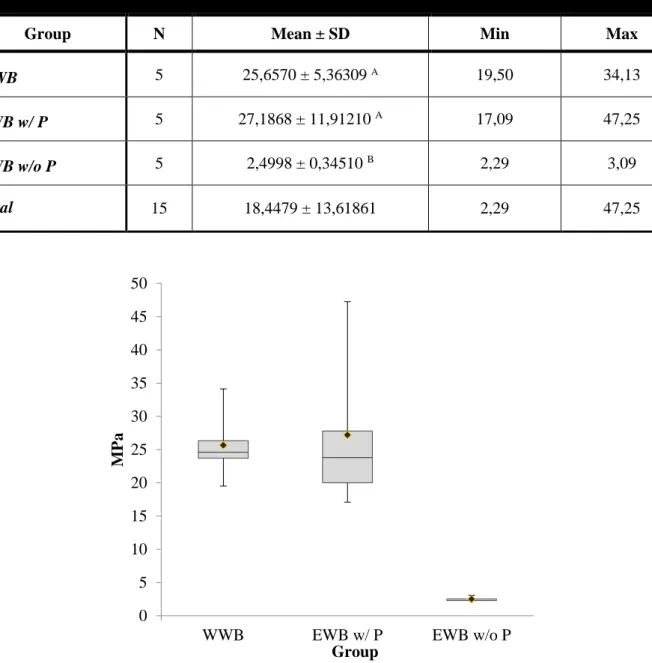
The mean values in MPa and standard deviations (SD) of the microtensile bond strength for each group are listed in Table 3 and shown in Figure 15. The number of specimens (N), minimum (Min), maximum (Max) are also summarized.
Table 3 - Descriptive statistics of the µTBS in MPa for the three experimental groups tested. Mean values that
are not significantly different from one another share the same upper case letter at p<0,05.
Group N Mean ± SD Min Max
WWB 5 25,6570 ± 5,36309 A 19,50 34,13
EWB w/ P 5 27,1868 ± 11,91210 A 17,09 47,25
EWB w/o P 5 2,4998 ± 0,34510 B 2,29 3,09
Total 15 18,4479 ± 13,61861 2,29 47,25
Figure 15 - Box-whisker plots of the µTBS in MPa for the two different experimental groups tested. The median µTBS is represented by the central line. The box represents the interquartile
range. The mean µTBS is represented by the diamond mark (♦). 0 5 10 15 20 25 30 35 40 45 50
WWB EWB w/ P EWB w/o P
MP
a
The highest mean µTBS was obtained when bonding was performed using EWB with primer (27,1868 ± 5,32725), followed by WWB (25,6570 ± 2,39845). The lowest was obtained when bonding was done using EWB without primer (2,4998 ± 0,15433) (Figure 16).
After verifying the normality of the data distribution and the equality of the variances, data from µTBS were analyzed using one-way ANOVA, which revealed that there was a significant difference among groups (p = 0,000) (Table 4).
Table 4 – One-way ANOVA test
Sum of Squares df Mean Square F Sig. Between Groups 1913,411 2 956,706 16,806 0,00 Within Groups 683,120 12 56,927 Total 2596,531 14
There is much difference between the two Mean Squares (956,706 and 56,927), resulting in a significant difference (F = 16,806; Sig. = 0,000). This means that the null hypothesis must be rejected. Tukey’s HSD test was applied to statistically evaluate the Figure 16 – Mean µTBS value for each group. Mean values that are not significantly
different from one another share the same upper case letter at p<0,05.
A A B 0 5 10 15 20 25 30
WWB EWB w/ P EWB w/o P
MPa
difference in the mean bond strength of the experimental groups (Table 5). The significance level was set at α = 0,05 for all tests.
Table 6 – Tukey HSD test. Mean µTBS values in MPa for groups in
homogeneous subsets are displayed.
Subsets for α=0,05 Group N 1 2 EWB w/o P 5 2,4998 WWB 5 25,6570 EWB w/ P 5 27,1868 Sig. 1,000 0,945
Tukey’s test revealed that mean µTBS values obtained with EWB without primer were significantly lower than those obtained with approaches using both WWB and EWB with primer (p=0,001). There are no statistically significant differences among the two other groups (WWB and EWB with primer) (p=0,945) (Table 5 and 6). Table 6 represents, in a more intuitive way, which groups are statistically similar, since they were listed in the same subset.
Table 5– Tukey HSD Post Hoc Test (α=0,05)
(I) Group (J) Group Mean Difference (I-J) SE Sig.
WWB EWB w/ P -1,52980 4,77186 0,945 EWB w/o P 23,15723 4,77186 0,001 EWB w/ P WWB 1,52980 4,77186 0,945 EWB w/o P 24,68703 4,77186 0,001 EWB w/o P WWB -23,15723 4,77186 0,001 EWB w/ P -24,68703 4,77186 0,001
Failure mode distribution
Failure mode distribution of the debonded specimens per tested group is shown in Table 7 and 8, Figures 17 and 18. Four failure patterns were depicted: adhesive (A), mixed (M), cohesive in dentin (CD) and cohesive in composite (CC).
Fracture analysis revealed that failure pattern was predominantly adhesive in all groups tested (86,7%). Mixed failures were observed in 4,4% of the specimens. Cohesive failures in composite and dentin were observed in 6,2% and 2,7% of the specimens, respectively.
Table 7 - Number of specimens according to the failure mode and premature failures of the
three different experimental groups tested.
Mode of failure Pretesting
failures A M CC CD Total Group WWB (1) 100 14 18 9 141 29 EWB w/ P (2) 131 6 10 3 150 25 EWB w/o P (3) 159 0 0 0 159 156 Total 390 20 28 12 450 210 70,9 9,9 12,8 6,4 87,3 4 6,7 2 100 0 0 0 0 10 20 30 40 50 60 70 80 90 100 A M CC CD %
WWB EWB w/ P EWB w/o P
Figure 17 - Distribution of the specimens according to the failure mode of the three different experimental groups tested.
Table 8 - Number of specimens according to the failure mode and
percentages of all experimental groups tested.
Mode of Failure N % Adhesive (A) 390 86,7 Mixed (M) 20 4,4 Cohesive in composite (CC) 28 6,2 Cohesive in dentin (CD) 12 2,7 450 100 0 10 20 30 40 50 60 70 80 90 A M CC CD 87,6 4,4 6,2 2,7 %
Figure 18- Distribution of the specimens according to the failure mode of all experimental groups tested.
Discussion:
EWB is a philosophic concept which tries to be overcome the problems associated with current adhesive systems. The ultimate purpose of this approach is to improve the clinical performance of the contemporary adhesives and extend the longevity of the restorations. This seems ambitious and attractive because the loss of restorations is a great problem in dentistry.
The present study, as previously stated, advocates a hybrid protocol, which comprised the use of ascending ethanol concentrations (70% and 96% only once, for 30 seconds each) during 60 seconds and the use of an experimental hydrophobic primer. This new approach has the aim of being implemented in the everyday clinical practice. Sadek et al., 2008 and Ayar and al., 2014 showed that full chemical dehydration protocol obtained promising results; nevertheless they are time-consuming, rendering it clinically impracticable. Liu et al., 2011 and Khoroushi et al., 2014 showed that simplified protocol is technique sensitive and produces lower bond strengths when compared to the previous protocol; nevertheless, this protocol is more user-friendly and has more clinical acceptance. Thus, the new approach proposed in this study represents an attempt to overcome the complications of both protocols, embracing their strengths: promoting strong adhesion through a simple and user-friendly protocol.
After an extensive literature search, the authors outlined their study. Rendering a user-friendly protocol implies that easily available materials should be preferred. Thus, it seems only rational to use commercial adhesive systems. In this study, the phosphoric acid and the composite are not from the same manufacturer as the primer and adhesive. This has the purpose of avoiding possible bias in µTBS values when using all materials from the same manufacturer. The same user-friendly rationale was used to select the ethanol concentrations applied. This is the first study to perform EWB with two ethanol concentrations easily available at a supermarket: 70% and 96%. This contrasts with the full chemical dehydration protocol, which makes use of five different ethanol concentrations. Besides their availability, these two ethanol concentrations have the advantage of being less volatile than higher concentrations, allowing a gradual and effective water displacement (Pashley et al., 2007; Li et al, 2012).
In the literature search, it became evident that there was not a consensus about primer application. While some authors applied the commercial primers (Khoroushi et al., 2014), application of an experimental hydrophobic primer was advocated by several authors (Sadek et al, 2008; Sadek et al, 2010a; Sauro et al., 2011; Pei et al., 2012; Araújo et al., 2013). Yet, Mortazavi et al., 2012 did not use any primer; instead, the commercial adhesive was applied after the excess ethanol was absorbed. This last protocol seemed interesting because if ethanol removed water effectively, then there seems to be no need to apply a primer composed by hydrophilic monomers. Theoretically, it makes sense to assume that hydrophobic monomers, such as Bis-GMA, present in the adhesives, are able to be coaxed into ethanol-saturated dentin (Nishitani, et al., 2006). In order to access the effect of primer application on the EWB technique, the present study compares the application of an experimental hydrophobic primer to the application of no primer in the proposed EWB approach. Then, the two variables chosen for this study are defined.
The experimental hydrophobic primer was obtained by diluting the Adper Scotchbond® MultiPurpose Adhesive with 50 wt% 96% ethanol. The aim of this procedure was to produce a water-free bonding resin with similar composition of the hydrophilic adhesive employed in the water-wet protocol (Sadek et al, 2010a). The primer from this adhesive system is water-based and has HEMA and water in its composition. EWB displaces water within dentin, so it seems to be counterproductive to use a water-based primer whenever using a EWB approach.
However, the literature also exhibits a high variability in ethanol application protocols in terms of the required application time. In the quest of a simplified and yet effective protocol, reduced application times should be preferred. As EWB represents an additional step, ethanol must be applied during the minimum time required to be effective Sauro et al, 2010 stated that similar results arise when ethanol was applied during 300 seconds (5 minutes) or 60 seconds. Sadek et al., 2010a reported that 30 seconds were not enough for complete replacement of water within the dentin by ethanol. Hence, an application time of 60 seconds was chosen.
Contrary to most studies and despite the manufacturer recommendations, the authors increased the curing time of the resin composite. Each composite increment was polymerized during 40 seconds. In fact, Silva, 2008 and Pequeno, 2009 suggested that the 20 seconds recommended by the manufacturer was inferior to the ideal. Perdigão et
al., 2006 and Proença et al., 2007 obtained less cohesive fractures in composite when the resin composite was cured was cured twice over (4 seconds) the recommended time.
The first null hypothesis was rejected, as immediate bond strength varied, depending on the bonding approach. The results of this study showed that there are not statistically significant differences between the WWB and EWB w/ P. However statistically significant differences were observed between EWB w/o P and the other two bonding approaches, which rejects the second null hypothesis.
Despite the great variability found in protocols, the results of this study are in agreement with previous reports proving that there are not statistically significant differences between WWB and EWB, after 24 hours (Hosaka et al., 2009; Guimarães et al., 2012; Ekambaram et al., 2014; Manso et al., 2014; Yesilyurt et al., 2015).
When solely comparing EWB with WWB, there are no similar studies, since this is the first study that compares 70% and 96% ethanol concentrations. Nevertheless, studies in which commercial etch-and-rinse adhesives, such as Adper Scotchbond® MultiPurpose and Optibond FL, are used were conducted by several authors (Sadek et al, 2010a; Mortazavi et al., 2012; Pei et al., 2012; Khoroushi et al., 2014 and the work of Araújo et al., 2012) must be highlighted. Araújo et al., 2012 used two ethanol concentrations (50% and 100% for 20 seconds each) and the commercial etch and rinse adhesive Adper Scotchbond MultiPurpose, concluding that there are not statistically significant differences between EWB and the gold standard etch-and-rinse protocols, at 24h. According to the literature, same results could be obtained when booth commercial adhesive systems (Yesilyurt et al., 2015; Manso et al., 2014) and experimental primers and adhesives (Hosaka et al., 2009; Ekambaram et al., 2014) were used.
The lack of statistically significant differences between EWB and WWB at 24 hours, despite the adhesive system used, can be purportedly explained by the solubility theory. As stated by Nishitani et al., 2006, when etched dentin was ethanol-saturated, the Hoy’s solubility parameter of the collagen matrix was brought closer to those of the ethanol-solvated resins. It is speculated that optimal wetting of collagen fibrils by these solvated resins occurs when the polar surface-free energy components are similar (Barton, 1991). Hence, the significant relationships between resin hydrophilicity and µTBS may be due to the degree of wetting and penetration of acid-etched ethanol-saturated dentin by the resins (Rosales et al., 1999; Asmussen & Peutzfeldt, 2005). It is also conjectured
that relatively hydrophobic resins produces higher bond strengths over time (Brackett et al., 2005; Ito et al., 2005; Nishitani et al., 2006. The adhesive used, Adper Schotchbond MultiPurpose Adhesive, is solvent-free and relatively hydrophobic, despite having HEMA in its composition. As described by Tay et al., 2007, the sequential steps in EWB technique allow for improved miscibility of the solvated adhesive and the collagen matrix thereby enabling the solvated hydrophobic resin blend to infiltrate an ethanol-saturated collagen matrix. It is proposed that diluting this relatively hydrophobic resin represented a crucial step towards the success of this technique. The adhesive polar forces were brought closer to those of the ethanol-saturated dentin, enabling resin monomers infiltration, achieving a relatively homogeneous distribution of hydrophobic resins within the hybrid layer. (Nishitani et al., 2006; Liu et al., 2011). EWB technique exhibited statistically similar results to WWB, despite the last being accomplished through the use of a gold standard adhesive. Thus, it was conjectured the adhesive elution was responsible for the success of resin impregnation (Souza Júnior, 2015).
Some authors studied different protocols and achieved different results, registering statistically significant differences when comparing the two bonding approaches at 24 hours (Osorio et al., 2010; Huang et al., 2011; Ayar, 2014). These results may be partially explained due to the technique sensitivity inherent in the EWB bonding philosophy (Osorio et al., 2010; Huang et al., 2011). Tay et al., 2007 have found that any residual water molecule in the collagen fibril network interfered with the infiltration of hydrophobic adhesive monomers, since resin monomers are less soluble in water. Besides that, as proved by Osorio et al., 2010, the etched dentin surfaces exhibited topographical changes depending on the protocol applied. EWB produced smoother surfaces, narrow fibrils and wider interfibrillar spaces, when compared to WWB. These differences may account for variability in bond strengths.
The role of adhesive elution can be further proven since the group where the EWB was applied without a primer, exhibited statistically significant differences when compared to the other two groups. Only three sticks were left for testing, which proves that adhesion was not successfully achieved. Apparently, without the use of a primer, stiff monomer Bis-GMA was not able to fully diffuse into ethanol-saturated dentin. It is suggested that solvating the bonding resin in ethanol was responsible for decreasing the surface activity of the resin, allowing better wetting and penetration (Ounsi et al., 2009). It should be noted that although ethanol-saturated dentin might be a better substrate for
adhesive infiltration, the ethanol is highly volatile due to its vapor pressure being greater than that of water, thus compromising the wettability of etched dentin after a short period time (Li et al., 2012). Hence, adhesive should be applied within the time window when the dentin matrix is fully saturated with ethanol. On the other hand, Cadenaro et al., 2009 and Sauro et al., 2001 proposed that Bis-GMA contains hydroxyl groups that can bond with ethanol and that the residual presence of ethanol decreases the initial reaction rate but enhances degree of conversion of resins after 60 seconds exposure. Hence, it is postulated that, without the primer, the ethanol application time must be superior to 60 seconds. These results are not in agreement with the results from the only study in which primer was not applied (Mortazavi et al., 2012). In this study, the adhesive applied was OptiBond FL. OptiBond FL does not have the same composition as Adper Schochbond MultiPurpose, which may explain the different outcomes observed.
In the present study, there were no differences in the distribution of fractures across all tested groups. In fact, the most predominant failure pattern was adhesive in WWB, EWB w/ P, EWB w/o P groups (70,9%, 87,3% and 100%, respectively) (Figure 18). This is in accordance with previous studies in literature reporting that, when performing a microtensile bond strength test, more adhesive failures than cohesive failures are expected (Pashley et al., 1995; Schreiner et al., 1998). In fact, an accurate assessment of the strength of an adhesive material is best determined when the failure occurs within the material itself and does not involve dentin or composite (Sano et al., 1994; Ghassemieh et al., 2008). Sano et al., (1994) also states that composite cohesive failures during in vitro tests are not representative of clinical situations, limiting the interpretation of μTBS.
One of the limitations present in this study relates to the scarce literature data available. This limited the experimental study design, since there is little information available to support the study. Besides, as previously stated, there is a plurality of heterogeneous protocols described, which limited the comparisons to the present study.
Another limitation of this study has to do with the lack of long-term water storage. The present study did not evaluate the effects of ageing in the hybrid layers produced by EWB approaches, which is a crucial topic when evaluating the advantages associated with the EWB technique. With this in mind, future studies should analyze the effect of long-term water storage on the in vitro performance of a EWB approach.
It is suggested that further investigation should be made. Besides the long term studies, EWB must be study on tertiary dentin, since in a clinical scenario adhesion, adhesion procedures are performed in teeth which were affected by caries. Nanoleakage tests should also be performed.
It is important to state that the in vitro nature of this study does not allow direct extrapolation of the results to an in vivo situation, so whether the same results would be obtained in vivo should be the object of further investigation.
Conclusion:
Within the limitations of the present laboratory study, it may be concluded that, when applied with a primer, the proposed EWB showed similar bonding effectiveness to WWB, after 24 hours. On the other hand, when EWB was applied with no primer, it showed lower bonding effectiveness, when compared to the other two groups.
This has an enormous impact in the adhesive field, since it suggests that contemporary commercial etch and rinse adhesives, considered the gold standard, can be deeply improved. Improving the durability of a restoration affects not only the clinical practice (less failures, less restorations substituted) but also has a deep economic effect on the patients and on the manufacturers.
On the other hand, it can be concluded that some modifications must be made in order to directly bond the ethanol-saturated dentin without using a primer.
Since the EWB approach is relatively new, there is little information in the literature about the in vitro performance of this approach. Future studies should analyze the effect of long-term water storage on the in vitro performance of the proposed protocol.
Besides that, further studies on different substrates, such as tertiary dentin and nanoleakage studies, in association with in vivo tests are needed to assess the long-term clinical behavior of this protocol to support its application.
Clinical significance: Similar bonding effectiveness of the tested ethanol wet bonding
APPENDIX I
Materials, Manufacturers, Components and Batch Numbers
Table 9 – Materials, Manufacturers, Components and Batch Numbers
Material Manufacturer Composition Batch
Number
37,5% Phosphoric Acid Gel Etchant
Kerr Italia S.r.I. Scafati (SA), Italy
Phosporic acid Pigments Lot 4907232 Exp 07/2016 Adper Scothbond MultiPurpose Primer 3M ESPE Neuss, Germany HEMA (35–45%) Water (40–50%) Copolymer of acrylic and itaconic
acids(10–20%) Lot N547824 Exp 01/2017 Adper Scothbond MultiPurpose Adhesive 3M ESPE Neuss, Germany BISGMA (60–70%) HEMA (30–40%) Triphenylantimony (<0.5%) Lot N655294 Exp 02/2018 Herculite™ XRV Ultra™ Dentine
Kerr Italia S.r.I. Scafati (SA), Italy
ethoxylated Bisphenol A-dimethacrylate; 2,2-ethylenedioxydiethyl
Dimethacrylate;
3Methacryloxypropyltrimethoxysilane bisphenol A-glycidyl methacrylate
(BIS-GMA)
Lot 5629580 Exp 05/2018
Álcool 70% Vol Continente,Portugal
Álcool etílico 70% vol 0,25% cetrimida
Lot 15001003 Exp 11/2020
Álcool 96% Vol Continente Portugal
Álcool etílico 70% vol 0,25% cetrimida
Lot 15001130 Exp 12/2020
Ribeiro S
APPENDIX II
Table 10 – Number of sticks obtained in each tooth
Group Specimens Obtained
sticks Debonded sticks Lost sticks Tested sticks Tested sticks by group 1 WWB 1.1 25 9 2 14 111 1.2 32 6 2 24 1.3 34 1 0 33 1.4 32 5 2 25 1.5 26 8 3 15 2 EWB w/ P 2.1 36 8 0 28 125 2.2 28 5 0 23 2.3 32 2 0 30 2.4 31 4 0 27 2.5 23 6 0 17 3 EWB w/o P 3.1 31 31 0 0 3 3.2 27 26 0 1 3.3 24 22 0 2 3.4 47 47 0 0 3.5 30 30 0 0 Total 459 210 10 239 239
References:
Araújo JF, Barros TAF, Braga EMF, Loretto SC, Souza PARS, MHS Souza Júnior. One-Year Evaluation of a Simplified Ethanol-Wet Bonding technique:A Randomized Clinical Trial. Brazilian Dental Journal 2013;24(3): 267–73.
Ayar MK. Ethanol Application Protocols and Microtensile Dentin Bond Strength of Hydrophobic Adhesive. Tanta Dental Journal 2014a; 11(3):206–12.
Ayar MK, Yesilyurt C, Alp CK, Yildirim T. Effect of Ethanol-Wet-Bonding Technique on Resin-Enamel Bonds. Journal of Dental Sciences 2014b; 9(1):16–22.
Ayar MK. A Review of Ethanol Wet-Bonding: Principles and Techniques. European Journal of Dentistry 2016; 10(1).
Barton, AFM. Surfaces and interfaces. In: Barton, AFM., editor. CRC handbook of solubility parameters and other cohesion parameters. 2nd ed.. Boca Raton, FL: CRC Press, Inc.; 1991. p. 583-629
Asmussen E, Peutzfeldt A. Resin composites: strength of the bond to dentin versus surface energy parameters. Dent Mater 2005;21:1039–1043.
Bertassoni LE., Orgel JPR, Antipova O, Swain MV. 2012. The Dentin Organic Matrix - Limitations of Restorative Dentistry Hidden on the Nanometer Scale. Acta Biomaterialia 2012; 8(7):2419–33.
Brackett WW, Ito S, Tay FR, Haisch LD, Pashley DH. Microtensile dentin bond strength of self-etching resins: effect of a hydrophobic layer. Oper Dent 2005;30:733–738.
Breschi L, Mazzoni A, Ruggeri A, Cadenaro M, Di Lenarda R, Dorigo EDS. Dental Adhesion Review: Aging and Stability of the Bonded Interface. Dental Materials 2008; 24(1):90–101.


