ABSTRACT
Push-out bond strength and SeM evaluation of a
new bonding approach into the root canal
Carlos Augusto CARVALHO1, Lorenzo BRESCHI2, Maria Fidela NAVARRO3, Maria Teresa ATTA4, Marco FERRARI5
1- Graduate student, Department of Operative Dentistry, Endodontics and Dental Materials, Bauru School of Dentistry, University of São Paulo, Bauru, SP, Brazil; University of Florence and Siena, Tuscany School of Dental Medicine, Florence, Italy.
2- Associate Professor, Department of Medical Sciences, University of Trieste, Trieste, Italy; IGM, Unit of Bologna, C.N.R. - IOR, Bologna, Italy.
3- Full Professor, Department of Operative Dentistry, Endodontics and Dental Materials, Bauru School of Dentistry, University of São Paulo, Bauru, SP, Brazil. 4- Associate Professor, Department of Operative Dentistry, Endodontics and Dental Materials, Bauru School of Dentistry, University of São Paulo, Bauru, SP, Brazil. 5- Full Professor, University of Florence and Siena, Tuscany School of Dental Medicine, Siena, Italy.
Corresponding address: Carlos Augusto Ramos de Carvalho - Rua Gonçalves Dias, 2525 - 30140-092 - Belo Horizonte - Minas Gerais - Brasil - Phone: 55-31-9232-3262 - e-mail: [email protected]
Received: May 23, 2011 - Modiication: July 14, 2011 - Accepted: September 16, 2011
O
bjective: This study evaluated the performance of different adhesive systems in iber post placement aiming to clarify the inluence of different hydrophobic experimentalblend adhesives, and of one commercially available adhesive on the frictional retention during a luting procedure. Material and Methods: One luting agent (70 Wt% BisGMA, 28.5%
TEGDMA; 1.5% p-tolyldiethanolamine) to cement iber posts into root canals was applied
with 4 different adhesive combinations: Group 1: The etched roots were rinsed with water for 30 s to remove the phosphoric acid, then rinsed with 99.6% ethanol for 30 s, and blot-dried. A trial adhesive (base to catalyst on a 1:1 ratio) was used with an experimental luting agent (35% Bis-GMA, 14.37% TeGDMA, 0.5% eDMAB, 0.13% CQ); Group 2: A trial adhesive (base to catalyst on a 1:2 ratio ) was luted as in Group 1; Group 3: One-Step Plus (OSP, Bisco Inc.) following the ethanol bonding technique in combination with the luting agent as in Group 1; Group 4: OSP strictly following the manufacturer’s instructions using the luting agent as in Group 1. The groups were challenged with push-out tests. Posted root slices were loaded until post segment extrusion in the apical-coronal direction. Failure modes were analyzed under scanning electron microscopy. Results: Push-out strength
was not signiicantly inluenced by the luting agent (p>0.05). No statistically signiicant
differences among the tested groups were found as Group 1 (exp 1 – ethanol-wet bonding technique)=Group 2 (exp 2 – ethanol-wet bonding technique)=Group 3 (OSP – ethanol-wet bonding technique)=Group 4 (control, OSP – water-wet bonding technique) (p>0.05). The dominating failure modes in all the groups were cohesive/adhesive failures, which were predominantly observed on the post/luting agent interface. Conclusions: The results of this study support the hypothesis that the proposal to replace water with ethanol to bond
iber posts to the root canal using highly hydrophobic resin is plausible, but this seems to
be more the proof of a concept than a clinically applicable procedure.
Key words: Root canal. Luting cement. Hydrophobic adhesives. Dentinal bonding.
INTRODUCTION
The process of hybrid layer formation in etch-and-rinse dentin bonding systems (DBS) involves the penetration of resin monomers into a delicate
layer of unsupported collagen ibrils exposed by
the etching agent (usually 35-37% phosphoric acid). The etching agent was inactivated, and removed by copious air/water spray. This is
because etch-and-rinse DBS impregnate the substrate in accordance with the “water-wet” bonding technique, i.e. collagen fibrils should remain wet to avoid excessive shrinkage due to desiccation that can impair resin impregnation.
The residual water within the ibrilar network was
hydrophilic resin impregnation into water-wet dentin collagen matrices consists in a passive diffusion mechanism20-23. Solvents also have an important role in monomer impregnation as they can reduce resin viscosity, and increase the water substitution rate, thus facilitating water displacement within the
demineralized collagen ibrils. The more common
solvents used for DBS are ethanol, acetone, and water14.
As hydrophobic resin blends showed higher stiffness, improved stability over time, and reduced water uptake11,15,16 when compared to more hydrophilic ones10, hydrophobic monomers should be preferred to produce a stable bond over time. However, if the adhesive blend is too hydrophobic, suboptimal impregnation occurs since the solvents cannot replace all the residual water
within the demineralized dentin collagen ibrils. This insuficient resin penetration leads to the
formation of a hybridoid layer characterized by voids and porosities with reduced sealing ability3,4,8,20. Areas of incomplete resin impregnation can result
in nanoleakage, and can be identiied using a
tracer (silver nitrate) under an scanning electron microscopy (SeM)19.
In order to coax hydrophobic monomers into demineralized dentin collagen matrices, the “ethanol-wet bonding technique” has recently been proposed16,22.This technique is characterized by sequential rinses with ethanol at ascending
concentrations to replace interibrillar water16,18,22. Since the ethanol-saturated dentin is more compatible with hydrophobic resin monomers, collagen shrinkage is prevented, and impregnation is facilitated6.This technique has been shown to produce adhesive interfaces with higher bond strength, reduced interfacial nanoleakage expression, and increased stability over time when compared to the “water-wet bonding technique”9. The use of the ethanol-wet bonding technique has also been shown to produce encouraging results when used to lute posts to intra-radicular dentin5. In addition, the possibility of using high hydrophobic resin possibly minimizes endogenous collagenolytic activities9.
Despite promising in vitro results, the ethanol-wet bonding technique is time consuming, as several ethanol rinses should be performed to completely replace the residual water within the dentin collagen, and to allow hydrophobic
monomers to iniltrate into a fully ethanol saturated dentin. Recently, a simpliied ethanol wet-bonding
procedure with the reduction of time of application has been proposed to bond to coronal dentin17.
However, no studies have clariied if the proposed “simpliied ethanol-wet bonding technique” could be beneicial when luting iber posts to intra-radicular
dentin.
The aim of the present study was to compare the bond strength and interfacial morphology created by an experimental or a commercially-available DBS in association with resin-based cements used
to lute iber posts within the endodontic space,
by using the simplified ethanol-wet bonding technique. The null hypotheses tested were that: (1) no differences exist between the push-out bond strengths of hydrophobic experimental resin blends and a commercially available two-step etch-and-rinse adhesive; (2) no differences exist between the
push-out bond strengths of a simpliied ethanol-wet
bonding technique and those of a commercially available two-step etch-and-rinse adhesive.
MATERIAL AND METhODS
Specimen preparation
Twenty single-rooted premolars showing a single-canal, and extracted for orthodontic reasons were selected for the study after informed consent was obtained under a protocol approved by the University of Siena (Siena, Italy). exclusion criteria were teeth shorter than 20 mm, apex larger than
a size 25 K-ile before instrumentation, presence of caries, root issures, or fractures.
The teeth were hand-scaled, and stored in 1% chloramine, T at 4°C, and used within 1 month after extraction. Crowns were removed cutting the teeth 2-mm over the cementum-enamel junction, using a slow-speed diamond saw (Micromet, Remet, Casalecchio di Reno, BO, Italy). Canals were shaped using nickel-titanium rotary instruments (size S1, S2 and F3; Protaper, Dentsply-Maillefer, Ballaigues, Switzerland). Root canals were prepared using the Protaper Universal System according to the sequence: S1 and SX for the 2/3 coronal third, and then, instruments S1, S2, F1, F2, and
F3 at the work length for the inal preparation in
accordance with the crown-down fashion to an ISO size 30/0.07 taper. Irrigation with 5% NaOCl was performed (Niclor 5; Dentale-Ogna, Milan, Italy) during instrumentation using a syringe with a 30G endodontic needle (Perio/endo Irrigation Needle Biaggio Switzerland). Removal of the smear layer was obtained after irrigation with 3 mL of 17% eDTA for 2 min, followed by 3 mL of saline.
After the inal rinse, root canals were completely
dried with air stream and absorbent paper points
(Dentsply-DeTrey, Konstanz, Germany) and illed by
lateral condensation of gutta-percha cones and a resin-based sealer (AH-26, Dentsply-DeTrey). The
illed roots were coronally sealed with glass ionomer
cement (Fuji VII, GC Corporation, Tokyo, Japan) as the coronal temporary restorative material,
and stored in 100% humidity in labeled white ilm
containers for 24 h at 37°C.
Groups Bond Strength (SD)
Number of Slices
Failure mode (%) A/M/PA/C
1. Experimental adhesive 1 + simpliied ethanol-wet bonding technique
6.9 (5.9)a 33 0/38.89/11.11/50
2. Experimental adhesive 2+ simpliied ethanol-wet bonding technique
6.7 (5.4)a 32 0/0/40/60
3. OSP + simpliied ethanol-wet bonding technique 6.8 (4.3)a 32 0/32/4/64
4. OSP + water-wet bonding technique (Control) 6.9 (5.1)a 31 7.14/25/0/67.86
Table 1- Median push-out bond strength values* (SD) expressed in MPa, number of specimens (N), and percentage of failure mode distribution recorded in the experimental groups. OSP= One-Step Plus
gutta-percha was removed in all groups using a low-speed universal drill provided by the manufacturer, and keeping at least 4 mm of apical seal. A standardized 7-mm post space was drilled in each root with the #2 drill that corresponded to RelyX Fiber Post size #2 (3M eSPe, St. Paul, MN, USA).
The n value was obtained after a power analysis of 80%, in order to calculate the minimum effective size that is likely to be detected in a study using a given sample showing that the sample size was adequate; teeth were equally (n=5) and randomly divided into 4 groups according to the adhesive procedure (Table 1). All monomers were purchased by Sigma-Aldrich, St. Louis, MO, USA, Group 1: experimental adhesive (35% Bis-GMA, 14.37% TeGDMA, 0.5% eDMAB, 0.13% camphoroquinone (CQ); co-monomers to ethanol on a 1:1 ratio)
applied in accordance with the simpliied
ethanol-wet bonding technique; Group 2: experimental adhesive (composition similar to Group 1 with co-monomers to ethanol on a 1:2 ratio) applied in accordance with the simplified ethanol-wet bonding technique; Group 3: One-Step Plus (OSP)
applied in accordance with the simpliied
ethanol-wet bonding technique; Group 4: OSP applied strictly following the manufacturer’s instructions (i.e. water-wet bonding technique; control group). Chemical compositions of all materials and systematic clinical procedures are described in Table 1. In brief, root canal walls were etched with 32% H3PO4 gel (Bisco Inc., Schaumburg, IL, USA) for 15 s, using an intracanal tip, then specimens of groups 1, 2 and 3 were rinsed with water for 15 s using an endodontic needle, and root canals
were illed with 99.6% ethanol (Sigma-Aldrich)
for 30 s. Canals were then gently dried with paper points leaving an evident visual aspect of ethanol-wet saturated surfaces. Adhesive blends of groups 1-3 were then immediately applied on ethanol-wet dentin, and light-cured using a conventional quartz-tungsten-halogen light (600-mW/cm² output; VIP;
Bisco Inc.). The inal ethanol rinse was avoided in
specimens of group 4 that were prepared applying OSP on water-wet dentin.
The conical epoxy resin posts size #2
(RelyX-Posts; 3M eSPe) were cleaned in ethanol, and surface-treated with a silane solution (Porcelain P r i m e r ; B i s c o ) u s i n g a d i s p o s a b l e b r u s h , and gently air-dried for 5 s. An unfilled resin (Scotchbond MP/3M eSPe) was used as a luting agent (2-hydroxyethylmethacrylate, bis-phenol, A diglycidylmethacrylate, photoinitiator; Table 1), and placed into the canal using a disposable syringe. All
iber posts were then seated under inger pressure,
and the excess of luting material was removed while maintaining a seal of the exposed dentin along the coronal part of the root. Light curing was performed using a conventional quartz-tungsten-halogen light (600-mW/cm2; VIP; Bisco Inc.) by placing the light tip perpendicularly on the post for 40 s. All bonded specimens were then placed in individually labeled containers in 100% humidity for 24 h at 37°C.
Preparation of specimens for the push-out strength test
After 24 h, the portions of the roots corresponding
to the bonded iber post were transversely sectioned
into 1-mm-thick serial slices using a slow speed diamond saw under water irrigation (Micromet M, Remet; Casalecchio di Reno, Bologna, Italy). The apical surfaces of the slices were marked with a permanent black-ink dot. The push-out load was applied using a universal testing machine (Controls S.P.A., Milano, Italy) at a crosshead speed of 0.5 mm/min. The apical surface displaying the ink dot was placed facing the punch tip ensuring that loading forces were introduced in an apical to coronal direction. With regard to the tapered design of the post, three different sizes of punches were used for the push-out testing. The diameter of the punch pin was 1.2 mm for the coronal slices, 1.0 mm for the middle slices, and 0.8 mm for the apical slices. This guaranteed that the strength was applied as more adequately as possible to the bonded area during the loading process.
Bond failure was manifested by the complete
dislodgment of the iber-post from the root section.
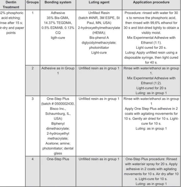
Figure 1- Chemical composition and application mode of the materials used in the study
Dentin Treatment
Groups Bonding system Luting agent Application procedure
32% phosphoric acid etching; rinse after 15 s; air-dry and paper
points
1 Adhesive
35% Bis-GMA, 14.37% TEGDMA, 0.5% EDMAB, 0.13%
CQ ligth-cure
Unilled Resin (batch #4NR, 3M ESPE, St
Paul, MN, USA) 2-hydroxyethylmethacrylate
(HEMA); Bis-phenol A diglycidylmethacrylate;
photoinitiator Light-cure
Procedure: rinsed with water for 30 s to remove the phosphoric acid, then rinsed with 99,6% ethanol for 30 s and blot-dried lightly to obtain a
visibly moist.
Mix Experimental Adhesive with Ethanol (1:1).
Light cured for 20 s. Luting: Apply unilled resin using a disposable syringe, then light cured
for 40 s.
2 Adhesive as in Group
1
Unilled resin as in group 1 Rinse with water/ethanol as in group 1.
Mix Experimental Adhesive with Ethanol (1:2).
Light-cured for 20 s Luting: as in group 1
3 One-Step Plus
(batch # 0500002430, Bisco Inc., Schaumburg, IL,
USA) Biphenyl dimethacrylate;
2-hydroxyethyl methacrylate; Acetone; amine; photoinitiator; dental
glass
Unilled resin as in group 1 Rinse with water/ethanol as in group 1
Apply One Step Plus adhesive in 2 coats with agitating movements for 10 s. Gently air dried for 10 s.
Light-cure for 10 s. Luting: as in group 1
4 One-Step Plus Unilled resin as in group 1 One-Step Plus procedure: Rinsed
with water/air spray for 20 s. Apply adhesive in 2 coats with agitating movements for 10 s. Air dry after 10
s. Light-cure for 10 s. Luting: as in group 1
of the upper and lower sides of each specimen were obtained, and the failure limits were traced with a closed line using image analysis software (Image Pro Plus 5.0; Media Cybernetics, Bethesda, MD, USA). Limits were then measured (after software calibration) in accordance with Ferrari, et al.7 (2009)
,and the thickness of the slice was individually measured using a digital caliper with 0.01-mm accuracy. SL was calculated as the lateral surface area of a truncated cone using the formula:
where R is the coronal post radius, r the apical post radius, and h the thickness of the slice.
Modes of failure where classiied as (A) adhesive
between dentin and cementing agent, (M) mixed, (PA) adhesive between post and cementing agent, or (C) cohesive if cementing agent failures were assessed with a stereomicroscope (Nikon SMZ645,
Nikon, Tokyo, Japan) at 30x magniication.
Statistical analysis
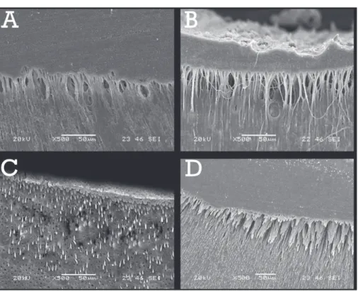
Figure 2- Scanning electron microscopy images (SEM) (magniication 500x, bar 50 μm) from a representative push-out
tested on dentin slice specimens: (A) Experimental Group 1; and (B) Experimental Group 2 showed an evident hybrid layer with long resin tags; (C) One Step Plus/Ethanol/Experimental Luting Agent resulted in the formation of short and discrete resin tags; (D) One Step Plus as the control group using the regular wet bonding technique demonstrated the formation of long, deep, and compact resin tags into the dentin
signiicance was set at p<0.05. The analyses were
performed using SigmaStat 3.5 (Windows Version; SPSS, Chicago, IL, USA).
SEM sample preparation
Representative fractured slices from each group were randomly assigned to scanning electron microscopy (SeM) analysis22.each slice was smoothened with wet silicon carbide paper of decreasing abrasiveness (up to 1200 grit). To analyze hybrid layer morphology and resin tag formation, the specimens were etched with silica free 32% H3PO4 (etching gel; Bisco) for 20 s, and, subsequently, immersed for 2 min in 2.5% NaOCl to remove the organic and mineral components of the dentin, rinsed with water, and dehydrated with 99.8% ethanol to analyze hybrid layer morphology, and resin tag formation. Specimens were then mounted on aluminum stubs, sputter coated with gold (Polaron Range SC7620, Quorum Technologies, Newhaven, england), and observed under a scanning electron microscope SeM (JSM 6060 LV, JeOL, Tokyo, Japan). Micrographs were
taken at different magniications in order to provide
an overview of each area, and to evaluate the type of micro morphologic pattern of the representative specimens.
RESULTS
Mean values and SDs expressed in MPa of push-out bond strength, and numbers of slices/group and failure modes (%) are summarized in Table 1. No premature failures were found during the cutting procedure or during the testing procedure.
No statistically signiicant differences were found
among the tested groups (p>0.05).
SEM evaluation
The majority of the specimens showed cohesive/ adhesive failures on the post/luting agent interface. By the images, Groups 1, 2, and 3 demonstrated a visually deeper penetration of the bonding material different from Group 4 (Figure 1). All groups formed a distinct hybrid layer.
DISCUSSION
The results of this study showed that experimental hydrophobic resin blends (Groups 1, 2) have similar bond strength values to OSP (Group 3) if applied in
accordance with the simpliied ethanol-wet bonding technique, and, thus, the irst null hypothesis
was accepted. In addition, similar bond strength was obtained when OSP was applied with the
simpliied ethanol-wet bonding technique (Group
thus, the second null hypothesis was also accepted. The rationale on the use of the ethanol-wet bonding technique is that hydrophobic monomers can better infiltrate the ethanol-saturated demineralized dentin due to reduced polarity of the collagen network to match the low polarity of high hydrophobic resins. Since hydrophobic resin blends have higher stiffness and stability than hydrophilic ones, bonding to dentin with hydrophobic resin blends in association with the ethanol-wet bonding technique showed excellent results both on immediate dentin bonding and on the longevity of the bond created on coronal dentin substrates2,9,13,18.
An essential pre-requisite for the achievement of the complete impregnation of the collagen
ibrils exposed by acid etching with hydrophobic monomers is that dentin interibrillar residual water
is fully replaced by ethanol, and, since ethanol is a water chasing solvent, it removes water from the tissues. If water remains within the collagen network, hydrophobic monomers cannot fully embed the demineralized dentin substrate, and water-rich domains remain within the hybrid layer, constituting areas of early degrading phenomena over time3.
The ethanol-wet bonding technique was initially
proposed by ive sequential rinses at ascending
ethanol concentrations (for 30 s) followed by absolute ethanol (re-applied three times) before the application of an hydrophobic DBS16.Because this procedure is time-consuming due to its extended
clinical application time, the support of a simpliied
dehydration protocol could make the ethanol-wet bonding technique more attractive. In a recent
study, it was shown that the use of a simpliied
ethanol wet-bonding technique applied on coronal dentin (a single 30 s application of absolute ethanol) results in a 50% bond strength reduction compared to the “standard” multi-step ethanol-wet bonding
technique after 6 months of aging in artiicial saliva,
and high interfacial nanoleakage expression17. It was speculated that the short application time of absolute ethanol is probably ineffective to completely replace water from the etched coronal dentin19 in which the simulated pulp pressure was applied. ethanol could rapidly evaporate or be replaced by water permeating from open and funnelled dentin tubules after smear-layer removal.
It can be speculated that the short dehydration
protocol proposed by the simpliied ethanol-wet bonding technique could be beneicial in luting iber
posts in endodontically treated teeth due to the absence of pulp pressure. Despite the absence of water permeating through the tubules, the results
of the present study support the indings of Sadek,
et al.17 (2010)showing that a single inal rinse of ethanol for only 30 s before the application of an
experimental hydrophobic adhesive blend does not improve push-out bond strength if compared to the bond produced by a commercially available DBS (OSP) if applied with the “standard” water-wet bonding technique. These data support the hypothesis that optimal impregnation of etched dentin cannot be achieved with the simplified technique probably due to the presence of inter
ibrilar water that needs multiple ascending ethanol
concentration rinses, and appropriate contact time
to allow complete interibrillar water replacement
by ethanol.
In this study, the analysis of the failure modes demonstrated that most failures occurred at the post/luting material interface, and this is in accordance with the results of a recently published investigation6. This type of fracture could be due to the lack of chemical union between the cured
epoxy resin matrix iber-post and the unilled resin
(HeMA, Bis-GMA; Figure 2).
Further studies are advisable to conirm the
supported hypothesis, and to evaluate the effect of concentration and application time of ethanol rinses to improve the bond to intraradicular dentin.
CONCLUSION
Within the limitations of this study, it can be
concluded that the simpliied ethanol-wet bonding technique is not suficient to enhance the push-out
strength in the root canal using the tested materials. The present study supports the hypothesis that the purpose of replacing water with ethanol to bond
iber post to the root canal using highly hydrophobic
resin is plausible, but this seems to be more the proof of a concept than a clinical applicable procedure.
REFERENCES
1- Ariyoshi M, Nikaido T, Foxton RM, Tagami J. Microtensile bond
strengths of composite cores to pulpal loor dentin with resin
coating. Dent Mater J. 2008;27:400-7.
2- Breschi L, Mazzoni A, Dorigo eDS, Ferrari M. Adhesion to intraradicular dentin: a review. J Adhesion Sci Technol. 2009;23:1053-83.
3- Breschi L, Mazzoni A, Ruggeri A Jr, Cadenaro M, Di Lenarda R, Dorigo eDS. Dental adhesion review: aging and stability of the bonded interface. Dent Mater. 2008;24:90-101.
4- Breschi L, Prati C, Gobbi P, Pashley DH, Mazzotti G, Teti G, et al.
Immunohistochemical analysis of collagen ibrils within the hybrid
layer: a FeISeM study. Oper Dent. 2004;29:538-46.
5- Carvalho CA, Cantoro A, Mazzoni A, Goracci C, Breschi L, Ferrari M. effect of ethanol application on post-luting to intraradicular dentine. Int endod J. 2009;2:129-35.
6- Carvalho CA, Monticelli F, Cantoro A, Breschi L, Ferrari M.
Push-out bond strength of iber posts luted with unilled resin cement.
J Adhes Dent. 2009;11:65-70.
7- Ferrari M, Carvalho CA, Goracci C, Antoniolli F, Mazzoni A,
Mazzotti G, et al. Inluence of luting material iller content on post
8- Hashimoto M. A review - micromorphological evidence of degradation in resin-dentin bonds and potential preventional solutions. J Biomed Mater Res B Appl Biomater. 2010;1:268-80. 9- Hosaka K, Nishitani Y, Tagami J, Yoshiyama M, Brackett WW, Agee KA, et al. Durability of resin-dentin bonds to water- vs. ethanol-saturated dentin. J Dent Res. 2009;88:146-51.
10- Ito S, Hashimoto M, Wadgaonkar B, Svizero N, Carvalho RM, Yiu C, et al. effects of resin hydrophilicity on water sorption and changes in modulus of elasticity. Biomaterials. 2005;26:6449-59. 11- Malacarne J, Carvalho RM, Goes MF, Svizero N, Pashley DH, Tay FR, et al. Water sorption/solubility of dental adhesive resins. Dent Mater. 2006;22:973-80.
12- Mumcu e, erdemir U, Topcu FT. Comparison of micro push-out
bond strengths of two iber posts luted using simpliied adhesive
approaches. Dent Mater J. 2010;29:286-96.
13- Nishitani Y, Yoshiyama M, Donnelly AM, Agee KA, Sword J, Tay FR, et al. effects of resin hydrophilicity on dentin bond strength. J Dent Res. 2006;85:1016-21.
14- Nunes MF, Swift eJ, Perdigao J. effects of adhesive composition on microtensile bond strength to human dentin. Am J Dent. 2001;14:340-3.
15- Ounsi HF, Salameh Z, Carvalho CA, Cantoro A, Grandini S, Ferrari M. Bond strength of composite core build-up materials
to iber-reinforced posts: a microtensile comparison between
conventional and wet-ethanol bonding systems. J Adhes Dent. 2009;11:375-80.
16- Pashley DH, Tay FR, Carvalho RM, Rueggeberg FA, Agee KA, Carrilho M, et al. From dry bonding to water-wet bonding to ethanol-wet bonding. A review of the interactions between dentin matrix and solvated resins using a macromodel of the hybrid layer. Am J Dent. 2007;20:7-20.
17- Sadek FT, Mazzoni A, Breschi L, Tay FR, Braga RR. Six-month evaluation of adhesives interface created by a hydrophobic adhesive to acid-etched ethanol-wet bonded dentine with
simpliied dehydration protocols. J Dent. 2010;38:276-83.
18- Sadek FT, Pashley DH, Nishitani Y, Carrilho MR, Donnelly A, Ferrari M, et al. Application of hydrophobic resin adhesives to acid-etched dentin with an alternative wet bonding technique. J Biomed Mater Res A. 2008;84:19-29.
19- Sano H, Takatsu T, Ciucchi, B, Horner J, Matthews WG, Pashley DH. Nanoleakage: leakage within the hybrid layer. Oper Dent. 1995;20:18-25.
20- Spencer P, Ye Q, Park J, Topp eM, Misra A, Marangos O, et al. Adhesive/dentin interface: the weak link in the composite restoration. Ann Biomed eng. 2010;6:1989-2003.
21- Tay FR, Gwinnett AJ, Pang KM, Wei SH. Resin permeation into acid-conditioned, moist, and dry dentin: a paradigm using water-free adhesive primers. J Dent Res. 1996;75:1034-44.