Chapter 11
Neurocognitive Improvement Through
Plant Food Bioactives: A Particular
Approach to Alzheimer
’
s Disease
Natália Martins and Isabel C.F.R. Ferreira
1
Introduction
Alzheimer’s disease (AD) is currently one of the most prevalent neurodegenerative
disorders, directly related to increasing rates of morbidity and autonomy impair-ment between worldwide citizens. Social and demographical changes are direct contributors; notwithstanding, modern lifestyle, oxidative stress and its related diseases, and, consequently, premature aging are also important triggering factors (Sun et al.2008; Ngo and Li2013).
Numerous drugs have been developed mainly to act as symptomatic agents, despite the serious side effects and increasing evidences of lack of effectiveness. Most of them were derived from plant-mimetic synthesis, but tenuous differences on their chemical structure and also the occurrence of synergisms in the pool of the whole plant phytochemicals are sufficient to provide considerable influences on the final biological potential (Ngo and Li 2013; Katalini et al. 2014; Ahmed et al. 2015). The use of medicinal plants, mainly through botanical preparations, is a millenary practice, which has been effectively used for a multitude of health con-ditions (Vanaclocha and Cañigueral 2003; Murray 2004; Murray and Pizzorno 2012). The interest for natural matrices is still increasing, not only to confirm its bioactive potential, but also to deepen knowledge on the modes of action, meta-bolism, bioavailability, bioefficacy, and active concentrations, aiming to develop upcoming and safer alternatives to the current ones. Among them, plant phyto-chemicals have shown to have promissory neurocognitive properties. In this sense, the present chapter aims to provide systematic information about the use of plant-food-derived bioactive molecules with evident in vitro and in vivo neuro-protective and neuroregenerative effects.
N. MartinsI.C.F.R. Ferreira (&)
Mountain Research Centre (CIMO), ESA, Polytechnic Institute of Bragança, Campus de Santa Apolónia, Apartado 1172, 5301-855 Bragança, Portugal e-mail: iferreira@ipb.pt
©Springer International Publishing AG 2017
M. Puri (ed.),Food Bioactives, DOI 10.1007/978-3-319-51639-4_11
2
Alzheimer
’
s Disease: Current Perspective
Human life cycle is a complex process, and numerous aspects still remain a mys-tery. Apart from the birth, growth, and maturation phases, the aging process is also an important focus among medical community and scientists. Memory decline and cognitive functions comprise the most evident signals of brain changes and activity impairment (Alza et al.2014).
In the last decades, an exponential increase of brain-related diseases, namely neurodegenerative diseases, has been observed. Taking into account social and demographic changes, in which older people assume the leadership, it is nor-mally conceived that age-related diseases tends to increase progressively. Notwithstanding, and disturbingly, a double prevalence every 20 years has been counted, increasing nearly to 115 million by 2050 (Vauzour 2014). Among the neurodegenerative diseases, AD and Parkinson’s (PD) diseases, also dementia,
comprise the most representative and alarming causes of morbidity and autonomy impairment among worldwide population (Vauzour2014; Ahmed et al.2015; Sun et al.2015). Genetic and human lifestyle has been pointed as the most pronounced triggering factors to the development of those complex medical conditions (Sun et al.2008; Vauzour2014; Ahmed et al.2015). One of thefirst features in patients with AD is the presence of brain lesions, commonly known as senile plaques: deposits of b-amyloid (Ab) protein between neurons (Kumar and Nisha 2014; Adewusi and Steenkamp 2015). Being a product from sequential proteolytic cleavage, Ab is abnormally accumulated in older individuals due to a clearance impairment and/or its overproduction. In particular, to Aboverproduction, genetic changes/mutations, namely from amyloid precursor protein (APP), are underlying, being related to early AD manifestation (Yoo and Park2012). High doses of Abare highly toxic, due to its ability of self-aggregation, forming, respectively,fibrillary or monomeric and then oligomeric forms. Particularly, Abaggregated as oligomers is highly harmful once acting as oxidative stress enhancer. Thus, and in association with free radical overproduction, Ab oligomeric forms largely determine the magnitude of cognitive damages, leading to synaptic dysfunctions, inflammation, and, consequently, organic dysfunctions (Yoo and Park 2012; Kumar and Nisha 2014). Abalso induces neuronal death and activates downstream c-Jun N-terminal kinase signal andN-methyl-D-aspartate-type glutamate receptor (NMDAR), which leads to synaptic loss and improves neuronal dysfunction (Yoo and Park 2012). Several studies indicate that the main components present in amyloid plaques of AD individuals range from 40 and 42 amino acid sequences (Patil et al. 2010; Kumar and Nisha 2014). Acetylcholine shortage is also a triggering factor for neuronal decline and AD progression (Yoo and Park2012). Despite all the phar-maceutical advances, acetylcholinesterase (AChE) inhibitors still remain the most popular prescribed drugs for symptomatic intervention, such as donepezil, galan-tamine, rivastigmine, and tacrine. Memantine is also used but acts asN-methyl-D -aspartic acid modulator (Ngo and Li2013). Notwithstanding, those drugs possess
numerous side effects and evidence a weak ability to block and even to reverse AD, acting only on clinical symptoms, but not on causal factors or prevention.
Botanical preparations are a complex pool of phytochemicals, being their ethnopharmacological potential mainly conferred by secondary metabolites (Spencer et al.2012). Particularly to AD, a multitude of studies have shown that they act as prominent brain health improvers, being useful not only to prevent but also to block and even avert neurological dysfunctions (Marco and Carreiras2006; Essa et al.2012; Smid et al.2012; Spencer et al.2012; Konrath et al.2013; Ahmed et al.2015). Furthermore, it is convenient to highlight that the majority of chemical drugs are plant-derived mimetic; notwithstanding, in the whole plant, different proportions of phytochemicals, their synergisms, antagonisms, and polyvalent reactions improve the bioactive potential and also block some harmful substances. Apart from those aspects, a correct and active lifestyle, which includes physical and intellectual activity, and balanced diet are also well-established aspects that provide important benefits to preserve, improve, and even block memory and cognition impairment (van Praag 2009; Murray and Pizzorno 2012). Regarding dietary aspects, not only correct food choices, but also nutritional supplementation confers prominent influences. Functional foods and nutraceuticals have the ability to act as bioactives by simply enriching daily diet (Murray and Pizzorno 2005, 2012). Overall, the use of plant-derived preparations, as part of a healthy lifestyle, might have a great impact on life expectancy and health improvement.
3
Plant-Food-Based Formulations: An Integrative
Approach
Natural matrices are an extremely rich source of bioactive molecules. Through photosynthesis, numerous organic compounds mainly derived from primary (lipids, proteins, carbohydrates, and chlorophyll) and secondary (such as, terpenes, steroids, alkaloids, and phenolic compounds) metabolites are daily produced by higher plants (Nelson and Cox2000). They possess crucial biological functions, most of them related to their proper survival, optimum growth, nutrition, and protection against invaders (Nelson and Cox 2000). Notwithstanding, increasing evidences confirm phytochemical, human health benefits, when integrated as part of daily routine (Cowan 1999; Fernandez-Panchon et al. 2008; Kaushik et al. 2010; Goodman et al.2011).
Several plant-derived preparations can be used according to thefinal biological potential and/or selected phytochemical(s). For example, berries (such as blueberry, cranberry, bilberry, grapes, barberry, and strawberry) have been related to antiaging benefits, mainly due to their richness in phenolic compounds, including antho-cyanins (Larrosa et al.2010; Tulipani et al. 2011; Hoggard et al.2013; Norberto et al. 2013; McKay et al. 2015). They can be directly consumed as fresh fruits, or even through infusions/decoctions, capsules, drops, and extracts, among others.
Despite the health-improving effects, the abundance of bioactive compounds in those matrices is doubtless different, and therefore, thefinal phytopharmacological potential will considerably vary. Apart from this aspect, the bioavailability and related bioefficacy of plant phytochemical preparations will also differ (Holst and Williamson2008; Rein et al.2013; Velderrain-Rodríguez et al.2014). It means that despite the promissory bioactive properties of numerous natural products, it is important to select the most adequate extraction solvent, binomial time/temperature, or type of formulation/preparation, according to each specificfinality. All of the stated premises assume that those products are correctly controlled in order to possess the higher abundance in bioactive constituents.
Regarding plant-derived phytochemicals with documented in vitro and in vivo neuroprotective (Tables1,2,3,4,5,6and7) and neuroregenerative (Figs.1,2,3,4 and5) effects, methanol (MeOH), water (H2O), ethanol (EtOH), and mixtures of the
previous (MeOH: H2O; EtOH: H2O) extraction solvents, in different proportions,
are amongst to the most commonly used extraction solvents. Dichloromethane (DCM), ethyl acetate (EtOAc),n-butanol, chloroform, and DCM: MeOH have also been sporadically used, mainly for alkaloid, terpene, and saponin extraction.
It is convenient to highlight that the use of solvent mixtures, such as MeOH: H2O, has been increasingly adopted. In fact, recent studies show that specific
proportions of solvent mixtures improve the efficiency of extraction and conse-quently the richness on bioactive molecules in thefinal extract.
4
Plant Food Bioactive Molecules with Neuroprotective
Activity
4.1
Bioactive Compounds with In Vitro Neuroprotective
Activity
4.1.1 Commercial Molecules
Tables1and2show the major groups of compounds from commercial origin with reported in vitro neuroprotective effects.
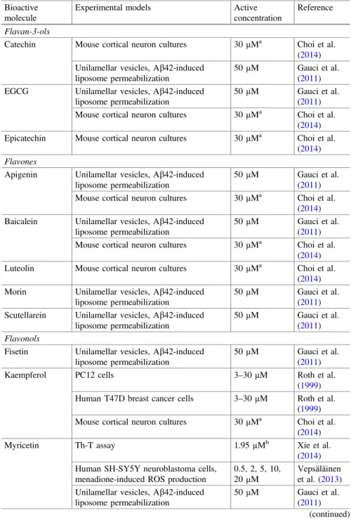
Among phenolic compounds (Table1), flavan-3-ols, flavones, flavonols,
iso-flavones, lignans, phenolic acids, and stilbenes comprise the most common classes. Quercetin (a flavonol), followed by myricetin (aflavonol) and kaempferol (a fl a-vonol), and then apigenin (flavone), baicalein (flavone), catechin (flavan-3-ols), and EGCG (flavan-3-ols) are the most studied phenolic compounds. In particular, quercetin has shown several neuroprotective effects, being its ability to reduce and/or to prevent reactive oxygen species (ROS) overproduction amongst to the most prominent; its active concentrations varied significantly according to the model used, but interestingly, as described by Vepsäläinen et al. (2013), at 0.5, 2.5, and 10µM a, pronounced reduction in ROS overproduction was observed in
Table 1 Phenolic compounds (from commercial origin) with reported in vitro neuroprotective effects
Bioactive molecule
Experimental models Active concentration
Reference
Flavan-3-ols
Catechin Mouse cortical neuron cultures 30µMa Choi et al. (2014) Unilamellar vesicles, Ab42-induced
liposome permeabilization
50µM Gauci et al. (2011) EGCG Unilamellar vesicles, Ab42-induced
liposome permeabilization
50µM Gauci et al. (2011) Mouse cortical neuron cultures 30µMa Choi et al.
(2014) Epicatechin Mouse cortical neuron cultures 30µMa Choi et al.
(2014)
Flavones
Apigenin Unilamellar vesicles, Ab42-induced liposome permeabilization
50µM Gauci et al. (2011) Mouse cortical neuron cultures 30µMa Choi et al.
(2014) Baicalein Unilamellar vesicles, Ab42-induced
liposome permeabilization
50µM Gauci et al. (2011) Mouse cortical neuron cultures 30µMa Choi et al.
(2014) Luteolin Mouse cortical neuron cultures 30µMa Choi et al.
(2014) Morin Unilamellar vesicles, Ab42-induced
liposome permeabilization
50µM Gauci et al. (2011) Scutellarein Unilamellar vesicles, Ab42-induced
liposome permeabilization
50µM Gauci et al. (2011)
Flavonols
Fisetin Unilamellar vesicles, Ab42-induced liposome permeabilization
50µM Gauci et al. (2011) Kaempferol PC12 cells 3–30µM Roth et al.
(1999) Human T47D breast cancer cells 3–30µM Roth et al.
(1999) Mouse cortical neuron cultures 30µMa Choi et al.
(2014) Myricetin Th-T assay 1.95µMb Xie et al.
(2014) Human SH-SY5Y neuroblastoma cells,
menadione-induced ROS production
0.5, 2, 5, 10, 20µM
Vepsäläinen et al. (2013) Unilamellar vesicles, Ab42-induced
liposome permeabilization
50µM Gauci et al. (2011)
(continued)
Table 1 (continued)
Bioactive molecule
Experimental models Active concentration
Reference
Mouse cortical neuron cultures 30µMa Choi et al. (2014) Quercetin Mouse cortical neuron cultures 30µMa Choi et al.
(2014) Primary cultures of astrocytes and
microglial cells
8µMb,c Elmann et al. (2013) Primary cultures of astrocytes 10µM Elmann et al.
(2014) Unilamellar vesicles, Ab42-induced
liposome permeabilization
50µM Gauci et al. (2011) Human SH-SY5Y neuroblastoma cells,
menadione-induced ROS production
0.5, 2, 5, 10µM
Vepsäläinen et al. (2013) Bovine serum albumin,D
-ribose-induced AGE formation
600lMb Ferchichi
et al. (2012)
Isoflavones
Genistein Unilamellar vesicles, Ab42-induced liposome permeabilization
50µM Gauci et al. (2011)
Lignans
NDGA Unilamellar vesicles, Ab42-induced liposome permeabilization
50µM Gauci et al. (2011)
Phenolic acids
3,5-di-O -Caffeoylquinic acid
Human neuroblastoma clonal SH-SY5Y Ab1–42-treated cells
20µM Han et al. (2010)
Caffeic acid Th-T assay 118µMb Kurisu et al.
(2013) Chlorogenic
acid
Bovine serum albumin,D -ribose-induced AGE formation
2000lMb Ferchichi et al. (2012) Ferulic acid Mutant human APP-overexpressing
murine neuron-like cells
1.563– 12.5µM
Mori et al. (2013)
p-Coumaric acid
PC12 cells, Ab25−35-induced toxicity 5, 25, 50µM Yoon et al.
(2014) Rosmarinic
acid
PC12 cells, Ab25-35-induced neurotoxicity
23.6µMd Na et al. (2010) Unilamellar vesicles, Ab42-induced
liposome permeabilization
50µM Gauci et al. (2011)
Stilbenes
Resveratrol Unilamellar vesicles, Ab42-induced liposome permeabilization
50µM Gauci et al. (2011)
aTested concentration to measure the percentage of LDH release after 20lM Ab
25–35exposure; bIC
50values;cvaried according to the used assay;dED50value
EGCGEpigallocatechin gallate;NDGAnordihydroguaiaretic acid;ROSreactive oxygen species;
Th-T assayThioflavin-T assay;AGEsadvanced glycation end-products
Table 2 Non-phenolic compounds (from commercial origin) with reported in vitro neuroprotec-tive effects
Bioactive molecule Experimental models Active concentration
Reference
Glycosides
Salidroside SH-SY5Y human neuroblastoma cells,
b-amyloid-induced oxidative stress
10, 50, 100µM
Zhang et al. (2010)
Iridoid glycosides
Geniposide N2a cell
formaldehyde-exposed
200µM Chen et al. (2014)
Phenylethanoids
Acteoside-tetramethylether Th-T assay >200µMa Kurisu et al. (2013)
Hydroxytyrosol 96µMa
Oraposide-tetramethylether >200µMa Quinones
1,4-Benzoquinone Insulin as amyloid model 50µM Gong et al. (2014) 1,4-Naphthoquinone
9,10-Anthraquinone 9,10-Phenanthraquinone Aloe-emodin
Chrysophanol Emodin
Terpenes
Asiatic acid Primary Sprague–Dawley rat cortical neurons
10µM Patil et al. (2010) Bilobalide Th-T assay 14.84%b Xie et al.
(2014) Carnosic acid U373MG human astrocytoma
cells
50µM Yoshida et al. (2014) Ginkgolide A Th-T assay 21.10%b Xie et al.
(2014) Ginkgolide B Th-T assay 13.56%b Xie et al.
(2014) Unilamellar vesicles,
Ab42-induced liposome permeabilization
50µM Gauci et al. (2011)
Ginkgolide C Th-T assay 13.92%b Xie et al.
(2014) Hyperforin Hippocampal neuron
cultures-amyloidfibrils and Aboligomer-induced damage
1µM Dinamarca et al. (2006)
(continued)
human SH-SY5Y neuroblastoma cells (with menadione as oxidative stress inducer). In the same line, myricetin also evidenced a similar potential, being together with quercetin theflavonols with the highest in vitro potential.
Two phenolic acids (ferulic acid and p-coumaric acid) and the flavonol (kaempferol) also showed interesting effects, directly related to their potent antioxidant activity. The active concentrations were 1.563–12.5 lM (ferulic acid), 5, 25, 50lM (p-coumaric acid), and 3–30lM (kaempferol). Other phenolic acids, such as rosmarinic (23.6lM) and 3,5-di-caffeoylquinic (20lM) acids, also evi-denced significant effects, followed by the flavan-3-ols (catechin, EGCG, and epicatechin) and the flavones (apigenin, baicalein, and luteolin), which were effective at 30lM. On the other hand, the flavones (morin and scutellarein), the
flavonolfisetin, the isoflavone genistein, the lignan NDGA, and, lastly, the stilbene resveratrol were only effective at 50lM. Finally, caffeic and chlorogenic acids proved to be the least effective, once the active concentrations were 118lM and 2000lM, respectively, which correspond to the IC50values.
By comparing the in vitro neuroprotective effects of phenolic compounds with other bioactive non-phenolic compounds (Table2), it is clearly evident that thefirst ones evidenced a greater effect, mainly the flavonols (quercetin and myricetin), being highly effective at lower doses. It is particularly convenient to highlight the effect of the following terpenes: hyperforin (1lM), ursolic acid (1, 10, 20lM), and asiatic acid (10lM) which presented the most prominent activity at lower con-centrations. Salidroside, a glycoside, several quinones (1,4-benzoquinone, 1,4-naphthoquinone, 9,10-anthraquinone, 9,10-phenanthraquinone, aloe-emodin, chrysophanol, and emodin), the terpenes (carnosic acid and ginkgolide B), and lastly purpurogallin trimethyl ether and rhein evidenced similar effects to those of phenolic compounds, which were effective at 50lM.
Overall, the phenolic compounds seem to present more significant in vitro neu-roprotective effects than non-phenolic molecules, which might be attributed to their
Table 2 (continued)
Bioactive molecule Experimental models Active concentration
Reference
Ursolic acid PC12 cells, Ab25−35-induced
toxicity
1, 10, 20µM Yoon et al. (2014) Withanolide A Primary Sprague–Dawley rat
cortical neurons
100µM Patil et al. (2010)
Others
Purpurogallin trimethyl ether
Unilamellar vesicles, Ab42-induced liposome permeabilization
50µM Gauci et al. (2011)
Rhein Insulin as amyloid model 50µM Gong et al. (2014)
aIC
50values;binhibition percentage of the tested compounds at the concentration of 100µM
Th-T assay and Thioflavin-T assay
Table 3 Plant-origin phenolic compounds with reported in vitro neuroprotective effects
Origin Plant part Bioactive extract/molecule Experimental models Active
concentration
Reference
Anthocyanins Anthocyanins Ribes nigrumL. Whole
plant
Anthocyanin-rich extract SH-SY5Y-APP751 cells,
staurosporine-induced apoptosis
4–31µg/mL Vepsäläinen et al. (2013)
Chalcones
Psoralea corylifoliaL. Seeds 4-Hydroxylonchocarpin Murine microglial cell line (BV-2), LPS-induced oxidative stress
10.2µg/mLa Lee et al. (2005)
Pulicaria incisa(Lam.) DC.
Aerial parts
Pulichalconoid B Primary cultures of astrocytes and
microglial cells
20µMa,b Elmann et al.
(2013)
Flavones Achillea fragrantissima (Forssk.) Sch.Bip. Whole plant
3,5,4′-trihydroxy-6,7,3′-trimethoxyflavone Primary cultures of astrocytes 8µM Elmann et al. (2014)
Calophyllum flavoramulumHend. & Wyatt-Sm.
Leaves Amentoflavone Bovine serum albumin,D-ribose-induced
AGE formation
0.05 mMa Ferchichi
et al. (2012)
Eragrostis ferruginea
(Thunb.) P. Beauv.
Aerial parts
7-Demethylageconyflavone A PC12 cells, Ab25-35-induced neurotoxicity >100µM c
Na et al. (2010)
Tricin 20.3µMc
Age-conyflavone A 58.7µMc
Rosmarinus officinalis
L.
Whole plant
Luteolin PC12 cells, corticosterone-induced
neurotoxicity
30, 40, 50µM
Sasaki et al. (2013)
Psoralea corylifoliaL. Seeds chromenoflavanone [7,8-dihydro-8-(4-hydroxyphenyl)-2,2-dimethyl-2H,6H -benzo-(1,2-b:5,4-b′)dipyran-6-one]
Murine microglial cell line (BV-2), LPS-induced oxidative stress
11.4µg/mLa Lee et al. (2005)
Table 3 (continued)
Origin Plant part Bioactive extract/molecule Experimental models Active
concentration
Reference
Flavonols Calophyllum flavoramulumHend. & Wyatt-Sm.
Leaves Quercitrin Bovine serum albumin,D-ribose-induced
AGEs formation
0.50 mMa Ferchichi et al. (2012)
Ginkgo bilobaL. Leaves Quercetin 3-O-b-D-rutinoside Th-T assay 33.02µMa Xie et al.
(2014) Quercetin 3-O-a-L-(b-D
-glucopyranosyl)-(1,2)-rhamnopyranoside
67.182µMa
Quercetin 3-O-a-(6′′′-p-coumaroyl glucopyranosyl-b-1,2-rhamnopyranoside)
32.56µMa
Kaempferol 3-O-b-D-rutinoside 30.89%d
Isorhamnetin 3-O-b-D-rutinoside 23.62%d
Kaempferol 3-O-a-L-(b-D -glucopyranosyl)-(1,2)-rhamnopyranoside
30.49%d
Kaempferol 3-O-a-(6′′′-p-coumaroyl glucopyranosyl-b-1,2-rhamnopyranoside)
34.98%d
Isoflavones
Flemingia macrophylla
L.
Aerial parts
Osajin Neuronal cells Ab-induced damage 5.01µMc Shiao et al.
(2005)
5,7,4′-Trihydroxy-6,8-diprenylisoflavone 11.25µMc
5,7,4′-Trihydroxy-6,3′-diprenylisoflavone 4.47µMc
Lignans
Eragrostis ferruginea
(Thunb.) P. Beauv.
Aerial parts
Nectandrin B PC12 cells, Ab25-35-induced neurotoxicity 44.1µMc Na et al.
(2010)
4-Ketopinoresinol 54.8µMc
(continued)
276
N.
Martins
and
I.C.F.R.
Table 3 (continued)
Origin Plant part Bioactive extract/molecule Experimental models Active
concentration
Reference
Magnolia fargesii
(Finet & Gagnep.) W. C.Cheng
Flower buds
(+)-Eudesmin BV-2 cells 30µMa Kim et al.
(2009)
(+)-Magnolin 20.5µMa
(+)-Yangambin 28.6µMa
Epimagnolin B 10.9µMa
Valeriana amurensis
P. Smirn. ex Kom.
Rhizomes and roots
Olivil-4′-O-b-D-glucopyranoside Ab1–42-induced PC12 cell neurotoxicity 5, 12.5,
25µM
Wang et al. (2014a) Lariciresinol-4,4′-di-O-b-D-glucopyranoside
Olivil-4-O-b-D-glucopyranoside
8-Hydroxylariciresinol-4’-O-b-D -glucopyranoside
Lariciresinol-4-O-b-D-glucopyranoside
Neoarctin A
Lariciresinol-4’-O-b-D-glucopyranoside
(−)-Massoniresinol 3a-O-b-D-glucopyranoside
(+) Pinoresinol-4,4’-di-O-b-D-glucopyranoside Ab25–35-induced PC12 cell death 5, 12, 25µM Wang et al. (2012) (+) Pinoresinol-8-O-b-D-glucopyranoside
8-Hydroxypinoresinol- 4,4′-di-O-b-D -glucopyranoside
Phenolic acids
Calophyllum flavoramulumHend. & Wyatt-Sm.
Leaves 3,4-dihydroxybenzoic acid Bovine serum albumin,D-ribose-induced
AGEs formation
0.50 mMa Ferchichi et al. (2012)
Rosmarinus officinalis
L.
Rosmarinic acid PC12 cells, corticosterone-induced
neurotoxicity
5, 15, 25µM Sasaki et al. (2013)
Salvia miltiorrhiza
Bunge
Salvianolic acid B PC12 cells, Ab25–35-induced cytotoxicity 10, 100,
200µg/mL
Zhou et al. (2011)
(continued)
11
Neurocognitive
Improvement
Through
Plant
Food
Bioactives
…
Table 3 (continued)
Origin Plant part Bioactive extract/molecule Experimental models Active
concentration
Reference
Stilbenes
Vitis viniferaL. Skin and seeds
Resveratrol PC12 rat pheochromocytoma cells,
Ab25-35-induced apoptosis
10, 20µM Kim et al. (2007)
Tannins
Paeonia suffruticosa
Andr.
Whole plant
1,2,3,4,6-Penta-O-galloyl-b-D-glucopyranose SK-N-SH cells 10µM Fujiwara
et al. (2009)
Xanthones
Calophyllum flavoramulumHend. & Wyatt-Sm.
Leaves 3-Methoxy-2-hydroxyxanthone Bovine serum albumin,D-ribose-induced
AGE formation
0.06 mMa Ferchichi et al. (2012)
aIC
50values;bvaried according to the used assay;cED50values;dinhibition percentage of the tested compounds at the concentration of 100µM Th-T assayThioflavin-T assay.AGEsadvanced glycation end-products;LPSlipopolysaccharide
278
N.
Martins
and
I.C.F.R.
Table 4 Plant-origin non-phenolic compounds with reported in vitro neuroprotective effects
Origin Plant part Bioactive molecule Experimental models Active
concentration
Reference
Alkaloids
Crinum macowaniiBaker Bulbs Hamayne HeLa cells line 10µg/mLa Kwon et al.
(2011)
Lycorine 5µg/mLa
Coumarins
Eleutherococcus senticosus (Rupr. et Maxim.) Maxim.
Rhizomes Isofraxidin Rat cortical neurons 1; 10µM Bai et al.
(2011) Isofraxidin 7-O
-glucoside
Curcuminoids
Curcuma longaL. Whole
plant
Calebin-A PC12 cells fromb-amyloid (b25-35;
b1-42) insults
1; 2µg/mLb,c
Park and Kim (2002)
Curcumin 7;
10µg/mLb,c
Demethoxycurcumin 4;
5µg/mLb,c
Bisdemethoxycurcumin 2;
3.5µg/mLb,c
Iridoids
Valeriana amurensisP. Smirn. ex Kom.
Rhizomes and roots
Xiecaoside E Ab1–42-induced PC12 cell
neurotoxicity
5, 12.5, 25µM
Wang et al. (2014a)
Quinones
Euclea crispasubsp.Crispa Roots Natalenone HeLa cells 50µg/mLa Kwon et al.
(2011) (continued)
11
Neurocognitive
Improvement
Through
Plant
Food
Bioactives
…
Table 4 (continued)
Origin Plant part Bioactive molecule Experimental models Active
concentration
Reference
Saponins
Eleutherococcus senticosus (Rupr. et Maxim.) Maxim.
Rhizomes Stigmasterol 3-O-b-D -glucopyranoside
Rat cortical neurons 1; 10µM Bai et al.
(2011)
Eleutheroside E Eleutheroside B
Terpenes
Croton yanhuiiY. T. Chang Twigs Crotonpene A PC12 cells 15µM Sun et al.
(2014) Crotonpene B
Euclea crispasubsp.Crispa Roots 3-oxo-oleanolic acid HeLa cells 10µg/mLa Kwon et al.
(2011)
Laurus nobilisL. Leaves Spirafolide DA-induced apoptosis in human
neuroblastoma SH-SY5Y cells
5.7µMb Ham et al. (2010) Rosmarinus officinalisL. Whole
plant
Carnosic acid PC12 cells, corticosterone-induced neurotoxicity
10, 20, 30µM
Sasaki et al. (2013)
Tussilago fárfaraL. Flower
buds
Tussilagone Murine microglial cells 8.67µg/mL
(NO)a 14.1µg/mL (PGE2)a
Lim et al. (2008)
Valeriana amurensisP. Smirn. ex Kom.
Rhizomes and roots
Heishuixiecaoline A Ab25–35-induced PC12 cells death 5, 12, 25µM Wang et al.
(2012) Heishuixiecaoline B
Heishuixiecaoline C Volvalerenal C aIC
50values;bED50values;cobtained results to anti-bA(25–35) and anti-bA(1–42) activities, respectively
280
N.
Martins
and
I.C.F.R.
Table 5 Ecdysterones, phenylethanoid glycosides, and other plant-derived bioactive compounds with in vitro neuroprotective effects
Origin Plant part Bioactive molecule Experimental models Active
concentration
Reference
Ecdysterones
Klaseopsis chinensis (S.Moore) L. Martins
Roots 2-O-Acetyl-20-hydroxyecdysone SH-SY5Y neuroblastoma cells, Ab42-induced cytotoxicity
50µM Yang et al. (2010) 3-O-Acetyl-20-hydroxyecdysone
25,26-Didehydroponasterone A
Stachysterone C Carthamosterone
Phenylethanoid glycosides Orobanche minor J. E. Smith.
Whole plant
Acteoside Th-T assay 8.9µMa Kurisu et al.
(2013)
Oraposide 3.6µMa
Others
Curcuma comosa Roxb.
(3R) 1,7-Diphenyl-(4E,6E)-4,6-heptadien-3-ol LPS-treated microglia 0.1, 0.5, 1µM
Thampithak et al. (2009) Curcuma longaL.
1,7-Bis(4-hydroxy-3-methoxyphenyl)-1,4,6-heptatrien-3-one
PC12 cells from
b-amyloid (b25-35;b1-42) insults
>50µg/mLb, c
Park and Kim (2002)
1-Hydroxy-1,7-bis(4-hydroxy-3-methoxyphenyl)-6-heptene-3,5-dione
30.7; 44.3µg/mLb, c
1,7-Bis(4-hydroxyphenyl)-1-hep- tene-3,5-dione 0.5;
1.0µg/mLb,c 1,7-Bis(4-hydroxyphenyl)-1,4,6-heptatrien-3-one >50µg/mLb,
c
1,5-Bis(4-hydroxy-3-methoxyphenyl)-1,4-pentadien-3-one
>50µg/mLb, c
(continued)
11
Neurocognitive
Improvement
Through
Plant
Food
Bioactives
…
Table 5 (continued)
Origin Plant part Bioactive molecule Experimental models Active
concentration
Reference
Cynanchum paniculatum (Bunge) Kitag.
Roots 2,3-dihydroxy-4-methoxyacetophenone HT22 cells, glutamate-induced neurotoxicity
10.94µMb (Weon et al.
2013) Dendrobium nobile Lindley 3-[[(6-Methoxy-10-methyl-1H,3H- benzo[h]furo [4,3,2-de]-2-benzopyran-1-yl)oxy]methyl]-5-methylnaphtho[2,3-b]furan-4,9-dione
PC12 cells, H2O2-induced cell death
5, 10µg/mL Yoon et al. (2011)
Eleutherococcus senticosus(Rupr. et Maxim.) Maxim.
Rhizomes b-Sitosterol 3-O-b-D-glucopyranoside Rat cortical neurons 1; 10µM Bai et al. (2011)
Eragrostis ferruginea(Thunb.) P. Beauv.
Aerial parts
Corylin PC12 cells, Ab25-35
-induced neurotoxicity
>100µMb Na et al. (2010)
Euphorbia lagascae Sprengel
Seeds Piceatannol PC12 rat
pheochromocytoma cells, Ab25-35-induced apoptosis
10, 20µM Kim et al. (2007)
Flemingia macrophyllaL.
Aerial parts
Aureole Neuronal cells
Ab-induced damage
12.09µMb Shiao et al. (2005)
Flemingichromone 31.43µMb
Rhodiola sachalinensisA. Bor
Roots Salidroside Rat pheochromocytoma,
Ab-induced neuronal damage on PC12 cells
1, 5, 10, 50µg/mL
Jang et al. (2003)
Salvia miltiorrhiza Bunge
Danshensu PC12 cells, Ab25–35
-induced cytotoxicity
10, 100, 200µg/mL
Zhou et al. (2011) aIC
50values;bED50values;cobtained results to anti-bA(25-35) and anti-bA(1-42) activities, respectively Th-T assayThioflavin-T assay;LPSlipopolysaccharide
higher antioxidant potential, namely as free radical scavengers and also as metal quenchers and hydrogen donators (Heim et al.2002; Grotewold2006; Li et al.2014).
4.1.2 Plant-Food-Derived Molecules
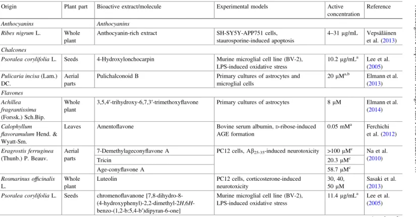
Tables3,4and5show the in vitro neuroprotective effects of plant-origin phenolic and non-phenolic molecules.
In relation to phenolic compounds (Table3), isoflavones, such as 5,7,4′
-tryhydroxy-6,3′-diprenyllisoflavone (4.47lM) and osajin (5.01lM) obtained from
Flemingia macrophyllaL., gave the most promissory effects, followed by the
lig-nan, olivil-4′-O-b-D-glucopyranoside (5; 12.5; 25lM) and (+) pinoresinol-4-4′ -di-O-b-D-glucopyranoside (5; 12; 25lM), both obtained from Valeriana amurensis P. Smirn. ex. Kom., and, lastly, the phenolic acid, rosmarinic acid (5; 15; 25lM), fromRosmarinus officinalisL.
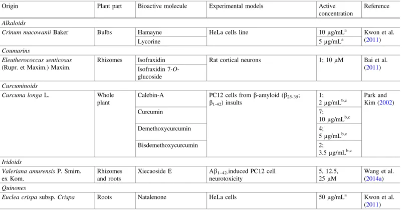
Despite the scarce results on in vitro neuroprotective effects of non-phenolic compounds (Table4), when compared with the first ones, they evidence a higher
Table 6 Bioactive compounds (from commercial origin) with reported in vivo
Bioactive molecule
Experimental models Active concentration
Reference
Flavones
Luteolin Male Sprague–Dawley rat model of chronic cerebral hypoperfusion
50, 100, 200 mg/kg b. w.
Fu et al. (2014)
Flavonols
Quercetin Wild-type adult zebrafish scopolamine-induced amnesia
50 mg/kg b.w. Richetti et al. (2011)
Rutin 50 mg/kg b.w.
Hydroxycinnamic acids
3,5-di-O -Caffeoylquinic acid
Male SAMP8 and SAMR1 mice 6.7 mg/kg b. w.
Han et al. (2010)
Ferulic acid Transgenic PSAPP mouse model of cerebral amyloidosis
30 mg/kg b.w. Mori et al. (2013)
Isoflavones
Genistein Male Wistar rats injected with Ab1–40 10 mg/kg b.w. Bagheri et al. (2012)
Terpenes
Hyperforin Male Sprague–Dawley rats injected with amyloidfibrils
6µM Dinamarca et al. (2006)
Phenylpropanoids
6-Shogaol Male ICR mice AbO1-42-induced
microglial cell activation
Table 7 Plant-origin bioactive compounds with in vivo neuroprotective effects
Bioactive molecule Origin Plant part Animal models Active
concentration
Reference
Anthocyanins
Anthocyanins Vitis viniferaL. Fruits Male Wistar rats,
scopolamine-induced memory deficits
200 mg/kg b. w.
Gutierres et al. (2014)
Curcuminoids
Bisdemethoxycurcumin Curcuma longaL. Rhizomes Drosophila melanogaster 1 mM Wang et al.
(2014b) Curcumin
Demethoxycurcumin
Flavonols
Kaempferol Brassica oleraceavar.
gemmifera
Sprouts Mouse model-Ab-induced memory deficit
12, 24 ppm Kim et al. (2013)
Tannins
1,2,3,4,6-Penta-O-galloyl-b-D
-glucopyranose
Paeonia suffruticosa Andr.
Whole plant
Tg2576 APPswe transgenic mice 8 mg/kg b.w. Fujiwara et al. (2009)
Others
2,3-Dihydroxy-4-methoxyacetophenone Cynanchum paniculatum(Bunge) Kitag.
Roots Amnesic mice scopolamine-induced
50 mg/kg b. w.
Weon et al. (2013)
284
N.
Martins
and
I.C.F.R.
and more promissory effect. Coumarins obtained from the rhizomes of
Eleutherococcus senticosus (Rupr. Et Maxim.) Maxim., namely isofraxidin and
isofraxidin 7-O-glucoside, and also saponins, which include stigmasterol 3-O-b-D -glucopyranoside and eleutheroside E and B, exert a higher effect than phenolic compounds. Its active concentrations were for both coumarins and saponins, 1 and 10lM.
Several terpenes, such as spirafolide (5.7lM) from the leaves ofLaurus nobilis L., and others obtained from the rhizomes and roots ofValeriana amurensis were also effective at lower concentrations (5; 12; 25lM), being able to reduce apoptosis in human neuroblastoma cells and PC12 cells.
The bioactive constituent, (3R) 1,7-diphenyl-(4E,6E)-4,6-heptadien-3-ol, obtained from Curcuma comosa Roxb., was highly effective (0.1; 0.5; 1lM) (Table5), being able to reduce lipopolysaccharide (LPS)-induced NO and PGE2
production, in a dose-dependent manner. Furthermore, the active concentrations
Fig. 1 Overview of the in vitro neuroregenerative effects of specific phytochemicals. Legend:
BDNFbrain-derived neurotrophic factor;ERKextracellular signal-regulated kinase;CREBcyclic AMP response element-binding protein;iNOSinducible nitric oxide synthase. The effects were observed by using the concentrations 0.1, 0.3, and 1µM of each one of the tested compounds in cerebral cells from the cortex of fetal Sprague–Dawley rats (Jeon et al.2010)
were also able to reduce inducible NO synthase and cyclooxygenase 2 (COX-2). Similarly, b-sitosterol 3-O-b-D-glucopyranoside (1; 10 lM) obtained from the rhizomes ofEleutherococcus senticosusstrongly inhibited neuritic atrophy induced
Zingiber purpureum Roscoe
Cis-3-(3’4’-dimethoxyphenyl)-4- [(E)-3’’,4’’-dimethoxystyryl]cyclohex-1-ene
Trans -3-(3’4’-
dimethoxyphenyl)-4-[(E)- 3’’,4’’-dimethoxystyryl]cyclohex-1-ene
Induce neurite sprouting
(30 µM)
Increase neurite length and number
(0.3–3 µM)
Protect against cell death
(3-30 µM)
Induce neurite sprouting
(10-30 µM)
Increase neurite length and number
(0.03–3 µM)
Protect against cell death
(30 µM)
Fig. 2 In vitro neuroregenerative effects of phenylbutanoid dimers obtained from the methanol extract ofZingiber purpureumRoscoe. roots.LegendProtection against cell death and induction of neurite sprouting was assessed by using PC12 cells, while the evaluation of neurite length and number improvement was carried out in cultured primary cortical neurons of rats (Matsui et al.
2012)
Fig. 3 Bioactive molecules from commercial sources with in vitro neuroregenerative effects.
LegendEffects offlavonoids on the mitochondrial function were assessed by using 1µM of each one in murine neuroblastoma N2a cells, and then, measure its activity at a level of ROS production, MMP and ATP levels (Dragicevic et al.2011); evaluation of Ab production was assessed by using 50µM of each one of the tested compounds in CHO 2B7 cells (Chen et al.
2006)
by Ab25–35, being clearly evident its protective effects against cognitive and
memory impairments. 2,3-Dihydroxy-4-methoxyacetophenone (10.94lM), iso-lated from the roots of Cynanchum paniculatum(Bunge), also evidenced a pro-nounced effect against neuronal damage and toxicity, in HT22 cells, induced by glutamate.
Overall, and comparing the efficacy and efficiency of the studied bioactive molecules in relation to its sources (commercial vs. plant origin), it is possible to conclude that plant-origin bioactive compounds possess a doubtless prominent potential. Although some of them were not studied and then compared from both sources, several examples should be highlighted. While for luteolin, derived from commercial (30lM) and plant (30, 40, and 50 lM) sources, similar active con-centrations were found, for rosmarinic acid a completely different situation was observed; the commercial molecule was effective at 23.5–50lM, while the one from plant origin was effective at 5, 15, and 25lM. Similarly, resveratrol isolated fromVitis viniferaL. was highly effective at 10 and 20lM, while the commercial
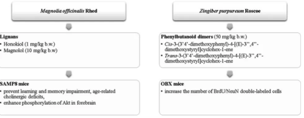
Fig. 4 Plant-origin bioactive molecules with in vivo neuroregenerative effects.Legend Magnolia officinalis Rhed plant (Matsui et al.2009) andZingiber purpureumRoscoe root (Matsui et al.
2012) methanol extracts
Fig. 5 Bioactive molecules (from commercial origin) with in vivo neuroregenerative effects.
1Dragicevic et al. (2011),2Chen et al. (2006)
molecule was active at 50lM. Moreover, carnosic acid obtained fromR. officinalis was effective at 10, 20, and 30lM, while the commercial one was active at 50lM. In the same line, the phenylethanoid glycosides, acteoside, and oraposide were also more effective when derived from natural sources (8.9 and 3.6lM) in comparison with the commercial molecules (>200lM). Finally, salidroside isolated from
Rhodiola sachalinensis A. Bor was effective at 1; 5; 10; 50lg/Ml, while the
commercial molecules were effective at 3; 15; 30lg/mL.
In summary, plant-food-derived bioactive molecules appear to be more effective than the commercial ones; notwithstanding, further studies are necessary to confirm this. Besides, the confirmation of the effective neuroprotective potential needs to be performed, mainly by in vivo studies, once numerous biochemical parameters influence thefinal bioactivity and active concentrations.
4.2
Bioactive Compounds with In Vivo Neuroprotective
Activity
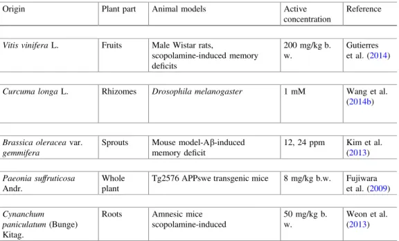
Tables6 and 7 show, respectively, bioactive molecules from commercial and plant origins with reported in vivo neuroprotective effects. Comparing with the previously discussed in vitro neuroprotective effects, only commercial quercetin, ferulic acid, genistein, and hyperforin and bisdemethoxycurcumin, curcumin, demethoxycurcumin, 1,2,3,4,6-penta-O-galloyl-b-D-glucopyranose, and 2,3-dihydroxy-4-methoxyacetophenone from plant food origin were also evaluated. Higher concen-trations were necessary to obtain the same effect on in vivo experiments. For example, hyperforin at 1lM was effective under an in vitro model, while 6lM was the active concentration in the in vivo experiment. Additionally, when compared with the in vitro reports, in vivo studies are significantly scarce. But despite this limitation, the obtained achievements need to be highlighted in order to systematize the knowledge on this area. Phenolic compounds seem to be the most studied phytochemicals regarding neurocognitive benefits. The effects offlavonols have been evaluated using both commercial and plant-origin molecules; flavones, hydroxycinnamic acids, and isoflavones were only studied in commercial forms, while anthocyanins and tannins were obtained from natural extracts (Vitis vinifera L. and Paeonia suffruticosa Andr., respectively). Commercial terpenes and phenylpropanoids, and curcumi-noids and other plant-origin biomolecules were also investigated for neuroprotec-tive effects.
Rats and mice, followed by wild-type adult zebrafish andDrosophila melano-gaster, including species with induced genetic variations, have been the most frequent animal models used. According to the selected animal model, several active con-centrations for each bioactive molecule have been observed. Moreover, none of the studied bioactive molecules were evaluated from both commercial and plant-origin sources, being difficult to make comparisons of their effectiveness/efficiency.
Among the tested commercialflavones, luteolin showed a dose-dependent effect, mainly acting as downregulator of NF-Kb and BACE-1, and decreased Ab depo-sition (Fu et al. 2014). On the other hand, by using a standard concentration of 50 mg/kg b.w., Richetti et al. (2011) observed that bothflavonols, quercetin and rutin, were able to prevent memory and cognitive impairments. Similarly, Mori et al. (2013) observed that ferulic acid (30 mg/kg b.w.) reversed transgene-associated behavioral deficits and decreased brain parenchymal and cerebral vascular
b-amyloid deposits and also hyperforin at 6µM (Dinamarca et al.2006).
Moon et al. (2014) proved that the phenylpropanoid, 6-shogaol, at 10 mg/kg b.w. was effective in reducing microgliosis and astrogliosis, ameliorating AbO1-42
-induced memory impairment and elevating NGF levels, and also pre- and postsy-naptic markers. In the same line, Bagheri et al. (2012) observed that genistein at the same concentration (10 mg/kg b.w.) leads to a significant inhibition of neurode-generation and Ab1-40-positive aggregate formation, alleviating consequently
extensive astrogliosis. The hydroxycinnamic acid, 3,5-di-caffeoylquinic acid (6.7 mg/kg b.w.), also showed a pronounced effect, being able to decrease in a significant manner escape latency time, by increasing PGK1 and mRNA expression, and also ATP production (Han et al.2010).
In relation to plant-origin bioactive molecules, all of them were able to prevent memory and cognitive impairments, in different proportions. For example, antho-cyanins from Vitis vinifera L. fruits extract (200 mg/kg b.w.) exerted beneficial effects mainly by preventing scopolamine-induced neurotoxic effects (Gutierres et al. 2014), while curcuminoids (bisdemethoxycurcumin, curcumin, and demethoxycurcumin) fromCurcuma longaL. rhizomes extract, at the concentration of 1 mM, rescued morphological defects onDrosophila melanogaster improving the movement coordination (Wang et al.2014b). In a similar manner, kaempferol (12 and 24 ppm), 1,2,3,4,6-penta-O-galloyl-b-D-glucopyranose (8 mg/kg b.w.), and 2,3-dihydroxy-4-methoxyacetophenone (50 mg/kg b.w.) obtained, respectively,
from Brassica oleraceae var. gemmifera, Paeonia suffruticosa Andr., and
Cynanchum paniculatum(Bunge) Kitag. markedly reduced escape latency time: the
first one by its contribution on the reduction in ROS production and consequently improvement in step-through latency time (Kim et al. 2013), the second one through the inhibition of Abfibril formation as also destabilization of the preformed Ab fibrils (Fujiwara et al. 2009), and the last one, related to NMDA receptor inhibition and breakdown of AChE (Weon et al.2013).
The above-described neuroprotective benefits, both in vivo and in vitro, incite the future use of bioactive molecules from plant food origin as leaders to the AD treatment. It is also important to highlight that despite its neuroprotective and preventive effects, and even treatment/alleviation of the symptomatic conditions, these phytochemicals also possess an interesting and underexplored neuroregen-erative potential, which needs to be studied in detail.
5
Plant Food Bioactive Molecules with Neuroregenerative
Activity
5.1
Bioactive Compounds with In Vitro Neuroregenerative
Activity
In addition to the previously stated promissory neuroprotective benefits of plant food bioactive constituents, its neuroregenerative effects have also been studied, but still have a low progression. Figures1and2show plant-origin bioactive molecules with prominent in vitro neuroregenerative potential, while Fig.3 shows results of commercial molecules. Once again, is clearly evident the scarcity of the studied plant species: Cinnamomum cassia Blume, Coptis chinensis Franch., Pueraria montana(Lour.) Merr.var. lobata(Willd.) Maesen & Almeida,Salvia miltiorrhiza Bunge andScutelaria baicalensisGeorgi (Fig.1), andZingiber purpureumRoscoe. Figure2 shows the currently recognized sources of bioactive molecules with in vitro neurodegenerative benefits. In particular, baicalein, cinnamic acid, epiberberine, genistein, tanshinone IIA, and wogonin, at low concentrations (0.1, 0.3, and 1µM), evidenced to act synergistically on the brain-derived neurotrophic factor (BDNF) release, mRNA, and protein expression, mainly by phosphorylation of extracellular signal-regulated kinase (ERK) and cyclic AMP element-binding protein (CREB), and inhibition of inducible nitric oxide synthase (iNOS) upregu-lation. Jeon et al. (2010) clarified those effects by using cortex cerebral cells from fetal Sprague–Dawley rats. On the other hand, Matsui et al. (2012) by using PC12 cells observed that both isolated phenylbutanoid dimers fromZ. purpureum(Fig.2) were able not only to induce neurite sprouting (10–30µM), but also to confer a significant protection against cell death (3–30µM). Furthermore, the authors observed a pronounced induction of the number and length of neurites (0.03–3 µM) by using primary cortical neurons of rats.
Apart from the studied plant food molecules, several commercial biomolecules were also investigated, mainly in what concerns to the ability to restore mito-chondrial functions and reduce Ab production (Fig.3). In general, phenolic com-pounds were the most frequently studied biomolecules toward to assess mitochondrial function restauration ability, while saponins have been studied to determine the effect on Ab production. Indeed, and as previously mentioned, phenolic compounds are widely recognized for their antioxidant potential (mainly as free radical scavengers, metal quenchers, and hydrogen donators) (Heim et al. 2002; Grotewold2006; Li et al. 2014). Commonly known as“powerhouse of the
cells,” “ATP reservoir”or“energetic factory,”mitochondria contribute not only to
the proper cellular function, but also to an intensive free radical production (one of the most important endogenous sources of ROS). In fact, brain cells need a higher and continuous demand for energy supply (Chaturvedi and Beal2013). Therefore, it is of the utmost importance not only to ensure a proper neuronal function, but also to avoid cellular damages, by discovering new and effective alternatives to restore the optimum mitochondrial functions (in case of injuries). Taking into account
those features, several biomolecules have been intensively investigated. It should be pointed that, by itself, a free radical overproduction is not only a triggering factor to AD development, but also promotes Ab production, which leads to an intense organic dysfunction and inflammation. As shown in Fig.3,flavanones (hesperitin, naringenin), flavan-3-ols (catechin, EGCG), flavones (acacetin, aminoflavone, apigenin, baicalein, chrysin, diosmetin, diosmin, luteolin, methoxyflavone, and methylflavone), flavonols (quercetin), and phenolic acids (ferulic acid, methyl cinnamate, and methyl gallate) are among the most prominent phenolic compounds with mitochondrial function restauration ability, at the concentration of 1µM, in murine neuroblastoma N2a cells. To evaluate the potential of those molecules, Dragicevic et al. (2011) used the levels of ROS, MMP, and ATP as positive indicators.
On the other hand, Chen et al. (2006) observed that saponins, at thefinal con-centration of 50µM, exerted considerable effects on Ab40 (ginsenoide Rb1, panaxadiol) and Ab42 (ginsenoide Rg1, pseudoginsenoside F11) and also on Ab40 and Ab42 (ginsenoide Rb2, Re, Rg3; notoginsenoside R1) reduction, by using CHO 2B7 cells. In spite of the great interest of these results, further studies are necessary to assess the in vivo effects, including security (mainly to saponins, which depending to the used doses, are often slightly toxic) and bioavailability, and also to establish the therapeutic doses.
5.2
Bioactive Compounds with In Vivo Neuroregenerative
Activity
Figures4and 5show the in vivo neuroregenerative effects of bioactive molecules from plant and commercial origins, respectively. Among the bioactive molecules from plant origin, only individual phytochemicals obtained fromMagnolia offi ci-nalisRhed andZingiber purpureumRoscoe were evaluated (Fig.4).M. officinalis lignans presented the better effect, namely honokiol (1 mg/kg b.w.) and magnolol (10 mg/kg b.w.) (Matsui et al. 2009), on the prevention of learning and memory impairments, age-related cholinergic deficits, as also on the improvement in Akt phosphorylation in SAM8 mice forebrain. The phenylbutanoid dimers (50 mg/kg b.w.) appear as the great contributors to OBX mice BrdU/NeuN double-labeled cell improvement (Matsui et al. 2012). In general, the authors concluded that the mentioned phytochemicals possess promissory neurotrophic effects; so, their further investigation is of the utmost importance, to be effectively used, in the near future, to threat and even to modify the course of several neurological disorders.
Among the commercial bioactive compounds (Fig.5), the flavan-3-ols with a renowned and doubtless antioxidant potential, namely EGCG (37.1 mg/kg b.w.) from green tea (Dragicevic et al. 2011), were the most studied, and also triterpenoid saponins that include ginsenoside Re, Rg1, and Rg3 (25 mg/kg b.w.) (Chen et al.2006).
In thefirst case, EGCG was able to improve the activity ofa-secretase and to reduce brain mitochondrial Ab and AbPP levels in AbPP/PS1 double-mutant transgenic mouse models. Otherwise, ginsenoside Re, Rg1, and Rg3 evidenced a higher potential to reduce not only Ab42, but also Ab40 brain levels in Tg2576 female transgenic mice models that overexpress the human APP gene.
Overall, and despite the described effects of the tested bioactive components, it is difficult to compare their efficacy and also efficiency once different biochemical parameters were assessed, and also biological antineuronal damage effects.
6
Neuromodulation and Neuroplasticity: Future Trends
to an Optimum Brain Health
The in vitro and in vivo promissory neurocognitive benefits evidenced by plant products clearly point to an upcoming approach as future leaders on neurodegen-erative diseases. In particular, their neuroprotective effects are pivotal once pre-vention comprises one of the most important key points on integrative interpre-vention. Notwithstanding, in some cases, considerable changes on learning and memory abilities, and also lagged perceptions and neuronal losses, are found, which requires a therapeutic intervention (van Praag2009; Essa et al.2012; Yoo and Park2012; Ahmed et al.2015).
Otherwise, along with the disseminated idea that brain cells (such as neurons, microglia, and other glial cells) are not able to regenerate, increasing evidences have shown that they possess an interesting ability to synapse remodeling and consequently to recover its synaptic plasticity, neuronal spine density, and, there-fore, cognitive improvement. Numerous signaling molecules, transporters, and codifying proteins, genes, and so on are involved on the mentioned complex pro-cess (McCoy et al.2009; van Praag 2009; Nadim and Bucher 2014; Wester and McBain2014). Plant-derived bioactive molecules, and also physical exercise, have shown to act as important contributors, once rest neurodegeneration, and improve neuroregenerative processes (van Praag 2009). In spite of their pivotal biological interests, the neuromodulatory and neuroplasticity abilities of numerous bioactive molecules remain poorly investigated, as also the involved modes of action. For example, it is convenient to highlight that 2,3,5,4′-tetrahydroxystilbene-2-O-b-D -glucoside (TSG), a phenolic compound derived from Polygonum multiflorum Thunb., evidenced potent cognitive improving and hippocampal synaptic plasticity promoting (Wang et al. 2011) abilities: facilitates the induction of hippocampal long-term potentiation (LTP) through activation of postsynaptic signal molecules and other signalling pathways, which contributes to the in vivo improvement of learning behavior, memory, and neuronal networks in rats. Liang et al. (2014) also showed that dihydromyricetin (DHM), aflavonoid compound, restores gephyrin (a postsynaptic anchor protein at GABAergic synaptic sites) levels, when adminis-tered in mouse models with AD. Gephyrin is directly involved on GABA receptor
functioning, once regulates its formation, plasticity and availability, as also from other signalling molecules (Liang et al. 2014). Furthermore, Zhan et al. (2014) observed that berberine, a plant alkaloid, was able to reverse synaptic deficits induced byD-galactose and rescued important intermediates (mRNA,Arc/Arg3.1) directly involved on normal synaptic plasticity.
Otherwise, and in association with the previous stated effects, it is also important to point out that hypothalamic neuromodulator systems are also affected/affect daily energy homeostasis. For example, under specific conditions (such as short-term fasting and other metabolic state changes), considerable synaptic circuits and respective (inter)neuronal controllers suffer from restructuration, which leads to physiological variations on energy homeostasis and may cause synaptic plasticity impairments (Horvath2006). Thus, the above-described mechanisms of action of bioactive molecules can be also extremely useful in other contexts, such as feeding and appetite controllers. For example, plant-derived cannabinoids (phytocannabi-noids) and endocannabinoids, in spite of the whole negative connotation attributed to cannabis, have shown greater contributive properties not only for physiological appetite and satiety controllers (Berry and Mechoulam 2002), but also for brain therapeutical purposes (i.e., neuromodulators, neuroprotective, neuroregenerative, and synapsis plasticity regulators) (Fisar2009).
References
Adewusi EA, Steenkamp V (2015) Medicinal plants and their derivatives with amyloid beta inhibitory activity as potential targets for drug discovery. Asian Pacific J Trop Dis 5:430–440.
doi:10.1016/S2222-1808(15)60810-6
Ahmed A, van der Marck M, van den Elsen G, Rikkert MO (2015) Cannabinoids in late-onset Alzheimer’s disease. Clin Pharmacol Ther 97:597–606. doi:10.1002/cpt.117
Alza NP, Richmond V, Baier CJ et al (2014) Synthesis and cholinesterase inhibition of cativic acid derivatives. Bioorganic Med Chem 22:3838–3849. doi:10.1016/j.bmc.2014.06.030
Bagheri M, Roghani M, Joghataei MT, Mohseni S (2012) Genistein inhibits aggregation of exogenous amyloid-beta 1–40 and alleviates astrogliosis in the hippocampus of rats. Brain Res 1429:145–154. doi:10.1016/j.brainres.2011.10.020
Bai Y, Tohda C, Zhu S et al (2011) Active components from Siberian ginseng (Eleutherococcus senticosus) for protection of amyloid b(25–35)-induced neuritic atrophy in cultured rat cortical
neurons. J Nat Med 65:417–423. doi:10.1007/s11418-011-0509-y
Berry EM, Mechoulam R (2002) Tetrahydrocannabinol and endocannabinoids in feeding and appetite. Pharmacol Ther 95:185–190. doi:10.1016/S0163-7258(02)00257-7
Chaturvedi RK, Beal MF (2013) Mitochondrial diseases of the brain. Free Radic Biol Med 63:1–29. doi:10.1016/j.freeradbiomed.2013.03.018
Chen F, Eckman E, Eckman CB (2006) Reductions in levels of the Alzheimer’s amyloid beta peptide after oral administration of ginsenosides. FASEB J 20:1269–1271. doi: 10.1096/fj.05-5530fje
Chen J, Sun M, Wang X et al (2014) The herbal compound geniposide rescues formaldehyde-induced apoptosis in N2a neuroblastoma cells. Sci China Life Sci 57:412–421. doi:10.1007/ s11427-014-4643-0
Choi S-M, Kim BC, Cho Y et al (2014) Effects of flavonoid compounds on b -amyloid-peptide-induced neuronal death in cultured mouse cortical neurons. Chonnam Med J 50:45–51
Cowan MM (1999) Plant products as antimicrobial agents. Clin Microbiol Rev 12:564–82 Dinamarca MC, Cerpa W, Garrido J et al (2006) Hyperforin prevents beta-amyloid neurotoxicity
and spatial memory impairments by disaggregation of Alzheimer’s amyloid-beta-deposits. Mol Psychiatry 11:1032–1048. doi:10.1038/sj.mp.4001866
Dragicevic N, Smith A, Lin X et al (2011) Green tea epigallocatechin-3-gallate (EGCG) and other
flavonoids reduce Alzheimer’s amyloid-induced mitochondrial dysfunction. J Alzheimer’s Dis 26:507–521. doi:10.3233/JAD-2011-101629
Elmann A, Telerman A, Erlank H et al (2013) Protective and antioxidant effects of a chalconoid from Pulicaria incisa on brain astrocytes. Oxid Med Cell Longev 2013:1–10. doi:10.1155/ 2013/694398
Elmann A, Telerman A, Mordechay S et al (2014) 3,5,4′-Trihydroxy-6,7,3′-trimethoxyflavone protects astrocytes against oxidative stress via interference with cell signaling and by reducing the levels of intracellular reactive oxygen species. Neurochem Int 78:67–75. doi:10.1016/j. neuint.2014.09.003
Essa MM, Vijayan RK, Castellano-Gonzalez G et al (2012) Neuroprotective effect of natural products against Alzheimer’s disease. Neurochem Res 37:1829–1842. doi: 10.1007/s11064-012-0799-9
Ferchichi L, DerbréS, Mahmood K et al (2012) Bioguided fractionation and isolation of natural inhibitors of advanced glycation end-products (AGEs) from Calophyllum flavoramulum. Phytochemistry 78:98–106. doi:10.1016/j.phytochem.2012.02.006
Fernandez-Panchon MS, Villano D, Troncoso AM, Garcia-Parrilla MC (2008) Antioxidant activity of phenolic compounds: from in vitro results to in vivo evidence. Crit Rev Food Sci Nutr 48:649–671. doi:10.1080/10408390701761845
Fisar Z (2009) Phytocannabinoids and endocannabinoids. Curr Drug Abuse Rev 2:51–75. doi:10. 2174/1874473710902010051
Fu X, Zhang J, Guo L et al (2014) Protective role of luteolin against cognitive dysfunction induced by chronic cerebral hypoperfusion in rats. Pharmacol Biochem Behav 126:122–130. doi:10. 1016/j.pbb.2014.09.005
Fujiwara H, Tabuchi M, Yamaguchi T et al (2009) A traditional medicinal herb Paeonia suffruticosa and its active constituent 1,2,3,4,6-penta-O-galloyl-b-D-glucopyranose have potent
anti-aggregation effects on Alzheimer’s amyloidbproteins in vitro and in vivo. J Neurochem 109:1648–1657. doi:10.1111/j.1471-4159.2009.06069.x
Gauci AJ, Caruana M, Giese A et al (2011) Identification of polyphenolic compounds and black tea extract as potent inhibitors of lipid membrane destabilization by Ab42 aggregates. J Alzheimer’s Dis 27:767–79. doi:10.3233/JAD-2011-111061
Gong H, He Z, Peng A et al (2014) Effects of several quinones on insulin aggregation. Sci Rep 4:1–8. doi:10.1038/srep05648
Goodman M, Bostick RM, Kucuk O, Jones DP (2011) Clinical trials of antioxidants as cancer prevention agents: past, present, and future. Free Radic Biol Med 51:1068–1084. doi:10.1016/ j.freeradbiomed.2011.05.018
Grotewold E (2006) The science offlavonoids. The Ohio State University, Colombus
Gutierres JM, Carvalho FB, Schetinger MRC et al (2014) Neuroprotective effect of anthocyanins on acetylcholinesterase activity and attenuation of scopolamine-induced amnesia in rats. Int J Dev Neurosci 33:88–97. doi:10.1016/j.ijdevneu.2013.12.006
Ham A, Kim B, Koo U et al (2010) Spirafolide from bay leaf (Laurus nobilis) prevents dopamine-induced apoptosis by decreasing reactive oxygen species production in human neuroblastoma SH-SY5Y cells. Arch Pharm Res 33:1953–1958. doi: 10.1007/s12272-010-1210-5
Han J, Miyamae Y, Shigemori H, Isoda H (2010) Neuroprotective effect of 3,5-di-O -caffeoylquinic acid on SH-SY5Y cells and senescence-accelerated-prone mice 8 through the up-regulation of phosphoglycerate kinase-1. Neuroscience 169:1039–1045. doi:10.1016/j. neuroscience.2010.05.049
Heim KE, Tagliaferro AR, Bobilya DJ (2002) Flavonoid antioxidants: chemistry, metabolism and structure-activity relationships. J Nutr Biochem 13:572–584. doi:10.1016/S0955-2863(02) 00208-5
Hoggard N, Cruickshank M, Moar K-M et al (2013) A single supplement of a standardised bilberry (Vaccinium myrtillusL.) extract (36% wet weight anthocyanins) modifies glycaemic response in individuals with type 2 diabetes controlled by diet and lifestyle. J Nutr Sci 2:1–9. doi:10.1017/jns.2013.16
Holst B, Williamson G (2008) Nutrients and phytochemicals: from bioavailability to bioefficacy beyond antioxidants. Curr Opin Biotechnol 19:73–82. doi:10.1016/j.copbio.2008.03.003
Horvath TL (2006) Synaptic plasticity mediating leptin’s effect on metabolism
Jang SI, Pae HO, Choi BM et al (2003) Salidroside from Rhodiola sachalinensis protects neuronal PC12 cells against cytotoxicity induced by amyloid-b. Immunopharmacol Immunotoxicol 25:295–304. doi:10.1081/IPH-120024498
Jeon SJ, Bak H, Seo J et al (2010) Synergistic increase of BDNF release from rat primary cortical neuron by combination of several medicinal plant-derived compounds. Biomol Ther 18:39–47. doi:10.4062/biomolther.2010.18.1.039
Katalini M, Bosak A, Kovarik Z (2014) Flavonoids as inhibitors of human butyrylcholinesterase variants. Food Technol Biotechnol 52:64–67
Kaushik G, Satya S, Khandelwal RK, Naik SN (2010) Commonly consumed Indian plant food materials in the management of diabetes mellitus. Diabetes Metab Syndr Clin Res Rev 4:21–40. doi:10.1016/j.dsx.2008.02.006
Kim HJ, Lee KW, Lee HJ (2007) Protective effects of piceatannol against beta-amyloid-induced neuronal cell death. Ann N Y Acad Sci 1095:473–482. doi:10.1196/annals.1397.051
Kim JK, Shin E-C, Kim CR et al (2013) Effects of brussels sprouts and their phytochemical components on oxidative stress-induced neuronal damages in PC12 cells and ICR mice. J Med Food 16:1057–1061. doi:10.1089/jmf.2012.0280
Kim JY, Lim HJ, Lee DY et al (2009) In vitro anti-inflammatory activity of lignans isolated from Magnolia fargesii. Bioorganic Med Chem Lett 19:937–940. doi:10.1016/j.bmcl.2008.11.103
Konrath EL, Passos CDS, Klein-Júnior LC, Henriques AT (2013) Alkaloids as a source of potential anticholinesterase inhibitors for the treatment of Alzheimer’s disease. J Pharm Pharmacol 65:1701–1725. doi:10.1111/jphp.12090
Kumar NS, Nisha N (2014) Phytomedicines as potential inhibitors ofb amyloid aggregation: significance to Alzheimer’s disease. Chin J Nat Med 12:801–818. doi:10.1016/S1875-5364 (14)60122-9
Kurisu M, Miyamae Y, Murakami K et al (2013) Inhibition of amyloidbaggregation by acteoside, a phenylethanoid glycoside. Biosci Biotechnol Biochem 77:1329–1332. doi:10.1271/bbb. 130101
Kwon HC, Cha JW, Park J-S et al (2011) Rapid identification of bioactive compounds reducing the production of amyloid b-peptide (Ab) from south African plants using an automated HPLC/SPE/HPLC coupling system. Biomol Ther 19:90–96. doi:10.4062/biomolther.2011.19. 1.090
Larrosa M, García-Conesa MT, Espín JC, Tomás-Barberán FA (2010) Ellagitannins, ellagic acid and vascular health. Mol Aspects Med 31:513–539. doi:10.1016/j.mam.2010.09.005
Lee MH, Kim JY, Ryu J-H (2005) Prenylflavones from Psoralea corylifolia inhibit nitric oxide synthase expression through the inhibition of I-kB-adegradation in activated microglial cells. Biol Pharm Bull 28:2253–2257. doi:10.1248/bpb.28.2253
Li A, Li S, Zhang Y et al (2014) Resources and biological activities of natural polyphenols. Nutrients 6:6020–6047. doi:10.3390/nu6126020
Liang J, Lindemeyer AK, Shen Y et al (2014) Dihydromyricetin ameliorates behavioral deficits and reverses neuropathology of transgenic mouse models of Alzheimer’s disease. Neurochem Res 39:1171–1181. doi:10.1007/s11064-014-1304-4
Lim HJ, Lee H-S, Ryu J-H (2008) Suppression of inducible nitric oxide synthase and cyclooxygenase-2 expression by tussilagone from farfaraeflos in BV-2 microglial cells. Arch Pharm Res 31:645–652. doi:10.1007/s12272-001-1207-4
Marco L, Carreiras MC (2006) Galanthamine, a natural product for the treatment of Alzheimer’s disease. Recent Patents CNS Drug Discov 1:105–111. doi:10.2174/157488906775245246
Matsui N, Kido Y, Okada H et al (2012) Phenylbutenoid dimers isolated from Zingiber purpureum exert neurotrophic effects on cultured neurons and enhance hippocampal neurogenesis in olfactory bulbectomized mice. Neurosci Lett 513:72–77. doi:10.1016/j.neulet.2012.02.010
Matsui N, Takahashi K, Takeichi M et al (2009) Magnolol and honokiol prevent learning and memory impairment and cholinergic deficit in SAMP8 mice. Brain Res 1305:108–117. doi:10. 1016/j.brainres.2009.09.107
McCoy PA, Huang HS, Philpot BD (2009) Advances in understanding visual cortex plasticity. Curr Opin Neurobiol 19:298–304. doi:10.1016/j.conb.2009.05.010
McKay DL, Chen C-YO, Zampariello CA, Blumberg JB (2015) Flavonoids and phenolic acids from cranberry juice are bioavailable and bioactive in healthy older adults. Food Chem 168:233–240. doi:10.1016/j.foodchem.2014.07.062
Moon M, Kim HG, Choi JG et al (2014) 6-Shogaol, an active constituent of ginger, attenuates neuroinflammation and cognitive deficits in animal models of dementia. Biochem Biophys Res Commun 449:8–13. doi:10.1016/j.bbrc.2014.04.121
Mori T, Koyama N, Guillot-Sestier MV et al (2013) Ferulic acid is a nutraceuticalb-secretase modulator that improves behavioral impairment and Alzheimer-like pathology in transgenic mice. PLoS ONE. doi:10.1371/journal.pone.0055774
Murray MT (2004) The healing power of herbs, 2nd edn. Random House, New York Murray MT, Pizzorno J (2012) The encyclopedia of natural medicine. Atria Books, New York Murray MT, Pizzorno J (2005) The Encyclopedia of healing foods. Atria Book, New York Na CS, Hong SS, Choi YH et al (2010) Neuroprotective effects of constituents ofEragrostis
ferrugineaagainst Ab-induced toxicity in PC12 cells. Arch Pharm Res 33:999–1003. doi:10. 1007/s12272-010-0704-5
Nadim F, Bucher D (2014) Neuromodulation of neurons and synapses. Curr Opin Neurobiol 29:48–56. doi:10.1016/j.conb.2014.05.003
Nelson DL, Cox MM (2000) Lehninger principles of biochemistry, 3rd edn. Worth Publishers Inc, New York
Ngo ST, Li MS (2013) Top-leads from natural products for treatment of Alzheimer’s disease: docking and molecular dynamics study. Mol Simul 39:279–291. doi:10.1080/08927022.2012. 718769
Norberto S, Silva S, Meireles M et al (2013) Blueberry anthocyanins in health promotion: a metabolic overview. J Funct Foods 5:1518–1528. doi:10.1016/j.jff.2013.08.015
Park SY, Kim DSHL (2002) Discovery of natural products from Curcuma longa that protect cells from beta-amyloid insult: a drug discovery effort against Alzheimer’s disease. J Nat Prod 65:1227–1231. doi:10.1021/np010039x
Patil SP, Maki S, Khedkar SA et al (2010) Withanolide A and asiatic acid modulate multiple targets associated with amyloid-b precursor protein processing and amyloid-b protein clearance. J Nat Prod 73:1196–1202. doi:10.1021/np900633j
van Praag H (2009) Exercise and the brain: something to chew on. Trends Neurosci 32:283–290. doi:10.1016/j.tins.2008.12.007
Rein MJ, Renouf M, Cruz-Hernandez C et al (2013) Bioavailability of bioactive food compounds: a challenging journey to bioefficacy. Br J Clin Pharmacol 75:588–602. doi: 10.1111/j.1365-2125.2012.04425.x
Richetti SK, Blank M, Capiotti KM et al (2011) Quercetin and rutin prevent scopolamine-induced memory impairment in zebrafish. Behav Brain Res 217:10–15. doi:10.1016/j.bbr.2010.09.027
Roth A, Schaffner W, Hertel C (1999) Phytoestrogen kaempferol (3,4′,5,7-tetrahydroxyflavone) protects PC12 and T47D cells fromb-amyloid-induced toxicity. J Neurosci Res 57:399–404. doi:10.1002/(SICI)1097-4547(19990801)57:3<399:AID-JNR12>3.0.CO;2-W
Sasaki K, El Omri A, Kondo S et al (2013) Rosmarinus officinalis polyphenols produce anti-depressant like effect through monoaminergic and cholinergic functions modulation. Behav Brain Res 238:86–94. doi:10.1016/j.bbr.2012.10.010