Tribocorrosion behavior of anodic titanium oxide films and assessment of cell-materials interactions for dental implants
152
0
0
Texto
(2)
(3)
(4)
(5) Acknowledgements Este trabalho representa o terminar de uma etapa da minha vida, durante a qual, resumidamente, “cresci”. O esforço, persistência, dedicação, empenho, a fé… são algumas das palavras que me caraterizam e que me acompanharam durante esta fase. Todas estas contribuíram para alcançar o patamar em que hoje me encontro. Em primeiro lugar gostaria de agradecer ao Professor Luís Rocha, que foi a pessoa que me concedeu a oportunidade de desenvolver este trabalho com toda a grandeza que o abrange. Obrigada pela constante disponibilidade, ajuda, orientação e incentivo nos diversos setores que fizerem parte da realização desta tese. Helena, não tenho palavras para te agradecer. Tu foste aquela que me “acolheste”, que me integraste na “casa”, que me ensinaste as primeiras coisas, em como dar os “primeiros passos”. Estiveste sempre disponível quando precisei. És fantástica! Todos vós, Alexandra, Fatih, Fernando, Helena, Maria João, fostes pessoas que me transmitiste o espirito de entreajuda que é fundamental existir num grupo de trabalho. Obrigada a todos por toda a disponibilidade e apoio prestado. Gostaria de agradecer à Professora Maria Helena da FMDUP por todo o apoio fornecido na realização dos ensaios biológicos e respetiva análise de resultados. Mónica, um especial obrigado por toda a ajuda e conhecimento transmitido. Agradeço também à Professora Mariana Henriques por todo o apoio prestado na execução deste trabalho. Obrigado Joaquim Barbosa por toda a ajuda e disponibilidade. Part of the experiments that allowed me to perform this thesis were done during my ERASMUS period in TEKNIKER-IK4 technological center, in Spain. In first place I wish to thank Amaya Igartua for the opportunity to work in this centre of merit. I am very grateful for the way as all the members of tribology department welcomed me. Thank you all. It is important to highlight that without the patience of my daily supervisor, this work would not be the same. Thanks Raquel for all of your support, patience and for all the advices and knowledge that you provided me. Marcello, one of the “italian guys” that crossed on my way! Thanks for all your interest on my work, because in all the opportunities you were there advising me and trying to help me in everything. Xana, thank you so much, you were a crucial element. Thank you for all your support and advices, for all your patience around that tribometer! Francesco, another “italian guy”, characterized by the absence of visible stress! Thank you for all your help and advices, for all your support on my experiments. Virginia, I wish thank you as well for all your assistance. I would like to thank also to Olatz for helping me always I needed. iii.
(6) Gemma, thanks for all your availability and help in this work. Juan, I wish to thank you as well for all your support. Vladimir and Marcus, thank you for all the moments at the company. Finally, Iván and Esther from metalography department, thanks for all your support in the polishment of my samples! Well, apart from the life inside TEKNIKER, I was granted with all your friendship and company. Stefano, the “stressed italian guy”, this period in Basque country would not be the same without you! Thank you for all the moments. Beatriz, Camille, Marcello, Marcus, Raquel, Vladimir… thank you all for everything! Thanks for all the people that I am not pointing the name here and that were part of my life during this period. Não poderia deixar de mencionar aqueles que, desde o início, me acompanharam. Marino, não tenho palavras para te descrever, és das pessoas mais puras que alguma vez conheci. Pimenta, Joana, Paulo, Philippe, Liliana…porque sem vós nada teria sido igual, obrigada. Diana, mana do coração. Foste aquela que me acompanhou desde o início até ao fim. Por todo o teu auxílio incondicional, por todas as palavras e gestos de conforto e de força, porque em todas as horas, quer eu estivesse longe ou perto, tu estiveste comigo. Obrigada por tudo o que és para mim. Obrigada a todas as pessoas que fizeram parte da minha vida durante este período, cujos nomes não são mencionados aqui. Por toda a força, por todo o carinho, por todas as palavras, obrigada. Quero dedicar um especial obrigado à minha segunda casa e a todos os amigos que dela fazem parte. Vocês sabem quem são. Sem o vosso apoio e proteção nada seria possível. Eu não seria o que sou e não conseguiria aquilo que consegui hoje. Obrigada especial à minha “mãe Filomena”. Obrigada por todos os ensinamentos. Agradeço a todos os meus familiares, cujos nomes não vou ditar pois são demasiados! Vós sabeis de quem falo! Obrigada por todo o apoio incondicional, por toda a força quer nos melhores ou piores momentos, quer eu estivesse perto ou longe. Adriana e Raquel, primas do coração. Obrigada por tudo aquilo que são para mim. Às minhas avós. Porque sois verdadeiros exemplos de vida para mim. Obrigada. Nuno, irmão, agradeço-te por tudo. Em todos os momentos estiveste e estás comigo. És aquele irmão em que não é apenas sangue, mas também coração. Finalmente, e por que “os últimos serão sempre os primeiros”. A vós pais… obrigada… por TUDO. Sem vós nada seria possível. Obrigada por toda a educação e aprendizagem. Agradeço por toda a paciência, dedicação e apoio incondicional. Por tudo o que representam para mim. Obrigada por todo o vosso amor. Obrigada por serem meus pais. É com orgulho, que vos dedico esta tese.. iv.
(7) Abstract Titanium (Ti) and its alloys are widely used for dental implant applications once have an excellent biocompatibility and corrosion resistance. Nevertheless, these materials display poor wear resistance. Despite the high success rate of dental implant systems a significant number of failures are still reported. The hostile oral environment can play a significant role on the degradation of dental implant material. During mastication, cyclic micro-movements can be generated at implant/bone interface leading to tribocorrosion phenomena characterized by the liberation of wear debris and/or corrosion products to peri-implant site, causing adverse biological reactions which can result in the implant loosening. The lack of osseointegration after implantation is also one of the most frequent causes of failure. Dental implant surface characteristics play a crucial role in the interactions between implant and bone tissue. Moreover, the surface features are known to influence the tribocorrosion performance of biomaterials in the corrosive biological environment. Thus, it is of crucial importance to use appropriate surface modifications to enhance wear/corrosion resistance and bone bonding ability of Ti dental implants for long-term clinical applications. The main objective of this work was to produce new multifunctional titanium surfaces with the ability to ensure an adequate tribocorrosion and biological performance. In this study, Ti surface features modification was performed by electrochemical techniques. Conventional anodizing allowed the growth of nanostructures. on. Ti surfaces. with. improved. tribocorrosion. performance.. Furthermore, the surface characteristics of Ti substrates were also modified by means of plasma electrolytic oxidation (PEO). According with PEO conditions, there was the formation of anodic oxide films on Ti surfaces with different features, namely, thickness, roughness, pore sizes, chemical composition and crystalline phases. The surface features existing on Ti samples after anodizing treatment at 25A/dm2 during 10 minutes, in a Ca- and P-based electrolyte, significantly improved the tribocorrosion resistance and biological performance of Ti specimens. This study helps to get a deeper insight about the surface features contributing for an enhanced performance of Ti dental implants, minimizing the risk of failure.. v.
(8) Resumo O Titânio (Ti) e respetivas ligas são materiais amplamente utilizados em implantes dentários, uma vez que possuem uma excelente biocompatibilidade e resistência à corrosão. Contudo, estes materiais exibem uma reduzida resistência ao desgaste. Apesar da elevada taxa de sucesso, um número significativo de falhas de implantes dentários é ainda relatado. O hostil ambiente oral pode desempenhar um importante papel na degradação do material do implante. Durante a mastigação, micro-movimentos cíclicos podem ser gerados na interface implante/osso, desencadeando fenómenos de tribocorrosão caracterizados pela libertação de partículas de desgaste e/ou de produtos de corrosão para os locais circundantes ao implante, provocando reações biológicas adversas que podem resultar na falha do implante. A falta de osteointegração após a colocação do implante é também uma das causas mais frequentes de falha. As características de superfície dos implantes dentários possuem um papel crucial nas interações entre o implante e o tecido ósseo, além de influenciarem o seu desempenho à tribocorrosão. Assim, é de extrema importância a realização de modificações superficiais que melhorem a resistência ao desgaste/corrosão e capacidade de osteointegração dos implantes dentários. O principal objetivo deste trabalho é a produção de superfícies multifuncionais de Ti com um adequado desempenho quer em termos de corrosão/desgaste, quer a nível biológico, por intermédio de técnicas eletroquímicas. A anodização convencional induziu a formação de superfícies nanoestruturadas com um melhor desempenho à tribocorrosão. Além disso, as características superficiais foram modificadas por oxidação eletrolítica por plasma. De acordo com as condições de anodização, deu-se o crescimento de filmes anódicos sobre os substratos de Ti com características distintas, nomeadamente, espessura, rugosidade, tamanhos de poros, composição química e fases cristalinas. As características superficiais nas amostras de Ti após a anodização realizada a 25A/dm2 durante 10 minutos, num eletrólito à base de Ca e P, melhoraram significativamente a respetiva resistência à tribocorrosão e desempenho biológico. Este estudo ajuda a ter uma visão mais profunda sobre as características de superfície que contribuem para um melhor desempenho dos implantes dentários de Ti, minimizando o respetivo risco de falha.. vi.
(9) Table of Contents Acknowledgements ................................................................................................. iii Abstract ................................................................................................................... v Resumo ................................................................................................................... vi Table of Contents ................................................................................................... vii List of Abbreviations................................................................................................ xi List of Figures ..........................................................................................................xv List of Tables .......................................................................................................... xxi. Chapter 1 General introduction 1.1. Dental implants ..........................................................................................................3 1.1.1. Causes of dental implant failure ......................................................................4 1.2. Titanium as a dental implant biomaterial ..................................................................8 1.3. Corrosion and mechanical wear of titanium in the oral cavity ..................................9 1.3.1. Titanium corrosion ...........................................................................................10 1.3.2. Titanium tribocorrosion....................................................................................11 1.4. Integration of titanium dental implants with bone .................................................15 1.4.1. Bone tissue .......................................................................................................15 1.4.2. Importance of implant surface features on osseointegration .........................16 1.5. Surface modification of titanium by anodizing for enhanced tribocorrosion behavior and improved biological response of dental implants .....................................17 1.6. Motivation and Objectives .......................................................................................21 1.7. Dissertation organization .........................................................................................22 References .......................................................................................................................23. Chapter 2 Tribocorrosion behavior of nano-scale oxide structures on titanium surfaces Abstract ...........................................................................................................................31 2.1. Introduction ..............................................................................................................32 vii.
(10) 2.2. Materials and methods ............................................................................................33 2.1.1. Anodizing treatments .......................................................................................33 2.1.2. Tribocorrosion experiments .............................................................................34 2.3. Results and discussion ..............................................................................................36 2.1.3. Surface characterization ...................................................................................36 2.1.4. Tribocorrosion results.......................................................................................38 2.3.1.1. Electrochemical behavior .........................................................................38 2.3.1.2. Open circuit potential vs Coefficient of friction .......................................40 2.3.1.3. Wear tracks characterization ....................................................................41 2.4. Conclusions...............................................................................................................42 References .......................................................................................................................43. Chapter 3 Tribocorrosion behavior of porous anodic oxide films produced on titanium by plasma electrolytic oxidation Abstract ...........................................................................................................................47 3.1. Introduction ..............................................................................................................48 3.2. Materials and methods ............................................................................................50 3.2.1. Anodizing treatments by PEO ...........................................................................50 3.2.2. Surface characterization ...................................................................................51 3.2.3. Tribocorrosion experiments .............................................................................52 3.2.4. Statistical analysis .............................................................................................54 3.3. Results and Discussion .............................................................................................55 3.3.1. Thickness and average roughness measurements ...........................................55 3.3.2. Surface morphology analysis ............................................................................57 3.3.3. Surface composition analysis ...........................................................................63 3.3.4. Tribocorrosion experiments .............................................................................66 3.3.4.1. Effect of the anodizing current density on tribocorrosion behavior of PEO treated samples ...............................................................................................................67 3.3.4.2. Effect of the anodizing process duration on tribocorrosion behavior of PEO treated samples .......................................................................................................75. viii.
(11) 3.3.4.3. Effect of the anodizing electrolyte composition on tribocorrosion behavior of PEO treated samples ....................................................................................80 3.3.5. X-Ray Diffraction analysis .................................................................................91 3.4. Conclusions...............................................................................................................93 References .......................................................................................................................95. Chapter 4 Effect of anodic titanium oxide films on osteoblastic cell response Abstract .........................................................................................................................101 4.1. Introduction ............................................................................................................102 4.2. Materials and methods ..........................................................................................103 4.2.1. Production of anodic oxide films by PEO .......................................................103 4.2.2. Surface characterization .................................................................................104 4.2.3. Cell culture ......................................................................................................105 4.2.3.1. SEM analysis............................................................................................105 4.2.3.2. CLSM analysis ..........................................................................................106 4.2.3.3. MTT assay ...............................................................................................106 4.2.4. Statistical analysis................................................................................................107 4.3. Results and Discussion ...........................................................................................107 4.3.1. Surface characterization .................................................................................107 4.3.2. Cell-materials interaction studies ...................................................................111 4.3.2.1. SEM and CLSM analysis...........................................................................111 4.3.2.2. Cell viability .............................................................................................115 4.4. Conclusions.............................................................................................................120 References .....................................................................................................................121. Chapter 5 Conclusions and future work 5.1. Main Conclusions ...................................................................................................127 5.2. Future work .............................................................................................................128. ix.
(12)
(13) List of Abbreviations. A. Anatase. Abs. Absorbance. AC. Alternating Current. Ag. Silver. AgCl. Silver Chloride. AS. Artificial Saliva. ASD. Anodic Spark Deposition. Al2O3. Alumina. BSA. Bovine Serum Albumin. C. Carbon. Ca. Calcium. CaCO3. Calcium Carbonate. CE. Commercial Electrolyte. CLSM. Confocal Laser Scanning Microscopy. CO2. Carbon Dioxide. CO32-. Carbonate. COF. Coefficient of Friction. CPE. Constant Phase Element. cp-Ti. commercially pure Titanium. CT2M. Centre for Mechanical and Materials Technologies. Cβ. Calcium- and Phosphate-based electrolyte. D. Depth. DMSO. Dimethyl sulfoxide. ECM. Extra-Cellular Matrix. EDS. Energy Dispersive X-Ray Spectroscopy. EDTA. Ethylenediaminetetraacetic acid. EIS. Electrochemical Impedance Spectroscopy. f. frequency. F-. Fluoride. xi.
(14) FBS. Fetal Bovine Serum. Fe. Iron. FE-SEM. Field Emission Scanning Electron Microscopy. FRA. Frequency Response Analysis. GPES. General Purpose Electrochemical System. HA. Hydroxyapatite. HF. Hydrofluoric Acid. HMDS. Hexamethyldisilazane. HNO3. Nitric Acid. K. Potassium. KCl. Potassium Chloride. MAO. Micro-Arc Oxidation. MEM. Minimum Essential Medium. Mg. Magnesium. MTT. 3-(4,5-dimethyl-2-thiazolyl)-2,5-diphenyl-2H-tetrazolium bromide. Na. Sodium. NaF. Sodium Fluoride. O. Oxygen. OCP. Open Circuit Potential. OH-. Hydroxide. P. Phosphorous. PBS. Phosphate Buffered Saline. PEO. Plasma Electrolytic Oxidation. PO43-. Phosphate. Pt. Platinum. R. Rutile. Ra. Average Roughness. Rs. Solution Resistance. SCE. Saturated Calomel Electrode. SD. Standard Deviation. SEM. Scanning Electron Microscopy. xii.
(15) SHE. Standard Hydrogen Electrode. Si. Silicon. SO42-. Sulfate. T. Thickness. TiO2. Titanium Dioxide. XRD. X-Ray Diffraction. Z. Impedance. xiii.
(16)
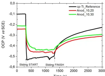
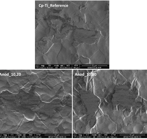
(17) List of Figures Chapter 1 General introduction Figure 1.1 – Schematic representation of a natural tooth (at left) and an endosseous dental implant attached to bone (at right) [13]. ...............................................................4 Figure 1.2 – Schematic illustration of the biomechanical factors acting in an endosseous dental implant system [12]. ..........................................................................5 Figure 1.3 – Schematic illustration of a sliding tribocorrosion mechanism. The counterbody is loaded (N) on Ti surface immersed in a corrosive environment, and rubbing action leads to third body particles formation in the contact area. ..................12 Figure 1.4 – Schematic representation of the four main groups of parameters affecting the sliding tribocorosion behavior under electrochemical control. Adapted from [49]. .........................................................................................................................14 Figure 1.5 – Schematic representation of a Ti anodizing treatment by PEO process. ... ....................................................................................................................................19. Chapter 2 Tribocorrosion behavior of nano-scale oxide structures on titanium surfaces Figure 2.1 – Schematic representation of the system used for anodizing treatments. ....................................................................................................................................34 Figure 2.2 – (a) Electrochemical cell used for tribocorrosion experiments, and (b) Experimental setup of tribocorrosion tests: A) Working electrode; B) Counter electrode; C) Reference electrode; D) PIN supporting the counter material. ..................................36 Figure 2.3 – SEM micrographs of cp-Ti_Reference, Anod,10,20 and Anod,10,30 samples surfaces..............................................................................................................36 Figure 2.4 – OCP evolution of non-anodized and anodized cp-Ti samples immersed in AS, before, during and after the reciprocating sliding tests (test conditions: 100 mN, 500 µm, 1 Hz, 1000 cycles). ............................................................................................38 Figure 2.5 – OCP and COF evolution during the reciprocating sliding time (test conditions: 100 mN, 500 µm, 1 Hz, 1000 cycles). Different groups of samples are. xv.
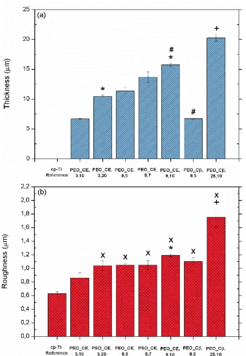
(18) properly identified in the pictures...................................................................................40 Figure 2.6 – SEM micrographs of the wear track of non-anodized and anodized cp-Ti samples after the reciprocating sliding tests (test conditions: 100 mN, 500 µm, 1 Hz, 1000 cycles). ...................................................................................................................41. Chapter 3 Tribocorrosion behavior of porous anodic oxide films produced on titanium by plasma electrolytic oxidation Figure 3.1 – PEO equipment used for cp-Ti anodizing treatments: (a) Power supply, (b) Electrochemical cell, and (c) Refrigerator system. ....................................................51 Figure 3.2 – (a) Electrochemical cell used for tribocorrosion experiments, and (b) Set-up of tribocorrosion tests: A) Working electrode; B) Thermometer; C) Ag/AgCl reference electrode; D) PIN supporting the counter material; E) Counter electrode of platinum...........................................................................................................................54 Figure 3.3 – (a) Thickness and (b) Ra values of cp-Ti_Reference specimens and of anodic oxide films grown onto cp-Ti surfaces during PEO experiments. Different groups of samples are properly identified below the pictures according with the PEO conditions at which were anodized. (*), significantly different from PEO_CE,3,10; (#), significantly different from PEO_CE,8,5; (+), significantly different from PEO_Cβ,8,5; (X), significantly different from cp-Ti_Reference samples; p < 0.05. ....................................55 Figure 3.4 – SEM micrographs of cp-Ti samples surface before and after PEO treatments. Different groups of samples are properly identified below the micrographs, according to the PEO conditions at which were anodized. The pictures are shown at lower and higher magnification (right upper corner). ....................................................61 Figure 3.5 – SEM micrographs of polished cross sections of cp-Ti samples before and after PEO treatments. Different groups of samples are properly identified below the figures, according to the PEO conditions at which were anodized. ................................62 Figure 3.6 – EDS spectra of the cross sections (inner part of the anodic films) of (a) PEO_CE,8,10 and (b) PEO_Cβ,25,10 samples. .................................................................65 Figure 3.7 – OCP evolution of non-anodized and anodized cp-Ti samples immersed in AS, before, during and after the reciprocating sliding tests (test conditions: 2 N, 3. xvi.
(19) mm, 2 Hz, 720 seconds). Different groups of samples are properly identified in the figure................................................................................................................................67 Figure 3.8 – OCP and COF evolution during the reciprocating sliding time (test conditions: 2 N, 3 mm, 2 Hz, 720 seconds). Different groups of samples are properly identified in the pictures. ................................................................................................70 Figure 3.9 – SEM micrographs of the wear track on non-anodized and anodized cp-Ti samples after the reciprocating sliding tests (test conditions: 2 N, 3 mm, 2 Hz, 720 seconds). Different groups of samples are properly identified below the micrographs. All the SEM pictures are shown at lower and higher magnification (right upper corner). ....................................................................................................................................72 Figure 3.10 – Wear volume and confocal images of the wear track of non-anodized and anodized cp-Ti samples after the reciprocating sliding tests (test conditions: 2 N, 3 mm, 2 Hz, 720 seconds). Different groups of samples are properly identified in the figure................................................................................................................................74 Figure 3.11 – OCP evolution of non-anodized and anodized cp-Ti samples immersed in AS, before, during and after the reciprocating sliding tests (test conditions: 2 N, 3 mm, 2 Hz, 720 seconds). Different groups of samples are properly identified in the figure................................................................................................................................75 Figure 3.12 – Evolution of the OCP and COF of PEO_CE,3,20 samples during the reciprocating sliding action (test conditions: 2 N, 3 mm, 2 Hz, 720 seconds). ...............77 Figure 3.13 - SEM micrograph of the wear track on PEO_CE,3,20 samples after the reciprocating sliding tests (test conditions: 2 N, 3 mm, 2 Hz, 720 seconds). The SEM picture is shown at lower and higher magnification (right upper corner). .....................78 Figure 3.14 – Wear volume and confocal images of the wear track of non-anodized and anodized cp-Ti samples after the reciprocating sliding tests (test conditions: 2 N, 3 mm, 2 Hz, 720 seconds). Different groups of samples are properly identified in the figure................................................................................................................................79 Figure 3.15 – OCP evolution of non-anodized and anodized cp-Ti samples immersed in AS, before, during and after the reciprocating sliding action (test conditions: 2 N, 3 mm, 2 Hz, 720 seconds). Different groups of samples are properly identified in the figure................................................................................................................................80 Figure 3.16 – OCP evolution of non-anodized and anodized cp-Ti samples immersed xvii.
(20) in AS, before, during and after the reciprocating sliding tests (test conditions: 5 N, 3 mm, 2 Hz, 1800 seconds). Different groups of samples are properly identified in the figure................................................................................................................................82 Figure 3.17 – Evolution of the OCP and COF during the reciprocating sliding tests (test conditions: 2 N, 3 mm, 2 Hz, 720 sec). Different groups of samples are properly identified in the pictures. ................................................................................................83 Figure 3.18 – Evolution of the OCP and COF during the reciprocating sliding action (test conditions: 5 N, 3 mm, 2 Hz, 1800 seconds). Different groups of samples are properly identified in the pictures...................................................................................85 Figure 3.19 – SEM micrographs of the wear track on anodized cp-Ti samples after the reciprocating sliding tests (test conditions: 2 N, 3 mm, 2 Hz, 720 seconds). Different groups of samples are properly identified below the micrographs. All the SEM pictures are shown at lower and higher magnification (right upper corner). ..............................86 Figure 3.20 – SEM micrographs of the wear track of non-anodized and anodized cpTi samples after the reciprocating sliding tests (test conditions: 5 N, 3 mm, 2 Hz, 1800 seconds). Different groups of samples are properly identified below the micrographs. All the SEM pictures are shown at lower and higher magnification (right upper corner). . ....................................................................................................................................88 Figure 3.21 – Wear volume and confocal images of the wear track on non-anodized and anodized cp-Ti samples after the reciprocating sliding tests (test conditions: 2 N, 3 mm, 2 Hz and 720 seconds). Different groups of samples are properly identified in the figure. (#), significantly different from PEO_CE,8,5; (X), significantly different from cpTi_Reference samples; p < 0.05. ......................................................................................90 Figure 3.22 – XRD spectra of (a) PEO_CE,8,10 and (b) PEO_Cβ,25,10 samples. .......92. Chapter 4 Effect of anodic titanium oxide films on osteoblastic cell response Figure 4.1 – SEM micrographs of cp-Ti samples surfaces, before and after PEO treatments. Different groups of samples are properly identified below the micrographs, according to the PEO conditions at which they were anodized. All the pictures are shown at lower and higher magnification (right upper corner). ..................................108. xviii.
(21) Figure 4.2 – Water contact angle measurements on cp-Ti_Reference and anodized samples. Different groups of samples are properly identified in the figure. (X), significantly different from cp-Ti_Reference samples; (+), significantly different from PEO_CE,8,10 samples; p < 0.05. ....................................................................................111 Figure 4.3 – SEM micrographs of the MG63 cells cultured on standard culture materials (control cultures), cp-Ti reference, PEO_CE,8,10 and PEO_Cβ,25,10 samples, after 1, 3 and 7 days of incubation. Different groups of samples are properly identified below the micrographs, according with the PEO conditions at which they were anodized. SEM micrographs at low magnification. .......................................................112 Figure 4.4 – SEM micrographs of the MG63 cells cultured on standard culture materials (control cultures), cp-Ti reference, PEO_CE,8,10 and PEO_Cβ,25,10 samples, after 1, 3 and 7 days of incubation. Different groups of samples are properly identified below the micrographs, according with the PEO conditions at which they were anodized. SEM micrographs at high magnification. ......................................................113 Figure 4.5 – CLSM micrographs of the MG63 cells cultured on cp-Ti_Reference, PEO_CE,8,10 and PEO_Cβ,25,10 samples, after 3 days of incubation. .........................115 Figure 4.6 – Viability of the MG63 cells cultured on standard culture materials (control cultures), cp-Ti reference, PEO_CE,8,10 and PEO_Cβ,25,10 samples, after 1, 3 and 7 days of incubation. For each culture time: (*), significantly different from control cultures; (#), significantly different from cp-Ti_Reference; (X), significantly different from PEO_CE,8,10 samples; p < 0.05. ...........................................................................115. xix.
(22)
(23) List of Tables Chapter 2 Tribocorrosion behavior of nano-scale oxide structures on titanium surfaces Table 2.1 – Composition of the Fusayama´s artificial saliva [88]. ..............................35 Table 2.2 – Ra values of non-anodized and anodized Ti samples. .............................38. Chapter 3 Tribocorrosion behavior of porous anodic oxide films produced on titanium by plasma electrolytic oxidation Table 3.1 – PEO conditions used for anodizing treatments. ......................................51 Table 3.2 – Composition of the Fusayama’s artificial saliva [88]. ..............................54 Table 3.3 – Surface elemental composition of non-anodized and anodized cp-Ti samples. Different groups of samples are identified in this table. .................................63 Table 3.4 – R/A ratio existing on anodic films of PEO_CE,8,10 and PEO_Cβ,25,10 samples. ...........................................................................................................................92. Chapter 4 Effect of anodic titanium oxide films on osteoblastic cell response Table 4.1 – Thickness and Ra values of Ti untreated specimens and of anodic oxide films grown on Ti surfaces during PEO experiments.....................................................109 Table 4.2 – Surface elemental composition of non-anodized and anodized cp-Ti specimens under different PEO process conditions. Different groups of samples are properly identified in the table. ....................................................................................110. xxi.
(24)
(25) Chapter 1 General introduction.
(26)
(27) Chapter 1. 1.1. Dental implants Dental implantation has increased significantly over the last years, being estimated that around two million implants are applied every year [1, 2]. In 2003, more than 1.3 million implantation were performed in Europe [3]. This number is expected to rise, both due to the aging of the population and to the success of dental implant therapies, with implant survival rates reported to be greater than 89 % after 10 – 15 years [2]. Despite high success rates, a significant number of failures in dental implant systems (1 – 20 %) have still been reported [2, 4, 5]. According to previous studies the rate of failure in the edentulous maxilla is higher in comparison with the mandible (10 – 20 % and 1 – 5 %, respectively) [5]. Dental implants are biocompatible metal anchors incorporated in the maxilla and/or mandible, underneath the gums, for the replacement of orofacial structures (e.g. tooth loss) as a result of decay, trauma, periodontal disease and/or congenital defects [4, 6, 7]. Sundry kinds of dental implant devices are available. These systems can be classified according to their shape and position as subperiosteal, transosteal and endosseous. Endosseous titanium dental implants are placed within the bone either in upper or lower jaw, and are the most widely used nowadays [8-10]. This dental implant system includes a wide range of sizes, shapes, and other prosthetic components [8]. Endosseous titanium dental implants with a screw design are the systems that will be addressed in this work. A screw design is one of the most widely used shapes, associated with the additional immediate fixation that a threaded implant can provide [8, 10]. Using a root form implant (e.g. with a screw design [11]), which is the closest in shape and size when compared with the natural tooth root, bone healing period usually varies from a few to six or more months, and during this period, osseointegration is expected to occur [6]. Figure 1.1 presents a schematic representation of an endosseous titanium dental implant with a screw design (screwtype implant). The implant system consists of an artificial tooth root that is screwed and anchored into the jaw bone to sustain an artificial crown where the natural tooth is missing [12, 13]. The abutment is the component that is screwed into the dental implant and responsible to support the crown, connecting both structures (Figure 1.1) [12].. Sofia Alves | Master Dissertation | 2012. 3.
(28) Chapter 1. Figure 1.1 – Schematic representation of a natural tooth (at left) and an endosseous dental implant attached to bone (at right) [13].. The clinical success of endosseous dental implants is directly related to early osseointegration, process that can be defined as the direct structural and functional connection between bone tissue and the surface of dental implant [1, 8, 14, 15]. This process is critical for implant stability and it is considered as a prerequisite for implant loading [8]. Osteointegrated implants are widely used not only in dental but also in maxillofacial and ear-nose-throat fields [15]. The biomaterials most commonly used to fabricate screw-type dental implants are commercially pure titanium (cp-Ti) and Ti alloys by virtue of an excellent combination of strength-to-weight ratio, excellent corrosion resistance and biocompatibility [16-18].. 1.1.1. Causes of dental implant failure Despite the high success of dental implant therapies, implant failure can still occur. Failure of a dental implant is mostly due to mechanical stresses, bacterial infection, rejection, accelerated bone loss and poor osseointegration [3, 19]. The. Sofia Alves | Master Dissertation | 2012. 4.
(29) Chapter 1. incidence of dental implant loss associated to the lack of osseointegration or to loss of integration after loading, has been well documented in numerous reports [5]. Once osseointegration is established, failures are usually divided into two major categories: biomechanical and biological [2, 20]. A dental implant is under a complex congregation of biomechanical solicitations and also exposed to a quite aggressive biological and chemical environment, which can influence its behavior and stability in the oral cavity and lead to its failure [3, 12].. Biomechanical environment Biomechanical failure can be associated with overloading conditions of implantbone interface due to application of high-level functional loads [20]. Cyclic stresses are often associated to premature mechanical failure of metallic biomaterials [21]. During mastication, occlusal forces (forces exerted on opposing teeth) can result in cyclic micro-movements at the implant-bone, implant-abutment and/or abutment-crown interfaces, as schematically illustrated on Figure 1.2 [12]. Occlusal forces produced during the masticatory function have been described to be in the range of 15N to 150N [6, 22].. Figure 1.2 – Schematic illustration of the biomechanical factors acting in an endosseous dental implant system [12].. During fixation or normal use of dental implants, they are submitted to wear mechanisms by shear forces [23]. The orientation of stresses transmitted to dental. Sofia Alves | Master Dissertation | 2012. 5.
(30) Chapter 1. implant systems during mastication is of crucial importance. The material of dental implant structures and bone tissue can be overloaded due to oblique loads transmitted during mastication, promoting failures by fatigue wear of the implant system [12]. In fact, as higher the relative displacement between these structural components as higher the tendency to occur tribological damages of the implant [12]. When the screw interface of the dental implant is submitted to external loads, micro-movements occur between the surfaces implant/bone which may lead to wear in the contact area [24]. These events can cause detachment of films existing on implant surface and wear debris generation leading to wear-mediated osteolysis process which is highly undesirable [23, 25]. The occurrence of implant fractures have been reported in numerous studies (0 – 16 %) and have been associated with overload conditions [20]. The liberation of wear debris to the peri-implant site and adjacent tissues can present a whopping health risk. The degradation of the material can induce to periimplant pathologies comprising inflammatory lesions (peri-implantitis) around functioning dental implants [12, 26]. Particles produced by wear at implant-bone interface can induce a foreign body inflammatory response, involving the presence of macrophages and the release of different cytokines, and consequently periprosthetic osteolysis and loosening of the implant [18, 27]. In fact, metallic wear debris have been shown to have a direct effect on cells including macrophages, fibroblasts and osteoblasts [25] which can affect the osseointegration process. Accumulation of Ti ions in tissues adjacent to implants has been reported in conditions not entirely attributed to wear [7]. When these events occur simultaneously in a corrosive environment (e.g. oral environment), wear and corrosion phenomena can take place leading to liberation of wear debris and corrosion ions to peri-implant site [7]. Ions released from the implant as a consequence of corrosion can also result in adverse biological reactions and as consequence, the mechanical failure of the implant device [7].. Chemical and Biological environment The oral environment is extremely complex and hostile and all parts present in the oral cavity are continuously bathed in saliva [6]. Human saliva is a complex fluid whose secretion is made by the major (parotid, sublingual and submandibular) and minor salivary glands and controlled by autonomic nervous system [28]. Saliva is an Sofia Alves | Master Dissertation | 2012. 6.
(31) Chapter 1. aqueous solution of chlorides, with varying amounts of sodium (Na), potassium (K), calcium (Ca), phosphate (PO43-), carbon dioxide (CO2) and sulphur compounds. The pH is normally between 5.5 and 7.5, however under plaque adhesion conditions, it can be low as 2, and the temperature is maintained around 36.5o [6]. Human saliva, besides glycoproteins (e.g. mucin) contains digestive enzymes (e.g. lipase and amylase) comprising also organic and inorganic substances and other components as immunoglobulins [28]. Oral environmental conditions can be strongly influenced by bacterial action through, for instance, the production of acidic metabolic products which can have a deleterious effect on the degradation of dental implant materials [12]. Infection is indentified as one of the main factors contributing to dental implants failure, generally associated with the microbial flora existing in the oral environment [2, 4]. Immediately after implantation, the dental implant will be in contact with body fluids such as blood and saliva, which contain proteins that can adhere to biomaterial surface acting as receptors to microbial attachment. Microbial adhesion is the first and most important step in implant infection. A lack of successful skin integration around the dental implant can be related with bacteria invasion [29]. Peri- implantitis, which can result in implant infection, is considered an inflammatory process which affects the function of surrounding tissues of an osseointegrated implant, resulting in loss of supporting bone [2, 4]. Microorganisms, once adhered, will generally colonize forming a biofilm, a complex structure where they are included [12]. According to previous studies, infected and failing implants show greater proportions of periodontal pathogens such as Peptostreptococcus micros and Prevotella intermedia [2]. Furthermore, Staphylococci are present in the oral cavity and their isolation from periimplant infections is significant [4]. Moreover, both Staphylococcus aureus and coagulase-negative Staphylococci are frequently responsible for infections associated with metallic biomaterials [4]. It is worth to note that biofilms can have a strong effect on the corrosion and/or wear behavior of dental implant material, influencing the implant stability in the oral environment [12]. Corrosion reactions can take place as a consequence of the hostile electrolytic biological environment. Thus, corrosion products may be released into the human body where they can remain during weeks or months. As reported in previous studies, during the corrosion process of an implant Sofia Alves | Master Dissertation | 2012. 7.
(32) Chapter 1. particles are released and, as a consequence, there is a recruitment of macrophages till the peri-implant tissue. These particles are phagocytosed by macrophages, stimulating the release of inflammatory intermediates, as cytokines, which contribute to bone resorption and implant failure [30].. 1.2. Titanium as a dental implant biomaterial Biomaterials are synthetic or natural materials, generally used in medical devices, which are intended to interact with biological systems [31]. Biocompatibility, defined as the ability of a material to establish an appropriate host response in a specific bioapplication [31], is a crucial feature that biomaterials should exhibit as well as possible. Metals, ceramics, polymers, glasses, carbons and composites materials can be included in the list of biomaterials [31]. In this work, metals, more specifically Ti, will be addressed as biomaterial for dental implant application. Chemically, Ti is a transition element in group IV and period 4 of Mendeleef´s periodic table. It has an atomic number of 22 and an atomic weight of 47.9 [10]. Nowadays, cp-Ti and Ti alloys are the most commonly used materials for dental implant applications mainly due to their excellent biocompatibility and corrosion resistance [1, 10, 32]. Cp-Ti exhibit different degrees of purity (graded from 1 to 4), which are established according to the oxygen (O), carbon (C) and iron (Fe) contents. The material strength increases from grade 1 to 4 [1]. The excellent corrosion resistance and improved biocompatibility of Ti are essentially owing to the dense, highly stable, well-adhering and protective oxide film (mainly TiO2) that is generated on its surface when exposed to oxygen in air or water (oxygen-containing environments). This layer provides an effective barrier to electron and ion transport which is associated with the superior corrosion resistance of this material. Due to the high reactivity of Ti, the reaction with the surrounding environment to form a TiO2 oxide film on its surface is almost spontaneous (nanosecond timescale) and generally it is characterized by a thickness of approximately 1.5 – 10 nm [3, 6, 16, 21, 33-35]. Those materials which have the ability to be protected from corrosion attack through the spontaneous formation of this oxide film onto its surface under oxidizing conditions, are named as passive [21, 36]. This. Sofia Alves | Master Dissertation | 2012. 8.
(33) Chapter 1. passive film avoid that the underlying bare metal surface is in direct contact with aggressive ions present in biological fluids such as sodium (Na+), magnesium (Mg2+), chloride (Cl-) and sulfate (SO42-) [21]. The passive oxide film that is formed adheres strongly to the surface of Ti substrate, and it is amorphous or poorly crystallized [6, 37]. The Ti thermodynamically stable oxide is TiO2 and can exist in three polymorphic forms: anatase (tetragonal), rutile (tetragonal) and brookite (orthorhombic) [16, 17, 38]. Anatase is a metastable phase and can be transformed to rutile at about 1100 K [39]. According to some previous studies, it is known that the presence of these phases on the oxide film grown on Ti surfaces confers higher hardness [40] and enhanced biological performance than the presence of an amorphous oxide film [25, 41, 42]. Furthermore, cp-Ti and Ti alloys have been widely used for dental implant applications by virtue of other features. The low elastic modulus of Ti-based materials is generally seen as a biomechanical advantage in the case of hard-tissue replacements, since it can result in a lower stress shielding, reducing bone resorption [10, 43]. Moreover, an excellent combination of strength-to-weight ratio and high melting temperature are also important features [18, 41]. Other properties that are related with the natural selection of Ti for implantation are the high osseointegration tendency that this material exhibit, which plays an important role in the long-term clinical success of osseointegrated implants as well as low its very low toxicity [7, 18, 43]. Besides all these advantages, it is known that Ti is a material with poor tribological properties [7, 44, 45] what can be associated with deleterious biological effects. The poor tribological behavior of Ti alloys is characterized by high coefficient of friction, severe adhesive wear and low abrasion resistance [46].. 1.3. Corrosion and mechanical wear of titanium in the oral cavity The degradation of a Ti dental implant in the oral cavity is an issue of main importance. Ti degradation can result in the release of particles into surrounding tissues by different mechanisms, including corrosion, mechanical wear, and mechanically accelerated electrochemical processes (e.g. stress corrosion, corrosion. Sofia Alves | Master Dissertation | 2012. 9.
(34) Chapter 1. fatigue and fretting corrosion). Cyclic loadings lead to generation of wear debris (contact body) which has been associated with adverse biological reactions, namely osteolysis, or accelerate articular wear if entrapped between the contacting surfaces compromising the implant performance [21, 47]. As already pointed out, during mastication, cyclic micro-movements between dental implant surface and bone can be generated. The fatigue of metals has been extensively studied once fluctuating stresses typical of fatigue can lead to the initiation and growth of a crack that, once reaching a critical size, leads to material fracture [21]. When biomaterials are exposed to a corrosive environment and simultaneously to a mechanical action, a degradation process named tribocorrosion takes place. Tribocorrosion is the irreversible transformation of a material due to the simultaneous action of corrosion and mechanical wear (adhesion, abrasion and fatigue) taking place in a sliding tribological contact [48]. The material degradation will be the result of a complex synergism established between chemical, electrochemical and mechanical processes, and so interactions taking place in a tribocorrosion system are very difficult to predict [36, 49].. 1.3.1. Titanium corrosion Corrosion is an electrochemical process that involves the transfer of electrons between a metal surface and its surrounding environment leading to a gradual degradation of the base material [30, 50]. Different corrosion processes can occur, namely uniform corrosion, galvanic and stress corrosion, cracking, pitting and fatigue corrosion [30, 51]. A corrosion rate of 2.5x10⁻⁴ mm/yr has been well accepted for metallic implant systems [51]. The gradual deterioration of biomaterials by electrochemical attack is of main concern essentially when a metallic implant is placed in a hostile electrolytic environment as the one existing in the human body (e.g. oral cavity) [30]. In the oral cavity, dental implants are subjected to an adverse chemical atmosphere where saliva tends to be highly corrosive and, once in this environment, the electrochemical behavior of metallic biomaterials is a function of many parameters including the composition of saliva in fluoride anion or organic compounds, such as proteins [52-54]. The corrosion process can be also strongly influenced by changes in pH values, which can be associated, for instance, to an imbalance in the biological system due to Sofia Alves | Master Dissertation | 2012. 10.
(35) Chapter 1. diseases, infections and other factors [7, 51]. Corrosion resistance of dental implant materials is an aspect of vital importance once it can adversely influence its biocompatibility and mechanical integrity [7, 53]. The interaction of the biomaterial and its chemical environment (corrosion) can lead to a loss of substance from the material, a change in its structural characteristics and loss of structural integrity [30]. In spite high corrosion resistance of Ti, there are evidences that in vivo conditions (non static conditions) can be promoted the modification of the stability of the passive film presents on its surface, enhancing the corrosion process [7, 51, 55, 56]. Under physiological conditions (e.g. under loading action), the passive film may not be sufficiently stable to withstand the mechanical action, and be degraded in the oral environment [56], and the corrosion resistance considerably decreased. The effect of chemical reactions between a dental implant and the oral environment can result in the release of unwanted metallic ions to its surroundings [30, 43, 57]. In addition, proteins can complex themselves to metal ions and transport them away from the implant surface contributing to further corrosion [51]. Analysis of retrieved implants pointed out a possible accumulation of ions in adjacent tissues to the implant. The dissolution of metal ions into the human body can interfere in the processes of life, inducing the release of osteolytic cytokines which can be involved in the implant loosening [37, 55]. After all these observations the demand for new treatments that decrease the corrosion susceptibility of Ti in the human body is of great importance. A wide collection of different surface treatments, such as mechanical treatments, anodizing and ion implantation, have already been studied in order to achieve enhanced surface properties [37, 55].. 1.3.2. Titanium tribocorrosion Tribology is the science and technology of friction, lubrication and wear. However, in several tribological systems the materials evolved in tribological contact are exposed to a corrosive environment, therefore they can be subjected simultaneously to chemical- electrochemical- mechanical effects, leading to their degradation by a process named tribocorrosion [36, 58-60]. The tribocorrosion effect was discovered by Edison, in 1875 and after this finding, tribocorrosion field has grown Sofia Alves | Master Dissertation | 2012. 11.
(36) Chapter 1. and is attracting the attention of researchers of all over the world mostly due to its practical importance and economic benefits [49]. Tribocorrosion science comprises the study of the combined effect of corrosion and mechanical wear in the degradation of materials in relative displacement (e.g. sliding or rolling contacts) [36, 59, 61]. Materials degradation due to a simultaneous action of mechanical and electrochemical processes is a result of a complex synergism established between both. Thus, the total material degradation differs from the simply adding of mechanical wear and corrosion measured separately, being usually higher [36, 48, 61, 62]. Sliding tribocorrosion leads to local damage or removal of the passive film (mechanical wear) existing onto Ti materials surface (e.g. by adhesion, abrasive or fatigue wear) as well the detachment of particulate debris to contact area acting as third body particles, which can affect seriously the friction and wear regime [36, 48]. It is well known that the damage and removal of the passive film existing onto Ti surfaces can lead to a strong increase in the local rate of corrosion (wear-accelerated corrosion) [36, 48]. The corrosive environment may change the mechanism of formation and the physical properties of third body particles. According with Landolt et al. [36] depending on the properties of these particles, corrosion can accelerate or slow down the rate of mechanical material removal from the substrate. During rubbing action, depassivation and repassivation phenomena of the oxide passive film can take place in the contact region. These processes involve charge transfer at the interface leading to the generation of dissolved metal ions and a solid oxide [48]. Corrosion products can be accumulated in the mechanical contact region, affecting the wear regime [7]. Thus, rubbing action leads to loss of material by mechanical and electrochemical mechanisms. Figure 1.3 represents schematically a sliding tribocorrosion mechanism.. Sofia Alves | Master Dissertation | 2012. 12.
(37) Chapter 1. Figure 1.3 – Schematic illustration of a sliding tribocorrosion mechanism. The counterbody is loaded (N) on Ti surface immersed in a corrosive environment, and rubbing action leads to third body particles formation in the contact area.. Endosseus Ti dental implants can be subjected simultaneously to electrochemical attack by the corrosive nature of oral environment (saliva) and mechanical sliding (e.g. due to cyclic micro-movements generated during mastication) at the implant-bone interface. These interactions may destroy and remove the protective film existing onto the Ti implant surface leading to tribocorrosion of the dental implant material [7, 61]. The disruption and removal of the protective oxide film from the metal surface lead to exposure of reactive metal atoms (susceptible to corrosion) to surrounding and aggressive environment. Consequently, the corrosion rate will increase and lead to an accelerated degradation of the contact region [7, 51, 61]. The resulting tribocorrosion products have been found to promote adverse biological effects [18, 27]. Figure 1.4 shows a schematic representation of the four main groups of parameters affecting tribocorrosion in the case of a sliding contact under electrochemical control (tribocorrosion system) [49]. This illustration depicts the large number of parameters affecting the tribocorrosion response. In the case of a tribocorrosion experiment, the selection of testing parameters depends on the application and researcher interest.. Sofia Alves | Master Dissertation | 2012. 13.
(38) Chapter 1. MECHANICAL/OPERATIONAL: -Norma Force; -Slidind velocity,; -Type of motion; -Shape and syze of contacting bodies; -Aligment; -Vibrations. ELECTROCHEMICAL:. SOLUTION:. -Applied potential;. -Viscosity;. -Ohmic resistance;. -Conductivity;. TRIBOCORROSION SYSTEM. -Repassivation kinetics; -Film growth; -Active dissolution.. -pH; -Corrosivity; -Temperature.. MATERIALS: -Hardness, plasticity; -Microstructure, inclusions; -Surface roughness; -Oxide film properties; -Wear debris, material transfer.. Figure 1.4 – Schematic representation of the four main groups of parameters affecting the sliding tribocorosion behavior under electrochemical control. Adapted from [49].. There are different electrochemical techniques to characterize the surface of a material once immersed in an electrolyte, namely, open circuit potential (OCP), polarization and electrochemical impedance spectroscopy (EIS) measurements. To perform a tribocorrosion test, corrosion and mechanical action responses have to be measured simultaneously. This way, the information obtained by corrosion techniques can be linked and related with in situ mechanical measurements, as coefficient of friction recorded during sliding action. This information can be also related with ex situ surface characterization techniques like scanning electron microscopy (SEM), X-ray diffraction (XRD), energy dispersive X-ray spectroscopy (EDS) and others, in order to gather a global insight on the degradation and protective processes taking place during tribocorrosion [59, 63]. Despite the high corrosion resistance and attractive toxicological properties of Ti and its alloys, the major disadvantage of these materials is their very poor wear resistance [35]. The poor tribological properties of Ti and Ti-based materials and also the hostile environment in which they are implanted can result in the release of. Sofia Alves | Master Dissertation | 2012. 14.
(39) Chapter 1. metallic wear particles and corrosion products which can promote adverse biological reactions and loosening of the implant. Despite all the aforementioned studies, just a few of them have focused the enhancement of the tribocorrosion behavior of Ti in biological systems. Thus, the demand of new techniques that allow the creation of a passive film with characteristics that improve the wear and corrosion resistance of Ti biomaterials for dental implant applications is an issue of greatest interest.. 1.4. Integration of titanium dental implants with bone The integration of a Ti dental implant into surrounding bone tissues is an issue of main importance. Some experiments indicate that clinical success of Ti oral implants depends on the degree and early osseointegration [64, 65]. The lack of bone formation around the biomaterial site after implantation is one of the most frequent causes of implants failure [3].. 1.4.1. Bone tissue Bones are complex structures constituted by important components such as blood, adipose tissue, bone marrow, nervous tissue, cartilage, and fibrous connective tissue. Moreover, bone is composed by a connective tissue based in a hard matrix consisting of organic (collagen and various protein-carbohydrate complexes such as glycosaminoglycans, proteoglycans and. glycoproteins) and. inorganic matter. (hydroxyapatite (Ca10(PO4)6(OH)2) that is a crystallized calcium phosphate salt, calcium carbonate (CaCO3) and few amounts of Mg, Na, K, fluoride (F-), SO42-, carbonate (CO32-) and hydroxide (OH-) ions). The production and organization of these components in the extra-cellular matrix (ECM) is essential to modulate the degree of cell attachment on biomaterials surface. In the case of dental implants, a mineralized ECM is necessary for osseointegration [66, 67]. The ECM is hardened by a process named mineralization, consisting on the deposition of calcium phosphate and other minerals. During this process, ions such as calcium and phosphate are taken from the blood plasma and deposited in bone tissue. Calcium phosphate crystals that are formed throughout this crystallization process become entrapped into collagen fibers produced by osteoblasts, which harden the matrix [66].. Sofia Alves | Master Dissertation | 2012. 15.
(40) Chapter 1. There are four principal kinds of cells being part of the bone tissue: 1) Osteogenic cells (Stem cells that arise from embryonic fibroblasts which are continuously being multiplied, and some of them differentiate into osteoblasts); 2) Osteoblasts (Non-mitotic and bone forming cells that synthesize the organic matter of bone matrix and also help to mineralize the bone); 3) Osteocytes (Osteocytes work as sensors once, when they detect strain on bone, they communicate this information to osteoblast cells.); 4) Osteoclasts (Bone-dissolving cells which are found on the bone surface) [66]. Immediately after implantation, the implant surface is in contact with body fluids interacting with a large number of proteins and different cell types [65], and osteoblastic cells interaction will take place through protein coatings [3]. Implant surface engineering has a challenge to attract, above all, osteoblastic cells producers of bone extracellular matrix, in order to ensure a higher bone-implant contact [65].. 1.4.2. Importance of implant surface features on osseointegration The biological fixation between the dental implant surfaces and the surrounding bone tissues should be considered a prerequisite for the long-term success of endosseous dental implants [68]. As already pointed out, the clinical success of oral implants is associated to their early osseointegration [1]. For the efficacy of dental implants It is fundamental to ensure a mechanically solid interface with complete fusion between the materials surface and the bone tissue without fibrous tissue formation at interface [69]. Although, the poor osteointegration is one of the most common causes of failure of dental implant systems [3, 65]. Several researches have been carried out showing that the surface features of an implant play a crucial role in implant osseointegration [1, 64, 68, 69]. It is known that implant surface topography, morphology, chemical composition and surface energy have a critical influence on proliferation, differentiation and osteoblastic ECM expression [23, 65, 67, 70]. These factors are critical for protein adsorption and cell adhesion both in early cell-material interactions as well during later and more chronic phases of the cell response [64, 65, 69, 71]. Cell adhesion is one of the initial critical stages to subsequent proliferation and differentiation [65, 72]. Surface topography can also affect cell shape influencing its proliferation, differentiation and function [71]. Sofia Alves | Master Dissertation | 2012. 16.
(41) Chapter 1. 1.5. Surface modification of titanium by anodizing for enhanced tribocorrosion behavior and improved biological response of dental implants A great number of Ti surface treatments have been applied targeting the improvement of dental implants osseointegration [1, 68, 73-75]. Modifications on surface properties by means of sand-blasting, acid etching, Ti plasma spraying of hydroxyapatite (HA), chemical and/or thermal treatments, and also electrochemical methods, have been widely used for enhancing biological response and bone tissue integration of Ti implants [73, 76-79]. Plasma-sprayed HA coatings have been widely used to improve bioactivity of Ti materials in order to enable a quicker healing. However, these coatings have shown poor long-term performance associated to problems concerning the adhesion of the coating to the substrate [73, 80]. The HA nucleation on biomaterials surface can act as a way to promote a direct and stronger implant-bone connection, without intermediate fibrous tissue as desirable [23, 81]. Moreover, the introduction of calcium and phosphate ions on Ti surface has been carried out as a surface treatment in order to improve its bioactivity promoting implant bond with bone [82, 83]. There is a growing interest in the formation of biocompatible and bioactive anodic films by incorporation of calcium and phosphorous into oxide film layers [84]. Bone-like apatite can be formed in a Ti anodic layer which has incorporated calcium and phosphate ions into its structure enhancing the implant adhesion with bone [82]. Besides the importance of developing surface treatments which enhance the biological performance of Ti dental implants, it is crucial these systems express simultaneously a good tribological and corrosion performance once exposed to the hostile environment of the human body. Surface modifications by techniques such as PVD, CVD, plasma immersion ion implantation, anodic oxidation, plasma and laser nitriding have been employed in order to enhance the tribological resistance of Ti and Ti-based materials [17, 45, 79]. As can be seen, a wide range of surface modification techniques have been investigated, however, there is a lack of exploration of more effective methods to improve the corrosion and tribocorrosion performance of Ti materials. Sofia Alves | Master Dissertation | 2012. 17.
(42) Chapter 1. Anodizing has attracted considerable attention as a simple and fast method for modifying Ti surface features [18, 76, 77]. Anodizing is an electrochemical process that promotes the formation of an oxide film onto Ti surface. In a conventional anodizing process, a voltage is applied between an anode (e.g. cp-Ti specimen) and a cathode (an unreactive or inert material, e.g. platinum), which are immersed in an anodizing bath. When the voltage is applied between the anode and the cathode is established a flux of electrons between both [85]. Anodic oxidation process includes electrode reactions and oxygen ion diffusion leading to the growing of an oxide film on anode surface [45, 85]. The anodizing solution can be constituted by different elements and depending on these, the oxide layer produced on Ti surface can have different characteristics [44], namely roughness, thickness and chemical composition. The anodizing technique is receiving a growing importance because of its convenience to create biologicallyinspired surfaces with different chemical compositions according to electrolyte which is being used during the process [32]. Many efforts have been done to develop nanoscale features on Ti substrates to mimic the micro/nanostructures of natural bone and so, increase implants biocompatibility [34, 86]. Surface profiles in this range have evinced play an important role in the adhesion of osteoblastic cells [1]. Researches have specifically showed that nanorough (created through electron beam evaporation), nanotubular and nanotextured Ti (created by anodization) can enhance osteoblast functions and minimize bacteria growth [87]. Furthermore, the surface modification of Ti materials at nano-level is believed to enhance wear and corrosion resistance of Ti. Faghihi et al. [27] studied the tribocorrosion behavior of nanostructured Ti substrates processed by high-pressure torsion and they concluded that these showed a better tribocorrosion performance compared to untreated samples. A wide range of fabrication methods have been used to form nanometer-sized TiO2 tubules, wires, dots and pillars. Among a wide range of techniques, anodic oxidation of Ti in fluorinated electrolytes is a simple method to synthesize nanoporous and nanotubular structures [88, 89]. Plasma electrolytic oxidation (PEO), micro-arc oxidation (MAO), or also named, anodic spark deposition (ASD), is a recent technology which has become one of the most important in surface engineering field [17, 90]. PEO is an electrochemical treatment based on the conventional anodizing, whose main principle is also to apply a Sofia Alves | Master Dissertation | 2012. 18.
(43) Chapter 1. voltage between an anode and a cathode that are immersed in an electrolyte, although, under the additional condition of being applied a high voltage capable to produce a plasma discharge phenomenon occurring at the electrode-electrolyte interface as a result of the dielectric breakdown [16, 76, 80, 91, 92]. In Figure 1.5 is schematically represented an anodizing treatment by PEO to a Ti substrate.. Figure 1.5 – Schematic representation of a Ti anodizing treatment by PEO process.. PEO is an effective technique which can be applied to Ti and its alloys in order to obtain a well adhered and porous surface layer enriched with some components, according with the electrolyte composition, by the conversion of the metal surface into Ti oxide [16, 17, 73, 91]. Roughness and porosity of Ti oxide films acquired after a PEO process are important features for improving Ti osseointegration [23, 39]. Furthermore, the formation of a TiO2 film firmly adhered to the substrate is also beneficial for the biological performance of implants [76]. Another great advantage of this technique is the possibility of incorporating bioactive components, such as Ca and P ions, into the anodic film in order to improve the bioactivity of implants promoting bone apposition [39, 76]. Several in vitro and in vivo researches have shown that the modification of Ti surface properties by electrochemical techniques leads to an improvement of the biological performance of Ti substrates [70, 73, 76]. Ravanetti et al. [73] researched about the in vitro cellular response and in vivo primary osteointegration of electrochemically modified Ti by ASD and concluded that the treatments enhanced the biological response in vitro, promoting an early osteoblast Sofia Alves | Master Dissertation | 2012. 19.
(44) Chapter 1. adhesion, and also the osteointegrative properties in vivo, accelerating the primary osteogenic response. Plasma discharge phenomenon affects the composition and the structure of the oxide film surface once the generation of many localized discharges at the Ti/electrolyte interface leads to local high temperature (as high as 2400 oC [93]) and pressure and to plasma chemical reactions at the materials surface [16, 92]. The high temperature reached during plasma discharges can result in conversion of the growing oxide film into crystalline phases which can confer higher hardness to the film than the amorphous oxide grown during conventional process [40]. Besides the high hardness and excellent bonding strength with the substrate, PEO films are also characterized by an improved corrosion and wear resistance [16, 39]. Investigations performed by Krishna et al. [94] reported that micro-arc oxidation enhanced the wear resistance of Al alloys under abrasion wear conditions, in comparison with samples anodized by conventional technique. The anodizing process is rather complex and depends on various parameters such as applied current density, concentration and composition of the electrolyte, the electrolyte temperature, duration of the process, the agitation speed of the solution during treatment and also the cathode to anode surface area ratios [95]. A wide range of investigations have already been carried out and demonstrated that the surface features of Ti oxide films promoted by PEO (e.g. porous structure, roughness, film thickness, hardness, wear and corrosion resistance) are strongly dependent on processing conditions [16, 23, 81, 84, 93]. PEO is quite promising in the scope of industrial use due to its easy and rapid operation, economic efficiency and its ecological friendliness [16, 45, 92]. Beyond this, PEO is a versatile technique and by this way can it be applied to components of almost any shape and size [13].. Sofia Alves | Master Dissertation | 2012. 20.
Imagem
![Figure 1.1 – Schematic representation of a natural tooth (at left) and an endosseous dental implant attached to bone (at right) [13]](https://thumb-eu.123doks.com/thumbv2/123dok_br/17803109.840969/28.892.210.708.111.547/figure-schematic-representation-natural-endosseous-dental-implant-attached.webp)
![Figure 1.2 – Schematic illustration of the biomechanical factors acting in an endosseous dental implant system [12]](https://thumb-eu.123doks.com/thumbv2/123dok_br/17803109.840969/29.892.207.672.668.969/figure-schematic-illustration-biomechanical-factors-acting-endosseous-implant.webp)


+7


Documentos relacionados
Varying the anodization time produces an increase both in surface area and in the amount of oxide generated, whereas the amount of incorporated anions
The current grew in the system when concentration of the activator (fluoride ion) increased; thus, velocities of oxide formation and dissolution processes in active centres
The curves demonstrated the deformation behavior of the films, indicating that the films elaborated with gelatin capsule waste of safflower exhibited the higher values for
The oxide films prepared in H2SO4 had greater contact angle, whereas there is no statistical significance between the values for the Ti anodic films produced in Na2SO4 and
It has been suggested that physiochemical and dielectric properties, crystal structure and surface morphology of titanium oxide films on dental implant surfaces play a crucial role
ing to this model, permanganate, which is absent near the metal/oxide interface, is not expected to have a significant effect on the corrosion resistance of the anodic oxide film on
The corrosion potential of the aluminum surfaces coated with poly- meric films shifted to less negative values, and the anodic current densities of these surfaces were lower than
The stability of anodic ilms grown on titanium materials was studied in a physiological electrolyte, up to 8.0 V, where thin titanium oxide ilms protect the surface of the
