Recebido em 04/2019. Aceito para publicação em 05/2019.
ANÁLISE DA PRODUÇÃO DE COLÁGENO EM TECIDO CARDÍACO HIPERTRÓFICO DE RATOS TRATADOS COM L-NAME E L-ARGININA ATRAVÉS DO PROCESSAMENTO DE IMAGENS
ANALYSIS OF COLLAGEN PRODUCTION IN HYPERTROPHIC CARDIAC TISSUE OF RATS TREATED WITH L-NAME AND L-ARGININE BY IMAGE PROCESSING
Vanessa Oliveira Colombo Marson1 Marcia Aparecida Silva Bissaco2 Luciano Ramos3 Silvia Cristina Martini4 Romildo Torres da Silva5 Fernando Francisco Pazello Mafra6 Michel Monteiro Macedo7 Patrícia Sardinha Leonardo8 Rodrigo Álvaro Brandão Lopes-Martins9
Resumo: Analisamos a produção de colágeno no coração de ratos hipertensos induzidos por L-name. Métodos: Imagem cardíaca de 5 grupos: NT, hipertensão induzida pelo name, name e 10 mg L-arginina, L-name e 30 mg L-arginina e L-name e 100 mg L-arginine. Quantificação de COL por histologia com picrosirius CFC_Image v. 2.0. Calibração do software selecionando valores de matiz, saturação e intensidade HSI na imagem de referência, uso do software nas imagens utilizadas. Resultados: As imagens não apresentaram diferenças estatísticas em nenhum dos conjuntos de HSI, indicando que a calibração do software é válida para o estudo. Doses maiores que 30 mg de L-arginina mostraram maior redução das áreas de colágeno, indicando maior proteção das estruturas cardíacas na hipertensão. Conclusão: O CFC_Image v. 2.0. A precisão demonstrada na quantificação da suplementação de COL, L-arginina promoveu efeitos protetores em modelos murinos de hipertensão.
Palavras-chave: Colágeno; processamento de imagem; tecido cardíaco; hipertrofia cardíaca; l-arginina.
Abstract: We analyzed the production of collagen in the heart of hypertensive rats induced by L-name. Methods: Cardiac imaging of 5 groups: NT, hypertension induced by name, name and 10 mg L-arginine, L-name and 30 mg L-arginine and L-name and 100 mg L-arginine. Quantification of COL by histology with picrosirius CFC_Image v. 2.0. Calibration of the software by selecting values of hue, saturation and HSI intensity in the reference image, use of the software in the images used. Results: The
1 Mestre - Universidade de Mogi das Cruzes, Programa de Pós-Graduação em Engenharia Biomédica, São
Paulo, Brasil. E-mail: [email protected].
2 Doutora - Universidade de Mogi das Cruzes, Programa de Pós-Graduação em Engenharia Biomédica, São
Paulo, Brasil. E-mail: [email protected].
3 Doutor em Farmacologia. Docente na Faculdade Estácio de Sá, Brasil. E-mail: [email protected]. 4 Doutora - Universidade de Mogi das Cruzes, Programa de Pós-Graduação em Engenharia Biomédica, São
Paulo, Brasil. E-mail: [email protected].
5 Doutor - Universidade de Mogi das Cruzes, Programa de Pós-Graduação em Engenharia Biomédica, São
Paulo, Brasil. E-mail: [email protected].
6 Doutor - Universidade de Mogi das Cruzes, Programa de Pós-Graduação em Engenharia Biomédica, São
Paulo, Brasil. E-mail: [email protected].
7Mestre Universidade de Mogi das Cruzes, Programa de Pós-Graduação em Engenharia Biomédica, São Paulo,
Brasil. E-mail: [email protected].
8Doutor - Universidade do Vale do Paraíba (UNIVAP), Biofotônica e Terapeutica Experimental, IP&D, São José
dos Campos - SP, Brasil. E-mail: [email protected].
images did not present statistical differences in any of the HSI sets, indicating that the calibration of the software is valid for the study. Doses greater than 30 mg of L-arginine showed a greater reduction in the collagen areas, indicating a greater protection of the cardiac structures in hypertension. Conclusion: CFC_Image v. 2.0. The accuracy demonstrated in the quantification of COL, L-arginine supplementation promoted protective effects in murine models of hypertension.
Keywords: Collagen; image processing; cardiac tissue; cardiac hypertrophy; L-arginine.
1 INTRODUCTION
Cardiac hypertrophy (CH) is a pathological condition generally considered as a compensatory mechanism which intends to normalize the increased ventricular tension resulting mainly from hypertension. However, CH is an important risk factor for cardiovascular morbidity and mortality. The disease itself can be caused by insufficient blood supply to the myocardium as a result of reduced coronary flow reserve (TSAI et
al. 2017).
Cardiac hypertrophy is also characterized by increased heart volume. In chronic conditions, pressure overload exerted on the heart is a pathological condition that leads to heart failure (CARMO et al., 2011; LIEBSON et al., 1993). The compensatory hypertrophy serves to reduce the stress of the heart wall; however, collagen content increases proportionally (PEREIRA; VIANNA; MANDARIM-DE-LACERDA, 1998). Endothelial cells play a crucial role in regulating vasomotor activity by releasing vasoactive substances such as the vasodilator nitric oxide (NO) and the vasoconstrictor endothelin-1 (ET-1). The effects of chronic inhibition of NO were first described by Zatz and De Nucci (1991) and characterized as a novel arterial hypertension experimental model by Ribeiro et al. (1992). In 1996, Moreno et al. demonstrated that chronic inhibition of NO biosynthesis causes cardiac ischemia associated with a mechanical dysfunction of the heart, similar to those seen in some patients suffering from chronic arterial hypertension.
According to the Brazilian Institute of Geography and Statistics, in 2004, there were about 17 million Brazilians with arterial hypertension, reaching 35% of the population after 40 years. In some Brazilian cities, the number of people with hypertension can vary from 22.3% to 43.9%, representing a serious health problem in Brazil and worldwide. The condition of arterial hypertension has a strong correlation with cardiac hypertrophy, which, in turn, leads to the other heart diseases that promote loss of cardiac function. In these conditions, 25% of the deaths due to coronary artery disease and 50% of the cases of end-stage renal disease are combined with diabetes (CALAIS; GARCIA; CALDEIRA, 2010). These data indicate that the health service cost of treating this disease increases over the years (CAETANO et al. 2008). Therefore, it is important to use intervention strategies capable of providing an alternative treatment of cardiac hypertrophy. In this sense, some studies indicate that the reduction in cardiac hypertrophy can be obtained by hypotensive drugs, such as ACE inhibitors, thiazides and calcium channel blockers (BRASIL, 2006). In addition, some studies
indicate that the use of L-arginine can attenuate cardiac hypertrophy in spontaneously hypertensive rats (MATSUOKA et al., 1996). Furthermore, (RAMOS, 2005) observed by spectroscopy that L-arginine presents excellent qualitative results in the treatment of cardiac hypertrophy, with a reduction in collagen production.
Therefore, considering the relationship between L-arginine and collagen production observed by (RAMOS, 2005), and that image processing programs are currently becoming indispensable tools in research or diagnostic laboratories and in clinics and hospitals (JUNQUEIRA; BIGNOLAS; BRETANI, 1979; MELO-JUNIOR et
al., 2006), the aim of the present study is to quantify, by image processing, the collagen
present in images acquired from histological slides containing a sample of ventricles of hypertensive rats treated with L-arginine at different doses.
2 MATERIAL AND METHODS
This is a secondary study based on the images produced by Ramos (2005), intending to validate image processing and the automated analysis using a new image processing methodology.
Images analyzed: a qualitative analysis was performed from histological samples, stained with picrosirius red, of the left ventricle of the heart of 30 male Wistar rats, young adults weighing between 250 and 350 grams, which were randomly divided into 5 groups with 6 subjects each, being: G1) control group (rats without treatment); and the other groups containing rats treated with: G2) only with L-Arginine methyl ester (L-name); G3) L-name and 10mg of L-Arginine; G4) L-name and 30mg of L-arginine; and G5) L-name and 100mg of L-arginine. The entire protocol was approved by the local ethics committee.
Calibration of the software for image interpretation: CFC_Image was calibrated for this set of images using a tone capture tool, which allowed the specific selection of the tonalities of the pixels, which represented the collagen in the images. This process was repeated 3 times for each image to obtain the mean of the HSI parameter. This way, the HSI parameters were standardized. Next, the option “Capture shades” on the interface of this software was selected to scan the image, paint it (with a previously selected color) and quantify all pixels corresponding to the collagen fibers.
Quantification of the Collagen Fibers: CFC_Image version v.2.0 was used, where the color model was implemented, which allows the user to select a single processing threshold, selecting the values of hue (H), saturation (S) and intensity (I) of the reference image as a form of software calibration for the set of images used. The images acquired with the 40x magnification were selected and they presented the representativeness of the sample (that is, absence of fat, colorless areas and blood vessels).
Statistical analysis: The results were presented as mean and standard deviation. The ANOVA test was applied to verify differences in the HSI parameters, as well as the evaluation by descriptive statistical analysis, being presented in percentage.
3 RESULTS
Figure 1 shows a typical image used in this study with and without the application of the processing parameters for the collagen disclosure. Collagen structures are shown in red as a result of red picrosirius staining (Figure 1A). With the analysis made by the software, there is a recognition of the pixels corresponding to the collagen structure, highlighting the blue structure (Figure 1B).
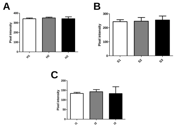
The calibration made in the program CFC_Image v.2.0 for the tonalities of the pixels representing the collagen in the hypertrophic cardiac tissue was performed using the mean of 3 parameter definitions performed in 3 different random images belonging to the previously selected group of images, totaling 9 definitions of parameters. As shown in Figure 2A-C, it is possible to observe that the definition of the calibration parameters for each condition of hue (H), saturation (S) and intensity (I) in different images do not present significant differences, indicating that the calibration performed is applicable to all samples. Thus, the parameters were defined as H: 345, S: 249 and I: 134.
After the image parameters were defined, the images included in the study were processed and the quantified collagen is shown in the graph of Figure 3.
Figure 1 – Analysis of the amount of collagen in rat cardiac tissue with Picrosirius red. A: histological section without the application of CFC_Image software parameters. B:
histological section with the application of CFC_Image software parameters.
Figure 2 - Calibration of hue (H), saturation (S) and intensity (I) parameters in different images. For each image, 3 calibrations were performed and the mean was calculated. A: mean of the analysis of hue in 3 images. H1 = 341.7 + 13.87; H2 = 350.3 + 14.57; H
3 = 342.7 + 33.50. p = 0.87. B: mean saturation analysis in 3 images. S1 = 243.7 + 23.54; S2 = 247.0 + 45.64; S3 = 255.3 + 49.22. p = 0.93. C: mean intensity analysis in
3 images. I1 = 133.7 + 9.866; I2 = 142.7 + 19.14; I3 = 133.0 + 61.29. p = 0.94. P ix e l in te n s it y H1 H2 H3 0 100 200 300 400 P ix e l in te n s it y S1 S2 S3 0 100 200 300 P ix e l in te n s it y I1 I2 I3 0 50 100 150 200
A
B
C
Source: Results obtained by image processing.
Figure 3 - Percentage of collagen quantification in comparison to the control group.
Co ll a g e n p e rc e n tu a l Con trol L-n am e 10 m g L-a rgin ine 30 m g L-a rgin ine 100 mg L-a rgin ine 0 50 100 150 200 250 L-name
Here we could observe that the chronic inhibition of Nitric Oxide synthesis by the administration of L-name resulted in an increase in collagen levels, corroborating with data already described in the literature (BERNÁTOVÁ et al., 2007).
It is possible to note that the inhibitory responses of L-arginine-mediated collagen production in the cardiac tissue becomes more effective as the amount of L-arginine administered increases, with an indicative of studies involving the administration of L-arginine in subjects with the risk of developing cardiac hypertrophy. It is important to highlight that these results were obtained with the digital processing of only 5 images, that is, in a quantitative way, they are compatible with the results presented by (RAMOS, 2005), which demonstrated the presence of collagen in rat hypertrophic cardiac tissue treated with L-name and L-arginine using a qualitative method.
4 DISCUSSION
The pressure-overload cardiac hypertrophy has arisen mainly due to an increase in blood pressure. The condition of congestive heart failure related to arterial hypertension is responsible for more than 1 million and 150 thousand hospitalizations (BOCCHI et al., 2009). These data indicate that the cost of treating this disease for public health is increasing over the years (CAETANO et al., 2008). Therefore, it is important to use interventions capable of providing treatment of cardiac hypertrophy.
According to the results, the application of L-arginine in doses higher than 10mg shows a significant reduction in the production of collagen in the cardiac tissue, indicating that this amino acid may help in the treatment of hypertrophy caused by arterial hypertension. The data presented in this work suggest an efficacy of oral administration of the amino acid L-arginine in reversing the cardiovascular effects induced by the production of nitric oxide. In addition, L-arginine may be used in the future as a cardiovascular risk prevention agent, under the protocol of cardiac rehabilitation or cardiovascular risk prevention in post-infarction or hypertensive patients (PEREIRA; VIANNA; MANDARIM-DE-LACERDA, 1998).
It is noteworthy that these authors analyzed the collagen of the cardiac interstitium of rats treated with L-name. However, the need for studies using more samples and long-term treatment should not be ruled out for better treatment responses using this amino acid. The scientific contribution of this work is the use of a quantitative imaging method that analyzes collagen production in the hypertrophic cardiac tissue of rats treated with L-name and L-arginine, which complements and corroborates the findings of (Ramos, 2005), who verified the effects of nitric oxide inhibition by oral administration of L-NAME on rat cardiac tissue and the possible reversal by L-arginine.
In fact, our results are very consistent with the original histological analysis performed by Ramos (2005) in his Master Thesis, which can validate the automated method presented here. We may conclude that the automated processing, at least in
our experimental model, was able to evaluate and quantify collagen contents in the heart tissue.
5 ACKNOWLEDGMENT
The authors are grateful to Fundação Valeparaibana de Ensino (FVE/UNIVAP), FAEP / UMC, and CAPES for financial support.
REFERENCES
BERNÁTOVÁ, I. et al. Chronic low-dose L-NAME treatment increases nitric oxide production and vasorelaxation in normotensive rats. Physiol. Res., v. 56, n. 2, p. S17, 2007.
BOCCHI, E. A. et al. III Brazilian Guideline for Chronic Heart Failure. Arq Bras Cardiol., v. 93, n. 1, p. 3-70, 2009.
BRASIL. Ministério da Saúde. Hipertensão arterial sistêmica para o Sistema Único de Saúde. Brasília: Ministério da Saúde, 2006. (Cadernos de Atenção Básica, n. 15) (Série A. Normas e Manuais Técnicos).
CAETANO, J. A. et al. Description of risk factors for cardiovascular disorders in a group of elderly people. Text. Context. Enferm., v. 17, n. 2, p. 327-35, 2008.
CALAIS, G. S. P.; GARCIA, G. C.; CALDEIRA, T. R. Hipertensão arterial. Saúde e Economia, ano. 2, n. 4, 2010. Available at:
http://portal.anvisa.gov.br/documents/33884/412285/Boletim+Sa%C3%BAde+e+Econo
mia+n%C2%BA+4. Accessed on: 12 Feb. 2019.
CARMO, E. C. et al. The association of anabolic steroids with physical-aerobic training leads to cardiac morphological changes and loss of ventricular function in rats. Ver. Bras. Med. Esporte, v. 17, n. 2, p. 137-141, Apr. 2011.
JUNQUEIRA, L.C.; BIGNOLAS, G.; BRENTANI, R. R. Picrosirius staining plus polarization microscopy, a specific method for collagen detection in tissue sections. Histochem. J., v. 11, n. 4, p. 447-455, 1979.
LIEBSON, P. R. et al. Echocardiographic correlates of left ventricular structure among 844 mildly hypertensive men and women in the Treatment of Mild Hypertension Study (TOMHS). Circulation, v. 87, n. 2, p. 476-486, 1993.
MATSUOKA, H. et al. Chronic L-arginine administration attenuates cardiac hypertrophy in spontaneously hypertensive rats. Hypertension, v. 27, n. 1, p. 14-18, 1996.
MELO-JÚNIOR, M. R. et al. Digital image analysis in pathology-the interface with Biomedical Engineering. Rev. Bras. Eng. Bioméd., v. 22, n. 3, p. 239-242, 2006. MORENO, H JR. et al. Chronic nitric oxide inhibition as a model of hypertensive heart muscle disease. Basic Res Cardiol., v. 91, n. 3, p. 248-55, 1996.
and myocardial stereology in hypertensive rats. Correlation with time of inhibition of nitric oxide synthesis. Arq. Bras. Cardiol, v. 70, n. 6, p. 397-402, 1998.
RAMOS, L. Estudo hemodinâmico e morfológico do miocárdio de ratos
hipertensos. Correlação com tratamento preventivo com L-arginina. São José dos Campos, SP, 2005. Dissertação (Mestrado em Ciências Biológicas) - Universidade do Vale do Paraíba, Instituto de Pesquisa e Desenvolvimento, São José dos Campos, 2005.
RIBEIRO, M.O. et al. Chronic inhibition of nitric oxide synthesis. A new model of arterial hypertension. Hypertension, v. 20, n. 3, p. 298-303, 1992.
TSAI, S. H. et al. Enhanced endothelin-1/Rho-kinase signalling and coronary
microvascular dysfunction in hypertensive myocardial hypertrophy. Cardiovasc Res., v. 113, n. 11, p. 1329-1337, 2017.
ZATZ, R.; DE NUCCI, G. Effects of acute nitric oxide inhibition on rat glomerular microcirculation. Am. J. Physiol., v. 261, n. 2, p. 360-363, 1991.