Cop
yright
© ABE&M t
odos os dir
eit
os r
eser
vados
.
Achalasia and thyroid disease:
possible autoimmune connection?
Acalasia e doença tireoidiana: uma conexão autoimune possível?
Ana Rosa P. Quidute1,2, Eduardo Vasconcelos de Freitas1, Tadeu Gonçalves de
Lima1, Ana Márcia Lima Feitosa1, Joyce Paiva dos Santos1, José Walter Correia1
SUMMARY
Many cases have been published showing a co-existence of autoimmune thyroid diseases (AITDs) and other autoimmune diseases. About a quarter of patients with achalasia have a con-current thyroid disease, most commonly associated with hypothyroidism. Although relatively rare, the association of achalasia and hyperthyroidism requires attention. The physiopathology of Grave’s Disease (GD) involves B- and T-mediator lymphocytes, which have an afinity for known thyroid antigens: thyroglobulin, thyroid-peroxidase, and thyrotrophin receptor. Curren-tly, however, the real physiopathogenesis of achalasia continues to be unknown. Some impor-tant indings are suggestive of an autoimmune mechanism: signiicant iniltration of the myo-enteric plexus by monocytes, presence of the class II-Human Histocompatibility Complex DQwl antigen and antibodies to myoenteric neurons. The present case reports a patient who, despite testing negative for Chagas’ disease, had achalasia, progressed to developing signiicant wa-sting and worsening of his quality of life, was later diagnosed with hyperthyroidism. After en-doscopic esophageal dilatation and radioiodine ablation of the thyroid gland, there was great improvement in the patient clinical condition. Arq Bras Endocrinol Metab. 2012;56(9):677-82
SUMÁRIO
Muitos casos têm sido publicados mostrando uma coexistência entre as doenças autoimunes da tireoide (DAIT) e outras doenças autoimunes. Cerca de um quarto dos pacientes com aca-lasia têm doenças da tireoide concomitantemente, sendo a mais comum a associação com hipotireoidismo. Apesar de ser relativamente rara, a associação da acalasia e hipertireoidismo requer atenção. A isiopatologia da doença de Graves (DG) envolve os linfócitos B e T-mediados, os quais têm ainidade pelos antígenos da tireoide: tireoglobulina, tireoperoxidase e recep-tor de tireotroina. Atualmente, a real isiopatogenia da acalasia continua desconhecida. No entanto, alguns importantes achados em análise são sugestivos de mecanismo autoimune: iniltração signiicativa do plexo mioentérico pelos monócitos, presença do antígeno-DQwl do Complexo Humano de Histocompatibilidade classe II e presença de anticorpos contra neurô-nios mioentéricos. Este presente caso aborda um paciente que, apesar de testes negativos para doença de Chagas, tem acalasia que progrediu para o desenvolvimento de signiicativa perda ponderal e piora da sua qualidade de vida, posteriormente, diagnosticado com hipertireoidis-mo. Após dilatação endoscópica esofágica e ablação da glândula tireoide com radioiodo, houve grande melhora na condição clínica do paciente. Arq Bras Endocrinol Metab. 2012;56(9):677-82
1 Internal Medicine Unit of the Teaching General Public Health Hospital Geral Dr. Cesar Calls (HGCC), Fortaleza, CE, Brazil 2 Endocrinology Unit of University Hospital Walter Cantídio, Universidade Federal do Ceará, Fortaleza, CE, Brazil
Correspondence to:
Ana Rosa P. Quidute Av. Padre Antonio Tomas, 3535 60190-020 – Fortaleza, CE, Brazil [email protected]
Received on July/28/2012 Accepted on Oct/2/2012
INTRODUCTION
A
utoimmune thyroidopathies affect in average 2% to 5% of the general population, with young adultCop
yright
© ABE&M t
odos os dir
eit
os r
eser
vados
.
are the most prevalent etiologies. Although genetics is well known to cause and inluence the progression of autoimmune diseases in approximately 79% (1), other environmental factors are known to be involved in the development of autoimmune thyroid diseases: quantity of ingested iodine, stress, drugs, pregnancy and changes in sexual hormones (1,2).
Graves’ disease is the most common type of autoim-mune hyperthyroidism, corresponding to approximate-ly 60%-80% of total cases, affecting women aged 20-50 years. Since close to 50% of GD cases do not manifest concomitant Graves’ ophthalmopathy, complete diag-nosis is less obvious (2,3).
The physiopathology of GD involves the B and T--mediator lymphocytes, which have an afinity for kno-wn thyroid antigens: thyroglobulin, thyroid-peroxidase and thyrotrophin receptor. However, the thyrotrophin receptor is by itself a primary autoantigen in GD and is responsible for the hyperthyroidism manifestations of the disease. Varied genes with signiicant susceptibili-ty to autoimmune diseases involving the thyroid gland have been identiied and classiied into two main clus-ters: gene immune-regulators (HLA, CTLA4, PTPN22,
CD40, CD25,and FCRL3), and speciic thyroid genes (thyroglobulin, TSH-receptor). It is well-known that they increase the susceptibility to other autoimmune diseases (1,4-6).
Many cases have been published showing a co--existence between autoimmune thyroid diseases (AITDs) and other autoimmune diseases for instan-ce: systemic erythematous lupus, Sjögren syndrome, pernicious anemia, primary biliary cirrhosis, vitiligo, and type 1 diabetes mellitus (4). It is important to note that the immune-pathophysiological process in-volving AITDs, with emphasis on GD, is similar to that identiied in other autoimmune diseases: cardinal participation of antibodies. Thus, as expected, target organs suffer signiicant lymphocyte iniltration in AI-TDs, with associated activated T and B-lymphocytes. It seems that GD pathogenesis results from both a central and peripheral dysfunctional tolerance home-ostasis of the immune system. Thus a continuous ini-tiated autoimmune process is propagated and main-tained by a clone of T and B-cells (3).
Esophageal achalasia is a rare primary dysfunction involving the physiology motility of the esophageal body and malfunction of the lower esophageal sphinc-ter (LES) characsphinc-terized by respective progressive dei-cient peristalsis and incomplete relaxation when
swallo-wing. Consequently, progressive obstruction develops at the esophageal-stomach junction, with associated proximal esophageal dilation (7,8).
Cop
yright
© ABE&M t
odos os dir
eit
os r
eser
vados
.
etiology of autonomic nervous system injury in patients with achalasia (14,15).
Storch and cols. demonstrated IgG-antibodies against Auerbach’s plexus by standard indirect im-munoluorescence. Antibodies to the cytoplasm of Auerbach’s plexus were found in 37 of 58 patients with achalasia at variable stages of the disease (I-IV) with a disease duration ranging from 1 to 20 years (16). Des-pite the fact that these indings link a possible autoim-mune mechanism to achalasia, further studies are nee-ded to establish if autoimmunity is a primary etiology or a co-factor in the pathogenesis of achalasia.
Frequently, patients with goiter refer to dysphagia which, in many cases, is attributed to mechanical factors caused by the enlarged gland. Unfortunately, clinical investigations usually ignore the existence of achalasia as the cause of dysphagia, despite the fact there is sufi-cient literature citations linking it mainly to Hashimoto thyroiditis and, to a lesser degree, to hyperthyroidism (17).
This case report the disease history of a patient who had achalasia, and was later diagnosed with hyper-thyroidism due to GD. Our main objective is to discuss such a rare association, citing the possible participation of autoimmunity and the conduct followed to improve the quality of life of the patient.
CASE REPORT
A.M.A., a 57-year-old male, coming from a metropolis non-endemic for Chagas’ disease, was admitted to our hospital reporting frequent vomiting and signiicant weight loss. Over the last 7 months, he had a sensation of ‘dry mouth’ and ‘burning’ in the anterior cervical re-gion, which extended up to the epigastric region. It was characteristically worse in the morning after waking up, but improved with the ingestion of refrigerated liquids. Three months before admission, daily post-prandial vo-miting (4 to 5 times) became frequent, besides conside-rable weight loss (35%; from 97 kg to 62.5 kg in three months), and progressive salivation, dysphagia, and ge-neral lethargy. No fever was reported during this pe-riod. During physical examination, low body weight was remarkable, together with painful abdomen upon supericial and profound palpation around the epigas-tric and mesogasepigas-tric region. The thyroid gland was painless and appeared diffusely enlarged, with its vo-lume approximately three times greater than normal and ibrous-elastic texture. This was later conirmed
by a cervical ultrasound that showed diffuse goiter. Other indings of the physical examination were un-remarkable.
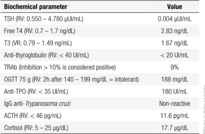
An upper digestive endoscopy and esophagogram were carried out, owing to the predominant dysphagy--related symptoms, both suggestive of esophageal acha-lasia involving the lower esophageal sphincter (LES). Due to the notable diffuse goiter upon physical exa-mination, we dosed thyroid-related hormones, and the results were compatible with primary hyperthyroidism (Table 1).
Laboratory indings and physical analysis were de-terminant for the indication of a thyroid scintigraphy followed by 131I ablation therapy. In relation to
signi-icant body weight loss and signs of malnutrition, we carried out endoscopic esophageal dilation, so as to im-prove his nutritional status. His epigastric pain owing to achalasia was treated conservatively, postponing Heller’s cardiomiotomy. As a result, the patient evol-ved with complete remission of his symptoms, inclu-ding the vomiting episodes. He was discharged from the hospital, and electively referred to the general sur-gery department, where he referred to heartburn and early abdominal satiety, but no vomiting since dilation was carried out. Furthermore, during this extra-hos-pital period, he recovered his body weight remarkably (62.5 kg to a current 92.6 kg, in one year), which led us to conclude that clinical treatment improved signii-cantly his nutritional status and quality of life. This was the reason for us to avoid Heller’s cardiomiotomy.
Table 1. Biochemical parameters at diagnosis
Biochemical parameter Value
TSH (RV: 0.550 – 4.780 µUI/mL) 0.004 µUI/mL
Free T4 (RV: 0.7 – 1.7 ng/dL) 2.83 ng/dL
T3 (VR; 0.79 – 1.49 ng/mL) 1.67 ng/dL
Anti-thyroglobulin (RV: < 40 UI/mL) < 20 UI/mL
TRAb (Inhibition > 10% is considered positive) 9%
OGTT 75 g (RV: 2h after 140 – 199 mg/dL = intolerant) 188 mg/dL
Anti-TPO (RV: < 35 UI/mL) 180 UI/mL
IgG anti-Trypanosoma cruzi Non-reactive
ACTH (RV: < 46 pg/mL) 11.6 pg/mL
Cortisol (RV: 5 – 25 µg/dL) 17.7 µg/dL
TSH, free T4, T3, anti-thyroglobulin, anti-TPO, ACTH and cortisol were quantiied by chemiluminescence; TRAB quantiied by radioceptor (RIA); OGTT by hexokinase; and IgG
Cop
yright
© ABE&M t
odos os dir
eit
os r
eser
vados
.
Figure 1. Esophagram: a characteristic addition image (‘Bird Beak’) seen at the inferior third of the esophagus. Note that there is a signiicant obstruction to the uniform low of radio-opaque liquid contrast at the eosophagus-stomach transition, resulting in signiicant increase in volume of the portion proximal to this point.
Figure 2. Pre-therapeutic scintigraphy (200MBq of 99mTc-Pertecnetate): large and topic thyroid exhibiting high capture of the radiotracer.
DISCUSSION
The cause for the inlammatory degeneration of eso-phageal neurons seen in achalasia is yet to be elucida-ted. The most accepted physiopathological hypothesis in idiopathic achalasia is a decrease in myenteric plexus neurons in the esophagus. This alteration in the myen-teric plexus has been explained by different etiopatho-genic mechanisms: infection, genetics, autoimmunity, ischemia, toxicicity etc. In recent years, the hypothesis of an autoimmune cause has acquired special relevance based on three aspects: histopathological indings (pre-sence of mononuclear and lymphocyte iniltration in the myenteric plexus in esophageal tissue samples of patients with achalasia), the association between class II antigen of the major complex of histocompatibility HLA-DQw1 and non-familial idiopathic achalasia; and the presence of autoantibodies against the myenteric plexus (AAM)
Cop
yright
© ABE&M t
odos os dir
eit
os r
eser
vados
.
Recently, Nunez and cols. described that variants in PTPN22 and IL23R genes confer predisposition to develop achalasia (22). Because these polymorphisms are common risk factors to several immunologic disor-ders, their results conirm that genetic variants that pre-dispose to these diseases may also confer susceptibility to achalasia. This inding supports the hypothesis that achalasia shares molecular pathways with other immu-ne-mediated diseases, which could explain the com-plexity of pathogenesis and progression of the disease (22). However, we could not conduct a genetic study, which we believe can provide a sample set of patient at high-risk of developing achalasia from the general population in the near future.
In our setting, the greatest prevalence of megae-sophagus is due to Chagas’ disease, thus this disease is the main differential diagnosis. Diagnosis in the chro-nic phase of Chagas’ disease is essentially serological; indirect hemagglutination tests, ELISA, and indirect immunoluorescence can determine the diagnosis in virtually 100% of cases (23). However, our patient tes-ted negative for Chagas’ disease and this etiology was ruled out. Furthermore, he denied any contact with the prevalent vector of the disease, came from a metropolis not endemic for Chagas’ and did not have previous his-tory of blood transfusions (23).
We are aware of varied causes of hyperthyroidism. The diagnosis of GD was determined by: suppressed TSH, increased free T4 and T3, a diffuse goiter determined by ultrasonography and the capture of the radiotracer by the thyroid gland in a diffuse pattern. These indin-gs reduced the chances of attributing thyroid disease to sub-acute thyroiditis or toxic uni- or multi-nodular goi-ter, reinforcing the autoimmune etiology. Furthermore, the fact that physical examination showed no Graves’ ophthalmopathy did not rule out the disease, since up to 50% of diagnosed patients do not manifest ocular disease in their presentation of hyperthyroidism (3). Intoleran-ce to glucose seen in our patient may be attributed to uncontrolled hyperthyroidism, since fasting levels fell to normal after treatment and outpatient follow-up (24).
The association between achalasia and AITDs has been described in literature. However, little is taken into consideration when dealing with a patient with goiter and who refers to achalasia-like symptoms, usu-ally due to the extrinsic compression of the hypertro-phied gland on the esophagus.
Emami and cols. (17), examining a group of 30 pa-tients (20 women and 10 men) with achalasia, whose ages varied from 30 to 70 years, found that this
esopha-geal anomaly was associated with AITDs in 23% of the cases (17). In this group, hypothyroidism due to Hashi-moto thyroiditis was the most common occurrence.
GD has been associated with achalasia in an infant who had to be subjected to Heller’s cardiotomy (25). In young patients, it is worth having Allgrove or Triple A syndrome (primary adrenal insuficiency, achalasia, and alagrimia) as a differential diagnosis (10). Despite the age, primary adrenal insuficiency was investigated in our patient and ruled out.
Usually, hyperthyroidism due to GD is diagnosed after a thorough symptomatic history and subsequent physical examination. However, our patient had no sig-niicant history, neither were the signs vivid, apart from a ‘goiter-form’ neck, which prompted us to investigate thyroid function. However, in their series, Emami and cols. (17) did not ind a direct correlation between the gravity of thyroid disease and the degree of achalasia symptoms. This inding was similar in our case, where the patient had more signiicant esophageal symptoms than those related to the thyroid disease. In our case, we opted for primary treatment with I131, since plasma
hor-mone concentrations were moderately high. Besides, the coexistence of untreated achalasia would be an impedi-ment to treat him with conventional anti-thyroid drugs, due to increased dificulty in absorption. This serves as an alert to clinicians and specialists when treating patients with GD with resistance to conventional treatment.
In conclusion, achalasia may be associated with va-ried autoimmune thyroid diseases (hyperthyroidism, hy-pothyroidism and thyroid nodular diseases) that ought to be considered when a new case of idiopathic acha-lasia is diagnosed. Additionally, patients presenting any autoimmune thyroid disease should be further inves-tigated for esophagus-related problems, ranging from characteristic esophageal dysmotility to more complex situations, such as excessive weight loss, that cannot be explained by thyroid disease, as occurred in our case.
Cop
yright
© ABE&M t
odos os dir
eit
os r
eser
vados
.
Disclosure: no potential conlict of interest relevant to this article was reported.
REFERENCES
1. Davies TF, Latif R, Yin X. New genetic insights from autoimmune thyroid disease. J Thyroid Res. 2012;2012:623852.
2. Weetman AP. Graves’ disease. N Engl J Med. 2000;343(17):1236-48. 3. Seigel SC, Hodak SP. Thyrotoxicosis. Med Clin North Am.
2012;96(2):175-201.
4. Weetman AP. Diseases associated with thyroid autoimmunity: explanations for the expanding spectrum. Clin Endocrinol (Oxf). 2011;74(4):411-8.
5. Sibarani RP. Genetics of Graves’ disease: the lost concept. Acta Med Indones. 2009;41(1):37-40.
6. Yin X, Latif R, Bahn RS, Davies TF. Genetic Proiling in Graves’ Disease: Further Evidence for Lack of a Distinct Genetic Contribu-tion to Graves’ Ophthalmopathy. Thyroid. 2012.
7. Eckardt AJ, Eckardt VF. Current clinical approach to achalasia. World J Gastroenterol. 2009;15(32):3969-75.
8. Francis DL, Katzka DA. Achalasia: update on the disease and its treatment. Gastroenterology. 2010;139(2):369-74.
9. Kraichely RE, Farrugia G, Pittock SJ, Castell DO, Lennon VA. Neural autoantibody proile of primary achalasia. Dig Dis Sci. 2010;55(2):307-11.
10. Kraichely RE, Farrugia G. Achalasia: physiology and etiopathoge-nesis. Dis Esophagus. 2006;19(4):213-23.
11. Wong RK, Maydonovitch CL, Metz SJ, Baker JR Jr. Signiicant DQw1 association in achalasia. Dig Dis Sci. 1989;34(3):349-52. 12. Verne GN, Hahn AB, Pineau BC, Hoffman BJ, Wojciechowski BW,
Wu WC. Association of HLA-DR and -DQ alleles with idiopathic achalasia. Gastroenterology. 1999;117(1):26-31.
13. Ghoshal UC, Daschakraborty SB, Singh R. Pathogenesis of acha-lasia cardia. World J Gastroenterol. 2012;18(24):3050-7.
14. Gockel I, Bohl JR, Doostkam S, Eckardt VF, Junginger T. Spectrum of histopathologic indings in patients with achalasia relects di-fferent etiologies. J Gastroenterol Hepatol. 2006;21(4):727-33. 15. Raymond L, Lach B, Shamji FM. Inlammatory aetiology of
pri-mary oesophageal achalasia: an immunohistochemical and ultrastructural study of Auerbach’s plexus. Histopathology. 1999;35(5):445-53.
16. Storch WB, Eckardt VF, Wienbeck M, Eberl T, Auer PG, Hecker A, et al. Autoantibodies to Auerbach’s plexus in achalasia. Cell Mol Biol (Noisy-le-grand). 1995;41(8):1033-8.
17. Emami MH, Raisi M, Amini J, Daghaghzadeh H. Achalasia and thyroid disease. World J Gastroenterol. 2007;13(4):594-9. 18. Domsic R, Fasanella K, Bielefeldt K. Gastrointestinal
manifesta-tions of systemic sclerosis. Dig Dis Sci. 2008;53(5):1163-74. 19. Booy JD, Takata J, Tomlinson G, Urbach DR. The prevalence of
autoimmune disease in patients with esophageal achalasia. Dis Esophagus. 2012;25(3):209-13.
20. Gockel HR, Schumacher J, Gockel I, Lang H, Haaf T, Nothen MM. Achalasia: will genetic studies provide insights? Hum Genet. 2010;128(4):353-64.
21. Santiago JL, Martinez A, Benito MS, Ruiz de Leon A, Mendoza JL, Fernandez-Arquero M, et al. Gender-speciic association of the PTPN22 C1858T polymorphism with achalasia. Hum Immunol. 2007;68(10):867-70.
22. Nunez C, Garcia-Gonzalez MA, Santiago JL, Benito MS, Mearin F, de la Concha EG, et al. Association of IL10 promoter polymorphis-ms with idiopathic achalasia. Hum Immunol. 2011;72(9):749-52. 23. Saúde SdVe, Saúde Md. Consenso Brasileiro em Doença de
Cha-gas. Rev Soc Bras Med Trop. 2005;38.