Cop
yright
© ABE&M t
odos os dir
eit
os r
eser
vados
.
Leptin: molecular mechanisms,
systemic pro-inlammatory effects,
and clinical implications
Leptina: mecanismos moleculares, efeitos pró-inlamatórios sistêmicos e implicações clínicas
Gilberto Paz-Filho1, Claudio Mastronardi1, Carina Bertoldi Franco2, Kevin Boyang Wang3, Ma-Li Wong1, Julio Licinio1
SUMMARY
Leptin, the adipokine produced mainly by the white adipose tissue, plays important roles not only in the regulation of food intake, but also in controlling immunity and inlammation. It has been widely demonstrated that the absence of leptin leads to immune defects in animal and human models, ultimately increasing mortality. Leptin also regulates inlammation by means of actions on its receptor, that is widely spread across different immune cell populations. The mo-lecular mechanisms by which leptin determines its biological actions have also been recently elucidated, and three intracellular pathways have been implicated in leptin actions: JAK-STAT, PI3K, and ERK 1/2. These pathways are closely regulated by intracellular proteins that decrease leptin biological activity. In this review, we discuss the molecular mechanisms by which leptin regulates immunity and inlammation, and associate those mechanisms with chronic inlam-matory disorders. Arq Bras Endocrinol Metab. 2012;56(9):597-607
Keywords
Leptin; immunity; inlammation; cytokines
SUMÁRIO
A leptina, uma adipocina produzida principalmente pelo tecido adiposo branco, tem um pa-pel importante não somente na regulação da ingestão alimentar, mas também no controle da imunidade e da inlamação. Já foi amplamente demonstrado que a ausência de leptina causa deiciências imunológicas em modelos animais e em humanos, levando ao aumento da morta-lidade. A leptina também regula a inlamação por meio da ação em seu receptor, amplamente distribuído em diversos tipos de células do sistema imunológico. Os mecanismos moleculares pelos quais a leptina determina suas ações biológicas foram recentemente elucidados, e três cascatas intracelulares são ativadas pela leptina: JAK-STAT, PI3K e ERK 1/2. Essas cascatas são reguladas por proteínas intracelulares, reduzindo as ações da leptina. Nesta revisão, são dis-cutidos os mecanismos moleculares pelos quais a leptina regula a imunidade e a inlamação, associando-os a enfermidades inlamatórias crônicas. Arq Bras Endocrinol Metab. 2012;56(9):597-607
Descritores
Leptina; imunidade; inlamação; citocinas
1 Department of Translational Medicine, The John Curtin School of Medical Research, The Australian National University, Canberra, Australia 2 Endocrinology and Diabetes Research Unit, Australian National University Medical School, Canberra, Australia 3 School of Medicine, Nursing and Health Sciences, Monash University, Melbourne, Australia
Correspondence to:
Gilberto Paz Filho Building 131, Garran Road Acton ACT 0200, Australia [email protected] Received on May/27/2012 Accepted on Oct/24/2012
INTRODUCTION
A
lthough initially considered as a simple storage of fat, overwhelming evidence led the adipose tissue to be conceptualized as an endocrine organ (1,2). The discovery of leptin has dramatically changed the under standing of the physiological importance of the adiposeCop
yright
© ABE&M t
odos os dir
eit
os r
eser
vados
.
licrelated factors, such as resistin and adiponectin, but it is also a source of cytokines and immune factors such as tumornecrosis factor alpha (TNFa), interleukin1 (IL1) alpha (IL1a), IL1β, IL1 receptor antagonist (IL1RA), and IL6 (1). All of these indings attracted attention to fat as an immunoendocrine organ that inte racts with several physiological systems of the organism. Leptin is a cytokine produced predominantly by the differentiated white adipose tissue, with various func tions in the immune and endocrine systems, including reproduction, glucose homeostasis, hematopoiesis, an giogenesis, osteogenesis, wound healing, and inlam mation (4). This hormone regulates caloric expenditure and intake, playing a central action in energy balance (5). Leptin levels are directly correlated with fat mass and are elevated in obese patients, who are also leptin resistant. Leptindeicient mice and humans are severely obese and have several metabolic and endocrine altera tions, such as hyperglycemia, insulin resistance, hyper triglyceridemia, hypogonadotropic hypogonadism, and central hypothyroidism (6,7).
Leptin plays a major role in the chronic proinlam matory state that is seen in obesity, metabolic syndrome, and their complications, such as atherosclerosis (8). By acting on the long isoform of the leptin receptor (Ob R), which is expressed by different immune cell types, leptin triggers inlammatory responses. Concomitantly, certain inlammatory and infectious stimuli, such as IL 1, lipopolysaccharide (LPS), and TNFa can also in crease leptin levels (9), which correlate with the level of inlammation. Therefore, the interactions between leptin and inlammation are bidirectional: proinlam matory cytokines increase the synthesis and release of leptin, which in turn helps to perpetuate the loop of chronic inlammation in obesity.
In this manuscript, we summarize the role of leptin in the immune system, the molecular links between leptin and inlammation, and the relationships between leptin and chronic inlammatory diseases.
Molecular mechanisms of leptin and modulation of immune cells
During states of normal nutrition, leptin signals to the brain that excess energy is available for several biological processes, including immune response. Under energe tic restriction, leptin levels are low, and immune respon se is impaired. Leptin exerts a link between the T helper (Th) 1 immune response and nutritional status, and inadequate body weight – both obesity and malnutri
tion – can importantly inluence immune cell function, both in innate and adaptive immune responses (10).
Leptin has structural homology with the cytokines of the longchain helical family that includes IL6, IL 11, IL12, and oncostatin M. Leptin receptor, ObR, has structural similarities with the members of the class I cytokine receptor (gp130) superfamily, which in cludes the receptor for IL6, leukocyte inhibitory factor (LIF), and granulocyte colonystimulating factor (G CSF). The ObR is expressed by neutrophils, mono cytes, macrophages, subpopulations of T cells and B cells, mast cells, dendritic cells (DC), and natural killer (NK) cells (11). There are at least six different isoforms of the leptin receptor in rodents; ObRa, ObRb, Ob Rc, ObRd, ObRe, and ObRf; these are all pro ducts of six alternatively spliced forms of the ObR gene (12). Of all of these isoforms, ObRb is the only long form, which contains a prominent cytoplasmic region containing several motifs required for signal transduc tion and is capable of activating the JAK–STAT path way. There are four truncated (short) forms, including ObRa, ObRc, ObRd, and ObRf, of which ObRa is regarded as a leptin transporter across the bloodbrain barrier and a leptin degrader; and a secreted form (Ob Re) that lacks both the intracellular and transmembrane domains, and serves as a plasmatic leptinbinding pro tein. These isoforms are involved in mediating leptin actions in the brain and peripheral organs. All isoforms, with the exception of ObRb, share the same identical extracellular, ligandbinding domains, but differ at the C terminus. Only ObRb has a long cytoplasmic region containing several motifs required for signal transduc tion, whereas the others have transmembrane domains, and lack some or all of these motifs. Transduction sig naling takes place by activation of conserved box 1 and box 2 motifs (intracellular amino acids 617 and 49 60, respectively). Cytokine receptor homology module 2 (CRH 2) is the main binding site for leptin on the ObR. The immunoglobulinlike domain (Iglike) and ibronectin III domain (FNIII) are critically involved in ObR activation. The role of CRH1 remains to be determined.
Cop
yright
© ABE&M t
odos os dir
eit
os r
eser
vados
.
putationalbased research studies predicted that leptin displayed tridimensional characteristics of a fourhelix bundled cytokine, such as those that activate the JAK STAT pathway (Figure 1) (13). Moreover, the cloning of the OB-R gene reported towards the end of 1995 (12) revealed that leptin receptor shared sequence ho mology with gp130, the signal transducing unit of the IL6 receptor that signals via the JAKSTAT pathway. The structural similarities that leptin and the OB-R
gene share with the IL6 cytokine family lead to func tional in vitro signaling studies that proved that leptin activated the JAKSTAT pathway (14). In vivo studies carried out in mice have shown that peripheral adminis tration of leptinactivated hypothalamic STAT3 in ob/ ob and WT mice, but not in db/db mice, indicating that the presence of a functional OB-R gene was required for leptininduced STAT3 activation within the brain.
Leptin controls other two key signaling path ways, namely the extracellular signalregulated ki nase (ERK)1/2 (Figure 2), and the activation of the phosphatidylinositol3kinase (PI3K) (Figure 3) (15). Janusactivated kinase phosphorylation of Tyr985 (or
phosphorylated JAK2 itself, independent of phosphor
ylated Tyr985) leads to recruitment and phosphoryla
tion of SHP2 (src homology 2containing tyrosine phosphatase). Then, phosphorylated SHP2 activates the ERK signaling pathway via growth factor recep tor binding2 (GRB2) (16). The ERK/MAPK path way is believed to be the main mechanism involved in regulatory T (Treg) cell regulation (11), by controlling the promoters for the transcription of c-fos, jun and
egr-1 genes, fundamental in cell proliferation and dif ferentiation (17). The ERK/MAPK pathway can be activated by both ObRa and ObRb leptin recep tors. It shares many similarities with the JAK2/STAT3 pathway as JAK2 is required for phosphorylation of the SH2domain containing protein tyrosine phosphatase (SHP2) via pathways both dependent and indepen dent of Tyr985 (18).
There is also one pathway that occurs independently of phosphorylation at tyrosine sites on ObRb, lead ing to the activation of phosphatidylinositol 3’kinase (PI3K). This pathway originates with JAK2 autophos phorylation, which leads to the recruitment and phos phorylation of insulin receptor substrate (IRS) proteins. Then, phosphorylated IRS proteins recruit phosphati dylinositol 3’kinase (PI3K) and activate downstream signals (15).
Figure 1. Leptin and the JAK-STAT pathway.
Leptin binds to the leptin receptor (Ob-Rb) and activates Janus-family tyrosine kinase 2 (JAK2) by auto- or cross-phosphorylation. JAK2 then phosphorylates the other tyrosine residues (Tyr985, Tyr1077, Tyr1138), which act as sites for the binding of intracellular signaling molecules. Signal Transducer and Activator of Transcription 3 (STAT-3) is a substrate of JAK 2 and subsequently binds to the phosphorylated Tyr1138. STAT-3 then undergoes dimerization and translocates into the cell nucleus to function as a promoter for gene transcription [4]. Members of the SOCS family (SOCS1 and SOCS3) suppress the action of leptin by binding to JAK 2 and tyrosine residues.
Leptin
Ob-Rb
JAK 2 JAK 2
STAT 3
STAT 3
STAT 3
Regulation of gene transcription P
P
SOCS 1/3
STAT 3 STAT 3
P P
P P
P
P
P Tyr985
Tyr1077
Tyr1138
Figure 2. Leptin and the PI3K pathway.
Both the leptin and insulin receptors play a role in the initial step of IRS 1/2 phosphorylation, which then binds to the regulatory p85 subunit of PI3K. The active p110 subunit then adds a phosphate to the 3’ position of PtdIns 4,5-diphosphate (PIP2) forming PtdIns 3,4,5-triphosphate (PIP3), which regulates proteins that contain the pleckstrin homology (PH) domain. These include the isoforms of protein kinase C, phosphoinositide-dependent kinase 1 (PDK1), and protein kinase B (Akt), which is activated by PDK1.
Insulin receptor
Ob-Rb
JAK 2
IRS 1/2
p85
PI3K
p110
PIP3 PDK1
EFFECT
PIP2
PTEN
AKt Rho/Rac
P P
Cop
yright
© ABE&M t
odos os dir
eit
os r
eser
vados
.
The PI3K pathway is an acute phase contributor of inlammation, in contrast to JAK2/STAT3, which is a slower process as it functions by altering gene tran scription (15). Both JAK2 and the insulin receptor participate in the phosphorylation of IRS, which then stimulate the p85 subunit of PI3K, thereby activat ing it. Once activated, the main mechanism of action of PI3K is intracellular phosphorylation of proteins. The chain of events involve phosphorylation of PtdIns 4,5diphosphate (PIP2) to form PtdIns 3,4,5triphos phate (PIP3) (19). Phosphatase and tensin homologue (PTEN) is an antagonist of this pathway and fosters the conversion of PIP3 back to PIP2 (Figure 3) (19). PIP3 is a key activator of phosphoinositidedependent kinase 1 (PDK1), which has many downstream effects, espe cially in regulating protein kinase B (Akt). Akt is a key participant in regulating neuropeptide Y and nitric ox ide (NO) mTOR via p70S6K (19). The PI3K pathway has also been implicated in controlling the Rho family of GTPases, which regulate apoptosis and remodel the actin skeleton within cells (15,19).
Soon after leptin discovery, it became evident in pre clinical and clinical experiments that obese rodents and
humans displayed leptin resistance. Thus, the search for the putative mechanisms leading to leptin resistance was taking place. In the mid90s, a new family of cytokine induced intracellular signaling inhibitors of the JAK STAT pathway were identiied, namely the supressors of cytokine signaling (SOCS 13) (20). SOCS3 plays a key role in providing negative feedback to this pathway, as it binds to Tyr985 and JAK2 to prevent phosphory
lation of SHP2, thus stopping the cascade. It is also believed to repress the activity of SHP2 (21). Since leptin also operates via JAKSTAT, Bjorbaek and cols. hypothesized that activation of SOCS genes could con tribute to leptin resistance (22). They demonstrated that peripherally administrated leptin rapidly increased the induction of hypothalamic SOCS3 in areas that are relevant for the control of food intake and ener gy expenditure. Moreover, they showed that SOCS3 was involved in negative regulation of leptininduced intracellular signal transduction. In rodent models, it was shown that SOCS3 could also be involved in leptin resistance that occurs during the aging process, and that lack of SOCS3 within the brain was effective to increase leptin sensitivity and to prevent dietinduced obesity (23).
Another intracellular mechanism that was shown to control leptin resistance is orchestrated by protein tyrosine phosphatase 1B (PTP1B) (24). This phospha tase acts by dephosphorylating JAK2 and, therefore, it prevents JAK2induced phosphorylation of the leptin receptor (24). Moreover, similar to mice deicient in SOCS3, neuronal deletion of PTP1B increased leptin and insulin sensitivity, preventing body weight gain in a dietinduced obesity animal model (25). Similar indings were previously shown in wholebody PTP1B knockout studies (26).
Leptin and innate immunity
Leptin binds to its receptor in macrophages and mo nocytes, improving phagocytosis by regulating oxidati ve stress. It also induces eicosanoid and nitric oxide syn thesis, acts as a chemoattractant, increases the secretion of cytokines, such as IL1RA, IL1, IL6, TNFa, and CCchemokine ligand, and prevents apoptosis (27). It also dosedependently increases the proliferation of cir culating cells, and stimulates the expression of activa tion markers, such as CD69 and CD25, among others (28). The increased number, and the activation of mo nocytes by the phorbol12 myristate 13acetate (PMA) or LPS is synergistically improved by leptin (29). Lep
Figure 3. Leptin and the ERK pathway.
Stimulation of Ob-Rb promotes phosphorylation of Tyr985 by JAK2. Both these units can phosphorylate SHP-2, which further modulates GRB2 and activates ERK1/2 of the MAPK family. ERK1/2 promotes the transcription of the c-fos and egr-1 genes. SOCS3 is an inhibitor of this pathway, with a mechanism similar to the JAK2/STAT3 pathway.
Leptin
Ob-Rb
JAK 2 JAK 2
SOCS 1/3 ERK1/2
c-fos egr-1
SHP-2 GRB2
P P
P P
P
P Tyr985
Tyr1077
Cop
yright
© ABE&M t
odos os dir
eit
os r
eser
vados
.
tin also activates macrophages by means of the mam malian Target of Rapamycin (mTOR) kinase pathway, which is an intracellular nutrientresponsedependent pathway that integrates growth factor and nutrientde rived signals to cellular growth rates, controlling cell growth and division. Leptin stimulates phagocytosis by stimulating phospholipase and increasing the produc tion of leukotriene B4, eicosanoids, NO, cholesterol acyltransferases1, and cyclooxygenase 2 (30). Leptin also increases the production of growth hormone by means of PKC and NOdependent pathways (31).
Leptin acts on several other immune cells. On eosi nophils, it induces the expression of adhesion molecules and CD18, increases chemokinesis, and stimulates the release of inflammatory cytokines IL1β, IL6, IL8, growthrelated oncogenea, and monocyte chemoat tractant protein1 (MCP1), which is a known chemo attractant for monocyte/macrophage infiltration (32). On DC, leptin increases the expression of cytokines, such as IL6 and TNFa; surface molecules, such as CD1a and CD80; and reduces apoptosis rates. Leptin also induces morphological and functional changes wi thin DC, directing them towards Th1 priming (33). Mast cells also express ObR, and it is probable that leptin acts on them both in a paracrine and/or auto crine fashion (34). On polymorphonuclear cells, leptin induces chemoattraction and the production of reac tive oxygen species (ROS) via mechanisms that might involve interaction with monocytes (35). Finally, leptin contributes to NK cell development, differentiation, activation, proliferation, and cytotoxicity (35).
Leptin and adaptive immunity
Leptin maintains the thymic parenchyma via its direct antiapoptotic effects on T cells by stimulating the me dullary thymic epithelial cell expression of IL7, a thy mocyte growth factor (36). Moreover, leptin affects the activation of T lymphocytes. Hypoleptinemia caused by starvation leads to thymic atrophy and higher fre quency of infections, and exogenous recombinant lep tin prevents the reduction in cortical doublepositive CD4+CD8+ thymocyte subpopulation (37). However,
leptin can only induce the proliferation and activation of mature human peripheral blood lymphocytes if it is coadministered with other general immunostimulants, such as concanavalin A (ConA) or phytohemagglutinin (PHA) (36). Importantly, the effect of leptin on lym phocyte proliferation is speciic to different subpopu lations. It inhibits the proliferation of memory T cells
(CD4+CD45RO+), but stimulates the proliferation of naive T cells (CD4+CD45RA+). This means that leptin polarizes Th0 cytokine production towards a proin lammatory (Th1, TNFa, IFNγ IL2, IL12, and leptin itself), rather than antiinlammatory phenotype (Th2, IL4, IL10) (38). It seems that leptin acts directly on circulating T lymphocytes when they are costimulated, since this effect is observed even in the lack of monocytes (38). On the subpopulation of Th1 cells, leptin promo tes IgG2a switching in B cells and induces TNFa and IFNγ production, while it exerts inhibitory actions on Th2 cells and IgG1 switching (39). Additionally, lep tin is also fundamental for Th2 cell development. For example, in an animal model of Th2dependent intesti nal inlammation, leptin deiciency was associated with decreased Th1 and Th2 polarization, and ob/ob mice were protected from intestinal inlammation. Indeed, after polarizing ob/ob naive and wildtype (WT) T cells in the Th2 direction, there was suppression of Tcell polarization in the cells of leptindeicient mice (27).
Furthermore, leptin is able to reduce the human CD4+CD25+ Treg cells, which are a small subset of CD4+ T cells that control the peripheral immune tole rance, and prevent inappropriate immune responses, such as allergy and autoimmunity. Commonly, Treg cells influence the activities of cells of the innate immune system and modulate effector T cell responses. In ani mal models of chronic leptin and ObR deficiencies, the percentage, absolute number, and activity of Treg cells is increased, resulting in resistance to autoimmune diseases. When leptin is replaced, the number of Treg cells returns to the same levels found in WT mice. In humans, leptin has a similar effect on Treg cells. Interest ingly, freshly isolated Treg cells can produce signiicant quantities of leptin, and express high density of the Ob R, which constrain their proliferation (28).
Besides the effects on CD4+ T cells, leptin adminis tration can stimulate the proliferation of B lympho cytes, NKT and CD8+ T cells, and increase cytokine responsiveness. The interactions between leptin and cy tokines are bidirectional, and both stimulate each other (Figure 3). Table 1 summarizes the leptin effects on innate and adaptive immunity.
LEPTIN AND CLINICAL CONDITIONS
Congenital deiciency of leptin
Cop
yright
© ABE&M t
odos os dir
eit
os r
eser
vados
.
to infections with elevated risk of death during chil dhood. Congenital deiciency of leptin is associated with a decrease in CD4+ T cell numbers, and an increase in
CD8+ T and B cells, as well as a pronounced reduction in the number of naive T cells, and an increase in me mory T cells. In addition, lymphocytes exhibit impaired proliferative response and reduced production of cytoki nes in response to several stimuli, with abolishment of IFNγ secretion, and a prevailing Th2 cytokine pheno type (40). However, these immune defects are not an obligatory characteristic of patients with mutation in the leptin gene, in whom the immune proile possibly de pends on the genotype and genetic expressivity (42). In leptindeicient children, leptin treatment increased pro liferation and cytokine response, particularly of IFNγ
levels, increased number of leukocytes, and improved recurrent dermatites and asthmatic crises (43).
Patients with leptinreceptor mutation present less prominent immune alterations than patients with con genital leptin deiciency (41). A reduction in the num ber and function of CD4+ T cells, a decrease in cytokine production after antigenic charge, and a compensatory in crease in the number of B cells have been described. These alterations are also restored with leptin replacement (41).
Hypoleptinemic states
Malnutrition or caloric deprivation produces immu nossupression in humans and animals. Acute hypolep tinemia caused by starvation is associated with reduced development of B cells in the bone marrow, and decre ased Tlymphocyte subpopulation with impairment of delayedtype hypersensitivity responses and thymic atro phy. These effects are more pronounced in patient sub mitted to chronic exposure to hypoleptinemia, and are prevented by replacement with exogenous leptin (44).
Women with hypothalamic amenorrhea due to chronic caloric restriction (e.g., strenuous exercise or low body weight) present hypoleptinemia and low le vels of soluble TNFa receptor that are normalized by the treatment with rmetHuLeptin (recombinant me thionyl human leptin) (45).
Congenital or acquired severe lipodystrophy is cha racterized by an almost complete lack of adipose tissue and low leptin levels. In general, these patients exhibit measurable abnormalities in immune markers at baseli ne, such as a higher percentage of B cells and reduction in TNFa production by peripheral blood mononuclear cells. Leptin replacement can increase CD4, CD8T, and NKT cell numbers, correct the disproportion of B cells, and improve TNFa release to physiological levels. Ho wever, patients with severe lipodystrophy surprisingly do not show clinical immune deiciency, and may present autoimmune diseases including autoimmune hepatitis, thyroidopathies, nephropathies, and type 1 diabetes (46).
Hyperleptinemic states: obesity and metabolic syndrome
Obesity, especially visceral adiposity, is considered an inlammatory disease, since the adipose tissue is able to produce inlammatory cytokines or collaborate with their production by other tissues (2). Also, obese pa tients exhibit higher level of inlammatory markers such as Creactive protein, TNFa, IL6, IL18, MIF (macrophage migration inhibitory factor), haptoglo bin, SAA (serum amyloid A), and plasminogen acti vator inhibitor1. Furthermore, type 2 diabetes, athe
Table 1. Leptin effects on innate and adaptive immunity
Innate Adaptive
Monocytes/ macrophages
↑Phagocytosis ↑Proliferation ↑IL1, IL6, TNF
Thymocytes ↑Maturation
↓Apoptosis
Monocytes ↑Expression of activation
markers ↑Expression of surface markers
Naive T cells ↑Proliferation ↓Apoptosis
Macrophages ↑Eicosanoids, NO, LB4, CTA1,
COX-2
Memory T cells
↓Proliferation
Neutrophils ↑Formation and release of ROS ↑Chemotaxis
↓Apoptosis ↑Expression of
CD11b
Th1 cell activation
↑IgG2a switch ↑TNF-a, ↑INF-γ
Eosinophils ↑Adhesion
molecules ↑Chemokinesis
↑Release of IL-1β,IL-6, IL-8
Th2 cell inhibition
↓IgG1 switch
Dendritic cells ↑ IL-1β, IL-6, IL-8, IL-12,
TNF-a ↑Maturation
↓Apoptosis
Treg cell ↑Anergy
↑Hyporesponsiveness
Natural killer ↑Proliferation, maturation ↑Differentiation,
activation, cytotoxicity
B cells NKT cells
↓Apoptosis ↑Proliferation
Cop
yright
© ABE&M t
odos os dir
eit
os r
eser
vados
.
rosclerosis and other diseases related to obesity are causally linked to inlammation.
Hyperleptinemia is correlated with proinlamma tory responses and with the chronic subinlammatory state observed in obesity. On the one hand, leptin en hances the production of inlammatory cytokines, and on the other hand, cytokines such as IL6 and TNFa
promote leptin production by the adipose tissue as well. Increased leptin resistance associated with high levels of free fatty acid and inlammatory cytokines may contribute to the reduction in lipid oxidation in insu linsensitive organs, leading to accumulation of lipids (lipotoxicity) and insulin resistance (47).
Moreover, leptin induces cholesterol uptake by mac rophages, angiogenesis, platelet aggregation, stimulates the oxidative stress in endothelial cells and inhibits va sorelaxation, increasing the risk of atherosclerosis (48). Indeed, ob/ob mice are resistant to atherosclerosis de spite all the metabolic risk factors, whereas apolipopro tein Edeicient mice develop atherosclerosis if treated with leptin (49).
In humans, leptin is an independent risk factor for coronary artery disease, and its levels are correlated with Creactive protein, plasma triglycerides, and fast ing plasma glucose levels (50). Leptin is associated with insulin resistance and metabolic syndrome independent of obesity via central obesity, possibly by impairment of insulin signaling at the insulin receptor substrate1 phosphorylation (51). Leptin is increased in patients with metabolic syndrome, but relative hypoleptinemia may be observed in cases of severe metabolic syndrome, possibly due to adipose tissue dysfunction (52).
Leptin and acute inlammation
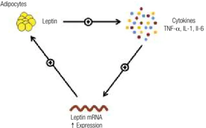
Leptin regulates the production of proinlammatory cytokines such as tumor necrosis factor alpha (TNFa), interleukin 1 (IL1), and interleukin (IL6). These cytokines also regulate the expression of leptin, which sustains a chronic proinlammatory state (Figure 4).
Many genes related to inlammation, including ge nes encoding acute phase response proteins such as tPA (tissue plasminogen activator), ibrinogenβ, lipoca lin2, PAP1, preprotachykinin, and MnSOD (manga nese superoxide dismutase) are induced by leptin (53). Leptin levels rapidly increase in acute inlammatory conditions, such as cholecystectomy, acute infection, and sepsis, particularly favored by cytokines, such as TNFa, IL6, and IL1β (36). Parenteral administra tion of LPS, commonly used to experimentally induce systemic inlammation, leads to a rise in plasma leptin.
A protective role of leptin in the clearance of pathogens is observed in leptindeicient ob/ob mice, which deve lop severe disease and die of infection with Klebsiella
more rapidly than WT mice. The ob/ob mice are also hi ghly susceptible to LPSinduced lethality, which can be reversed by the administration of leptin. The protective effects of leptin in these cases seem to occur by means of a modulation of TNFa and IL6 responses after en dotoxin priming (10).
Figure 4. Interaction between leptin and cytokines.
Leptin upregulates the production of pro-inlammatory cytokines, such as tumor necrosis factor alpha (TNF-a), interleukin 1 (IL-1), and interleukin (IL-6). These cytokines increase the expression of leptin mRNA, which subsequently sustains production of leptin. This is thought to be the mechanism for chronic inlammation.
Adipocytes
Leptin Cytokines
TNF-a, IL-1, Il-6
Leptin mRNA
↑ Expression
Elevated levels of leptin are found in sepsis and may be predictive of the severity of sepsis and increased sur vival (54). Leptin is a critical factor in host resistance and in its absence, sepsisinduced organ damage is increased, whereas neutrophil function is diminished. Furthermore, there is an important role of leptin in the central nervous system (CNS) in regulating survival and systemic im mune response in sepsis. Selective leptin administration into the CNS controls systemic immune response in a functionally relevant manner with signiicant protection from sepsis. A leptindependent neurocircuit in the CNS is required for eficient coordination of the immune re sponse in sepsis to limit organ damage and prevent mor tality (38). On the other hand, some studies have not found increased leptin levels in speciic inlammatory conditions, including acute experimental endotoxemia in humans, HIV infection, and newborn sepsis (30).
Leptin in chronic inflammatory diseases
Cop
yright
© ABE&M t
odos os dir
eit
os r
eser
vados
.
tin and increased risk of both chronic inlammatory and autoimmune disease. Serum leptin levels are elevated in many chronic inlammatory conditions (Table 2). In ex perimental animal models, the investigation of the effects of leptin on the susceptibility to autoimmunity has clearly indicated that leptin can promote autoreactivity. Addi tionally, leptindeicient ob/ob mice and leptin receptor deicient db/db mice are resistant to the development of several experimentallyinduced autoimmune diseases.
tal models of arthritis, ob/ob and db/db mice exhibited less severe disease, with lower levels of IL1β and TNFa in the synovial liquid, and T cells presented decreased pro liferative response induced by the antigen and a shift to ward a Th2 cytokine proile. On the other hand, clinical studies have reported controversial results when address ing the relationship between plasmatic and synovial leptin levels versus the activity or severity of the disease.
Leptin is involved in the pathogenesis of osteoar thritis via the production of matrix metalloproteinases and type 2 nitric oxide synthase. Moreover, the carti lage of patients with osteoarthritis present higher pro duction of leptin. Also, high leptin levels have been re ported in patients with systemic lupus erythematosus in whom the elevated leptin levels were positively corre lated with atherosclerosis and inlammatory biomarkers of atherosclerosis.
Leptin levels increased just before the commence ment of hyperglycemia and diabetes in spontaneous au toimmune nonobese diabetic (NOD) mice, a model of type 1 diabetes. In addition, exogenous administration of leptin to these animals enhanced interferonγ production by Tcells, and expanded the autoimmune destruction of
βcells, suggesting a possible proinlammatory inluence of leptin on Th1 cells in this disease. Children with gre ater weight gain in the irst year of life were more prone to develop type 1 diabetes in concordance with the evi dence that overnutrition and increased postnatal growth may be related to development of autoimmunity.
High levels of leptin were reported during acute exacerbations of chronic obstructive pulmonary dise ase, and were associated to serum IL6 and TNFa. Leptin is important to airway hyperresponsiveness as sociated with obesity, by the stimulation of cytokine and chemokine production in the lungs, but just in conjunction with other inlammatory agents and in su praphysiological levels.
Leptin may also be important in the cognitive im pairment observed in obesity and high fat diet, by means of inflammatory and oxidative pathways in microglia cells. Finally, it been suggested that leptin may be a link between adipose tissue and psoriatic inflammation. References (11,55,56) summarize the roles of leptin on different chronic inlammatory diseases.
Leptin as an anti-inlammatory cytokine
Leptin may be protective against type 1 diabetes by re ducing virusinduced insulitis (57). Besides, exogenous leptin may exert an antiinlammatory role in the exo
Table 2. Leptin and inlammatory diseases
Disease Effects of leptin
Obesity Pro-inlammation and atherosclerosis
Type 2 diabetes Pro-inlammation and atherosclerosis
Sepsis Higher levels associated with survival
Multiple sclerosis Activation of myelin-reactive T cells
Inlammatory bowel disease Pro-inlammation and anorexia
Osteoarthritis Increases matrix metalloproteinases
and type 2 nitric oxide synthase
Chronic obstructive pulmonary disease
Pro-inlammation Reduction of lung function
Asthma Pro-inlammation
Psoriasis Pro-inlammation
Cognitive impairment Pro-inlammation of glia cells
Systemic lupus erythematosus Pro-inlammation and atherosclerosis
Type 1 diabetes Dual effect (pro-inlammation and
anti-inlammation)
Pancreatitis Protection and anti-inlammation
Rheumatoid arthritis Pro-inlammation in animals
Prevention of erosive arthritis
Acute colitis Protection and anti-inlammation
In human studies on multiple sclerosis (MS), it has been reported that the secretion of leptin is increased in both the serum and the cerebrospinal luid (CSF) of naive patients with MS. This positively correlates with the se cretion of IFNγ in the CSF, and inversely correlates with the percentage of circulating Treg cells. Moreover, the in crease of leptin in the CSF is higher than in the serum, suggesting possible secondary in situ synthesis of leptin in the CNS and/or an increased transport across the blood brain barrier following enhanced systemic production.
Patients affected by Crohn’s disease exhibit in creased visceral adiposity with hypertrophic mesenteric fat, independent of the body mass index. In these pa tients, it was observed an overexpression of both leptin mRNA and leptin protein by the hypertrophic mesen teric fat, suggesting an inlammatory role of leptin.
Cop
yright
© ABE&M t
odos os dir
eit
os r
eser
vados
.
Adipocytes
Adipocytes APC
Naive T Cell
Th1 memory
TNF-a, IFN-γ, IL-2, IL-12
Th2 memory
IL-4, IL-5, IL-10 Macrophage/Monocyte
↑ Proliferation TNF-a, IL-1, IL-6 Leptin
Leptin
Thymocytes
↑Maturation
↓Apoptosis
crine pancreas, protecting rats against acute pancreatitis mediated by the nitric oxide pathway (58).
Leptin also played an antiinlammatory effect in an experimental model of acute colitis via a neutro phildependent mechanism that depends on the HPA (hypothalamicpituitaryadrenal) axis (59). Another study showed that endogenous leptin may locally pro tect the joints from the most severe form of erosive rheumatoid arthritis in humans (60). Table 2 summa rizes the role of leptin in clinical conditions.
CONCLUSIONS
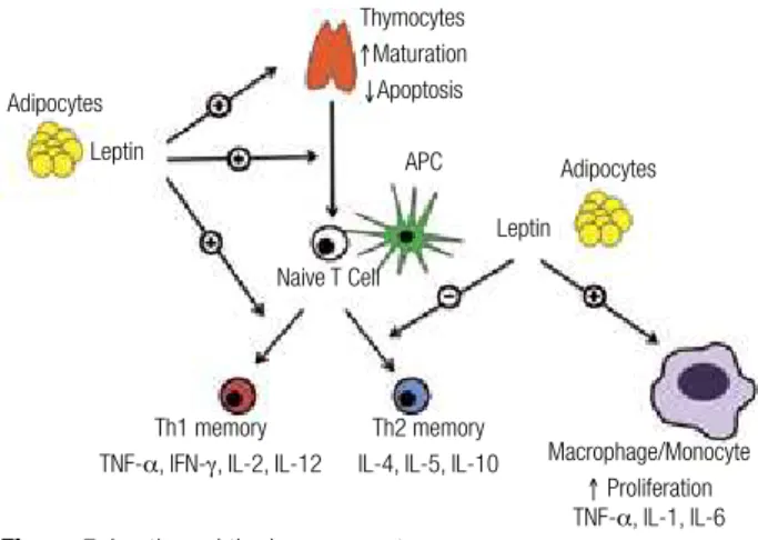
Leptin plays a major role in the regulation of the im mune system. It has a positive effect on thymocytes, leading to an overall increased level of T cells, and inhi bits the transformation of naive T cells into Th2 cells, which are antiinlammatory. Additionally, leptin in creases macrophage and monocyte proliferation rates, thereby increasing the levels of inlammatory cytokines (TNFa, IL1, IL6) (Figure 5). The absence of leptin leads to immune defects that ultimately translate into increased mortality due to infections. It is also a ma jor regulator of inlammatory response, with mainly proinlammatory actions. In excess, commonly seen in states of excess adiposity, leptin has been associated with several diseases where inlammation plays an im portant role in morbidity, such as cardiovascular disea ses, rheumatoid arthritis, and cancer.
Leptin acts by activating its receptor, which in turn trig gers different molecular pathways, namely the JAKSTAT, PI3K, and ERK/MAPK pathways. Within these path ways, several molecules modulate their activation, such as SOCS3. Better understanding of the molecular mecha nisms by which leptin regulates immunity and inlamma tion might lead to the development of therapeutic targets to treat diseases associated with leptin deiciency or excess.
Disclosure: no potential conlict of interest relevant to this article was reported.
REFERENCES
1. Kershaw EE, Flier JS. Adipose tissue as an endocrine organ. J Clin Endocrinol Metab. 2004;89:2548-56.
2. Wajchenberg BL, Nery M, Cunha MR, Silva ME. Adipose tissue at the crossroads in the development of the metabolic syndrome, inlammation and atherosclerosis. Arq Bras Endocrinol Metabol. 2009;53:145-50.
3. Friedman JM, Halaas JL. Leptin and the regulation of body weight in mammals. Nature. 1998;395:763-70.
4. Kelesidis T, Kelesidis I, Chou S, Mantzoros CS. Narrative review: the role of leptin in human physiology: emerging clinical applica-tions. Ann Intern Med. 2012;152:93-100.
5. Boguszewski CL, Paz-Filho G, Velloso LA. Neuroendocrine body weight regulation: integration between fat tissue, gastrointestinal tract, and the brain. Endokrynologia Polska. 2011;61:194-206. 6. Paz-Filho G, Mastronardi C, Delibasi T, Wong ML, Licinio J.
Con-genital leptin deiciency: diagnosis and effects of leptin replace-ment therapy. Arq Bras Endocrinol Metabol. 2010;54:690-7. 7. Paz-Filho G, Wong ML, Licinio J. Ten years of leptin replacement
therapy. Obes Rev. 2011;12:e315-23.
8. Conde J, Scotece M, Gomez R, Lopez V, Gomez-Reino JJ, Lago F, et al. Adipokines: biofactors from white adipose tissue. A com-plex hub among inlammation, metabolism, and immunity. Bio-factors. 2011;37:413-20.
9. Maury E, Brichard SM. Adipokine dysregulation, adipose tissue inlammation and metabolic syndrome. Mol Cell Endocrinol. 2010;314:1-16.
10. Bernotiene E, Palmer G, Gabay C. The role of leptin in innate and adaptive immune responses. Arthritis Res Ther. 2006;8:217. 11. La Cava A, Matarese G. The weight of leptin in immunity. Nat Rev
Immunol. 2004;4:371-9.
12. Tartaglia LA, Dembski M, Weng X, Deng N, Culpepper J, Devos R, et al. Identiication and expression cloning of a leptin receptor, OB-R. Cell. 1995;83:1263-71.
13. Madej T, Boguski MS, Bryant SH. Threading analysis suggests that the obese gene product may be a helical cytokine. FEBS let-ters. 1995;373:13-8.
14. Ghilardi N, Ziegler S, Wiestner A, Stoffel R, Heim MH, Skoda RC. Defective STAT signaling by the leptin receptor in diabetic mice. Proc Natl Acad Sci U S A. 1996;93:6231-5.
15. Donato J Jr, Frazao R, Elias CF. The PI3K signaling pathway medi-ates the biological effects of leptin. Arq Bras Endocrinol Metabol. 2010;54:591-602.
16. Banks AS, Davis SM, Bates SH, Myers MG. Activation of down-stream signals by the long form of the leptin receptor. J Biol Chem. 2000;275:14563-72.
Figure 5. Leptin and the immune system.
Cop
yright
© ABE&M t
odos os dir
eit
os r
eser
vados
.
17. Torii S, Nakayama K, Yamamoto T, Nishida E. Regulatory mechanisms and function of ERK MAP kinases. J Biochem. 2004;136:557-61. 18. Munzberg H, Bjornholm M, Bates SH, Myers MG Jr. Leptin
recep-tor action and mechanisms of leptin resistance. Cell Mol Life Sci. 2005;62:642-52.
19. Fruhbeck G. Intracellular signalling pathways activated by leptin. Biochem J. 2006;393:7-20.
20. Starr R, Willson TA, Viney EM, Murray LJ, Rayner JR, Jenkins BJ, et al. A family of cytokine-inducible inhibitors of signalling. Na-ture. 1997;387:917-21.
21. Bjorbak C, Lavery HJ, Bates SH, Olson RK, Davis SM, Flier JS, et al. SOCS3 mediates feedback inhibition of the leptin receptor via Tyr985. J Biol Chem. 2000;275:40649-57.
22. Bjorbaek C, Elmquist JK, Frantz JD, Shoelson SE, Flier JS. Identi-ication of SOCS-3 as a potential mediator of central leptin resis-tance. Molecular Cell. 1998;1:619-25.
23. Mori H, Hanada R, Hanada T, Aki D, Mashima R, Nishinakamura H, et al. Socs3 deiciency in the brain elevates leptin sensitivity and confers resistance to diet-induced obesity. Nat Med. 2004;10:739-43. 24. Myers MP, Andersen JN, Cheng A, Tremblay ML, Horvath CM, Pa-risien JP, et al. TYK2 and JAK2 are substrates of protein-tyrosine phosphatase 1B. J Biol Chem. 2001;276:47771-4.
25. Bence KK, Delibegovic M, Xue B, Gorgun CZ, Hotamisligil GS, Neel BG, et al. Neuronal PTP1B regulates body weight, adiposity and leptin action. Nat Med. 2006;12:917-24.
26. Zabolotny JM, Bence-Hanulec KK, Stricker-Krongrad A, Haj F, Wang Y, Minokoshi Y, et al. PTP1B regulates leptin signal trans-duction in vivo. Dev Cell. 2002;2:489-95.
27. Batra A, Okur B, Glauben R, Erben U, Ihbe J, Stroh T, et al. Leptin: a critical regulator of CD4+ T-cell polarization in vitro and in vivo. Endocrinology. 2010;151:56-62.
28. Matarese G, Procaccini C, De Rosa V, Horvath TL, La Cava A. Regu-latory T cells in obesity: the leptin connection. Trends Mol Med. 2010;16:247-56.
29. Lord GM, Matarese G, Howard JK, Baker RJ, Bloom SR, Lechler RI. Leptin modulates the T-cell immune response and reverses starvation-induced immunosuppression. Nature. 1998;394:897-901.
30. Iikuni N, Lam QL, Lu L, Matarese G, La Cava A. Leptin and inlam-mation. Curr Immunol Rev. 2008;4:70-9.
31. Dixit VD, Mielenz M, Taub DD, Parvizi N. Leptin induces growth hormone secretion from peripheral blood mononuclear cells via a protein kinase C- and nitric oxide-dependent mechanism. Endo-crinology. 2003;144:5595-603.
32. Rafail S, Ritis K, Schaefer K, Kourtzelis I, Speletas M, Doumas M, et al. Leptin induces the expression of functional tissue factor in human neutrophils and peripheral blood mononuclear cells through JAK2-dependent mechanisms and TNFalpha involve-ment. Thromb Res. 2008;122:366-75.
33. Steiner AA, Romanovsky AA. Leptin: at the crossroads of energy balance and systemic inlammation. Prog Lipid Res. 2007;46:89-107. 34. Otero M, Lago R, Gomez R, Dieguez C, Lago F, Gomez-Reino J, et
al. Towards a pro-inlammatory and immunomodulatory emerg-ing role of leptin. Rheumatology (Oxford). 2006;45:944-50. 35. Martin-Romero C, Santos-Alvarez J, Goberna R,
Sanchez-Mar-galet V. Human leptin enhances activation and proliferation of human circulating T lymphocytes. Cell Immunol. 2000;199:15-24. 36. Fernandez-Riejos P, Najib S, Santos-Alvarez J, Martin-Romero C,
Perez-Perez A, Gonzalez-Yanes C, et al. Role of leptin in the activa-tion of immune cells. Mediators Inlamm. 2010;2010:568343. 37. Howard JK, Lord GM, Matarese G, Vendetti S, Ghatei MA, Ritter
MA, et al. Leptin protects mice from starvation-induced lymphoid atrophy and increases thymic cellularity in ob/ob mice. J Clin In-vest. 1999;104:1051-9.
38. Tschop J, Nogueiras R, Haas-Lockie S, Kasten KR, Castaneda TR, Huber N, et al. CNS leptin action modulates immune response and survival in sepsis. J Neurosci. 2010;30:6036-47.
39. Siegmund B, Lehr HA, Fantuzzi G. Leptin: a pivotal mediator of in-testinal inlammation in mice. Gastroenterology. 2002;122:2011-25. 40. Farooqi IS, Matarese G, Lord GM, Keogh JM, Lawrence E, Agwu
C, et al. Beneicial effects of leptin on obesity, T cell hyporespon-siveness, and neuroendocrine/metabolic dysfunction of human congenital leptin deiciency. J Clin Invest. 2002;110:1093-103. 41. Farooqi IS, Wangensteen T, Collins S, Kimber W, Matarese G,
Ke-ogh JM, et al. Clinical and molecular genetic spectrum of congeni-tal deiciency of the leptin receptor. N Engl J Med. 2007;356:237-47. 42. Paz-Filho GJ, Delibasi T, Erol HK, Wong ML, Licinio J. Cellular im-munity before and after leptin replacement therapy. J Pediatr En-docrinol Metab. 2009;22:1069-74.
43. Gibson WT, Farooqi IS, Moreau M, DePaoli AM, Lawrence E, O’Rahilly S, et al. Congenital leptin deiciency due to homozygos-ity for the Delta133G mutation: report of another case and evalua-tion of response to four years of leptin therapy. J Clin Endocrinol Metab. 2004;89:4821-6.
44. Tanaka M, Suganami T, Kim-Saijo M, Toda C, Tsuiji M, Ochi K, et al. Role of central leptin signaling in the starvation-induced altera-tion of B-cell development. J Neurosci. 2011;31:8373-80. 45. Chan JL, Moschos SJ, Bullen J, Heist K, Li X, Kim YB, et al.
Re-combinant methionyl human leptin administration activates sig-nal transducer and activator of transcription 3 sigsig-naling in pe-ripheral blood mononuclear cells in vivo and regulates soluble tumor necrosis factor-alpha receptor levels in humans with rela-tive leptin deiciency. J Clin Endocrinol Metab. 2005;90:1625-31. 46. Oral EA, Javor ED, Ding L, Uzel G, Cochran EK, Young JR, et al.
Leptin replacement therapy modulates circulating lymphocyte subsets and cytokine responsiveness in severe lipodystrophy. J Clin Endocrinol Metab. 2006;91:621-8.
47. Zhang H, Zhang C. Adipose “talks” to distant organs to regulate insulin sensitivity and vascular function. Obesity (Silver Spring). 2010;18:2071-6.
48. Rasouli N, Kern PA. Adipocytokines and the metabolic complica-tions of obesity. J Clin Endocrinol Metab. 2008;93:S64-73. 49. Anfossi G, Russo I, Doronzo G, Pomero A, Trovati M.
Adipocy-tokines in atherothrombosis: focus on platelets and vascular smooth muscle cells. Mediators Inlamm. 2010;2010:174341. 50. Wallace AM, McMahon AD, Packard CJ, Kelly A, Shepherd J, Gaw
A, et al. Plasma leptin and the risk of cardiovascular disease in the west of Scotland coronary prevention study (WOSCOPS). Circula-tion. 2001;104:3052-6.
51. Esteghamati A, Khalilzadeh O, Anvari M, Rashidi A, Mokhtari M, Nakhjavani M. Association of serum leptin levels with homeosta-sis model assessment-estimated insulin rehomeosta-sistance and metabol-ic syndrome: the key role of central obesity. Metab Syndr Relat Disord. 2009;7:447-52.
52. Paz-Filho GJ, Volaco A, Suplicy HL, Radominski RB, Boguszewski CL. Decrease in leptin production by the adipose tissue in obesity associated with severe metabolic syndrome. Arq Bras Endocrinol Metabol. 2009;53:1088-95.
53. Hekerman P, Zeidler J, Korfmacher S, Bamberg-Lemper S, Kno-belspies H, Zabeau L, et al. Leptin induces inlammation-related genes in RINm5F insulinoma cells. BMC Mol Biol. 2007;8:41. 54. Bornstein SR, Licinio J, Tauchnitz R, Engelmann L, Negrao AB,
Gold P, et al. Plasma leptin levels are increased in survivors of acute sepsis: associated loss of diurnal rhythm, in cortisol and leptin secretion. J Clin Endocrinol Metab. 1998;83:280-3. 55. Fantuzzi G. Adipose tissue, adipokines, and inlammation. J
Al-lergy Clin Immunol. 2005;115:911-9.
Cop
yright
© ABE&M t
odos os dir
eit
os r
eser
vados
.
57. Kruger AJ, Yang C, Lipson KL, Pino SC, Leif JH, Hogan CM, et al. Leptin treatment confers clinical beneit at multiple stages of virally induced type 1 diabetes in BB rats. Autoimmunity. 2011;44:137-48. 58. Konturek PC, Jaworek J, Maniatoglou A, Bonior J, Meixner H,
Konturek SJ, et al. Leptin modulates the inlammatory response in acute pancreatitis. Digestion. 2002;65:149-60.
59. Cakir B, Bozkurt A, Ercan F, Yegen BC. The anti-inlammatory ef-fect of leptin on experimental colitis: involvement of endogenous glucocorticoids. Peptides. 2004;25:95-104.