Cop
yright
© ABE&M t
odos os dir
eit
os r
eser
vados
.
Cardiovascular risk in
Japanese-Brazilian subjects
Risco cardiovascular em nipobrasileiros
Patricia Moreira Gomes1, Regina Célia Garcia de Andrade2,
Roberta Carvalho de Figueiredo3, Ana Emília Pace4, Amaury Lelis Dal Fabbro3,
Laércio Joel Franco3, Milton César Foss1, Maria Cristina Foss-Freitas1
ABSTRACT
Objective:To evaluate the prevalence of risk factors for cardiovascular disease in Japanese--Brazilian subjects. Subjects and methods: One hundred thirty-one residents of the Mombu-ca community were studied. StatistiMombu-cal analysis was based on the X2 test, Fisher’s Exact test,
Student’s t test, and ANOVA, at a 5% signiicance level. Results: The average age was 56.7 years-old; 76.3% had dyslipidemia, 24.4% pre-diabetes (PDM), 10.7% type 2 diabetes mellitus (T2DM), 46.6% hypertension, 52.7% abdominal obesity, and 35.8% metabolic syndrome (MS). There were signiicant correlations between HOMA-IR and MS diagnosis and obesity, while HOMA-b levels were decreased in T2DM and PDM. The ankle-brachial index was positive for pe-ripheral artery disease in 22.3% of the individuals. Electrocardiograms did not show increased evidence of myocardial ischemia. Conclusion: Subjects of this community are exposed to major cardiovascular risk factors, namely high prevalence of MS diagnoses and increased HOMA-IR.
Arq Bras Endocrinol Metab. 2012;56(9):608-13
Keywords
Metabolic syndrome; ankle-brachial index; HOMA; cardiovascular risk; Japanese-Brazilian subjects; Mombuca
RESUMO
Objetivo: Avaliar a presença de fatores de risco para doença cardiovascular em nipo-brasileiros. Sujeitos e métodos: Foram estudados 131 moradores de Mombuca. Utilizaram-se os testes do Qui-quadrado, Exato de Fisher, t de Student e ANOVA, com signiicância de 5%. Resultados: A média de idade foi de 56,7 anos; 76,3% tinham dislipidemia, 24,4% pré-diabetes (PDM), 10,7% diabetes melito tipo 2 (DM2), 46,6% hipertensão, 52,7% obesidade abdominal e 35,8% síndrome metabólica (SM). Houve correlação signiicativa do HOMA-IR com SM e obesidade, enquanto HOMA-b esteve reduzido na presença de DM2 e PDM. O índice tornozelo-braquial foi positivo para doença arterial periférica em 22,3% dos indivíduos. O eletrocardiograma não mostrou aumento de isquemia miocárdica. Conclusão: A comunidade está exposta aos fatores de risco maiores para doença cardiovascular, o que pode ser resumido pela alta prevalência de diagnós-tico de SM e valores elevados de HOMA-IR. Arq Bras Endocrinol Metab. 2012;56(9):608-13
Descritores
Síndrome metabólica; índice tornozelo-braquial; HOMA; risco cardiovascular; nipo-brasileiros; Mombuca
1 Department of Internal
Medicine, School of Medicine of Ribeirao Preto, Universidade de Sao Paulo (FMRP-USP), Ribeirao Preto, SP, Brazil
2 Ribeirao Preto Pharmacy School,
USP, Ribeirao Preto, SP, Brazil
3 Department of Social Medicine,
FMRP-USP, Ribeirao Preto, SP, Brazil
4 Ribeirao Preto Nursing School,
Ribeirao Preto, SP, Brazil
Correspondence to:
Patricia Moreira Gomes Departamento de Medicina, Faculdade de Medicina de Ribeirão Preto, Universidade de São Paulo Av. Bandeirantes, 3900
14049-900 – Ribeirão Preto, SP, Brazil [email protected]
Received on Feb/6/2012 Accepted on Aug/23/2012
INTRODUCTION
T
here is considerable evidence that type 2 diabetes mellitus (T2DM) has become an important publichealth problem in Japanese-Brazilian subjects(1,2). The mortality rate for T2DM in Sao Paulo is twice as high among irst-generation immigrants as that observed in Japan (1). Japanese people exposed to Wes tern culture
now are more affected by cardiovascular disease (CVD), and the disease is strongly associated with changes in the diet and level of physical activity (3).
Cop
yright
© ABE&M t
odos os dir
eit
os r
eser
vados
.
insulin resistance and central fat deposition. MS diag-nosis has become increasingly important because of its strong association with greater cardiovascular risk.
The leading cause of peripheral arterial disease (PAD) is atherosclerosis. In the Framingham study, it was ob-served that the prevalence of PAD increases with age and with risk factors for CVD (4). Atherosclerotic dise-ase of at least one coronary artery is identiied in about 60 to 80% of patients with PAD of the lower limbs. The ankle brachial index (ABI) is able to diagnose PAD at all stages of the disease, except for individuals with non-compressible arteries, making the index an interesting and simple tool to identify individuals at risk for CVD.
In the Mombuca community, Guatapara, located in the state of Sao Paulo, Brazil, there is a population that migrated from Japan in 1962, living as a semi-rural community that has retained much of their traditional habits. This adult population is mostly composed by
Isseis (people born in Japan and who migrated to
Bra-zil) and, in smaller proportions, by Niseis (children
of Isseis). Thus, considering the recent migration and
maintenance of the Japanese tradition in a consider-able degree, we aimed at determining the prevalence of CVD risk factors in the Japanese-Brazilian population of Mombuca/Guatapara.
SUBJECTS AND METHODS
The study was conducted in the community of Mom-buca/Guatapara from April to December 2005, and in-cluded 131 individuals (69.2% Isseis and 30.8% Niseis),
representing 66.8% of the adult population living in the community. In this group of patients, 54 (41.2%) were men and 77 were (58.8%) women, with a mean age of 57 ± 16 years. Clinical and laboratory evaluations were performed by trained professionals with adequate equipment. The Ethics Committee of the University Hospital of the School of Medicine of Ribeirao Preto--USP approved the study protocol, and all participants signed a written informed consent form to participate in the study.
Clinical history data was obtained by a standardized questionnaire. All participants were asked about their current and former health problems, and the diseases were classiied using the International Classiication of Diseases (ICD-10). Anthropometric evaluation was performed with the participants wearing light clothes and no shoes. The weight in kilograms was assessed in an electronic scale (Filizola®), and height, in
centime-ters, was measured with a portable stadiometer. Body mass index (BMI) was calculated by dividing weight (kg) by height (m) squared. Patients were categorized according to BMI values determined by the Interna-tional Obesity Task Force (IOTF) for the Asian popula-tion: between 23 and 24.9 kg/m2 for overweight, and
≥ 25 kg/m2 for obesity (5).
Waist circumference was measured midway between the lower edge of the last rib and the iliac crest, and used to determine the abdominal obesity (AO). An automated device (OMRON®, Itapevi, Brazil) with
the appropriate cuff was used to assess blood pressure (BP). Normal values were those recommended by the VI Brazilian Guidelines for Hypertension (2010)(6).
A fasting blood sample was obtained, and concen-trations of triglycerides, HDL cholesterol, basal insulin, and glucose were assessed using automated techniques. For measures of triglycerides (TG), total cholesterol (TC), and HDL-cholesterol (HDL-C) we used an au-tomated spectrophotometric method (Wiener lab, Ro-sario, Argentina). Values of LDL-cholesterol (LDL-C) were calculated using the Friedewald formula, except when TG values were ≥ 400 mg/dL. Glucose was mea-sured by an enzymatic method (hexokinase), and the diagnosis of DM was made according to the criteria of the World Health Organization (WHO), 1999. Im-paired fasting glucose (IFG) and imIm-paired glucose tole-rance (IGT) were grouped as pre-diabetes (PDM)(7).
Basal insulin was measured by radioimmunoassay (Coat-a-Count insulin, Siemens Healthcare Diagnos-tics, Los Angeles, CA USA). The HOMA-beta was cal-culated by the formula: [20xBasal Insulin (µU/mL)] / [fasting glucose (mmol/L) - 3.5], with reference values from 167 to 175, and the HOMA-IR by the formula: [fasting glucose (mmol/L) x Basal Insulin (µU/mL)] / [22.5], with normal values below 2.71(8).
MS was evaluated by the criteria of International Diabetes Federation (IDF)/2009, which proposes dif-ferent values of CA according to the ethnicity of the individual. In this study, we used the CA values deter-mined for the Japanese population, in which values of ≥ 90 cm for men and ≥ 80 cm for women are consid-ered high(9).
sys-Cop
yright
© ABE&M t
odos os dir
eit
os r
eser
vados
.
tolic blood pressure of each leg (posterior tibial artery or dorsalis pedis artery) by each lower limb (brachial artery). Peripheral artery disease (PAD) was diagnosed when ABI was less than 0.9 in any hemibody.
For statistical analysis, data were expressed as means and standard deviations for continuous variables, and absolute numbers and percentages for categorical vari-ables. Continuous variables were compared by Stu-dent’s t-test or ANOVA, and categorical variables by
χ2 test or Fisher’s Exact test. HOMA indices were
com-pared with the following variables, using Fisher’s exact test: blood glucose (normal, pre-diabetes, and diabetes mellitus), presence of DM, MS, overweight, and
obe-sity. The level of signiicance was 0.05. Data were ana-lyzed using the SAS® 9.1 software.
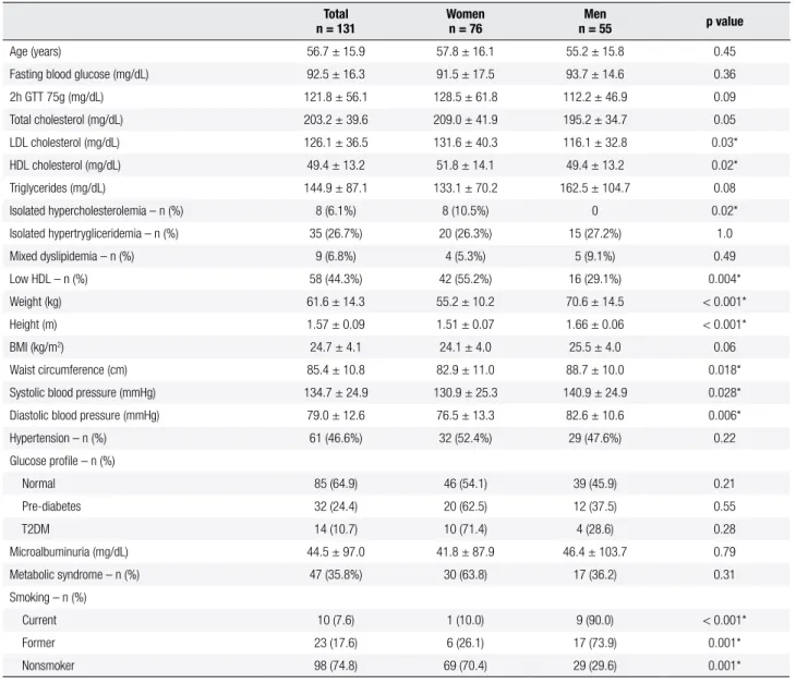
Table 1. Clinical and laboratory characteristics of the studied population
Total n = 131
Women n = 76
Men
n = 55 p value
Age (years) 56.7 ± 15.9 57.8 ± 16.1 55.2 ± 15.8 0.45
Fasting blood glucose (mg/dL) 92.5 ± 16.3 91.5 ± 17.5 93.7 ± 14.6 0.36
2h GTT 75g (mg/dL) 121.8 ± 56.1 128.5 ± 61.8 112.2 ± 46.9 0.09
Total cholesterol (mg/dL) 203.2 ± 39.6 209.0 ± 41.9 195.2 ± 34.7 0.05
LDL cholesterol (mg/dL) 126.1 ± 36.5 131.6 ± 40.3 116.1 ± 32.8 0.03*
HDL cholesterol (mg/dL) 49.4 ± 13.2 51.8 ± 14.1 49.4 ± 13.2 0.02*
Triglycerides (mg/dL) 144.9 ± 87.1 133.1 ± 70.2 162.5 ± 104.7 0.08
Isolated hypercholesterolemia – n (%) 8 (6.1%) 8 (10.5%) 0 0.02*
Isolated hypertrygliceridemia – n (%) 35 (26.7%) 20 (26.3%) 15 (27.2%) 1.0
Mixed dyslipidemia – n (%) 9 (6.8%) 4 (5.3%) 5 (9.1%) 0.49
Low HDL – n (%) 58 (44.3%) 42 (55.2%) 16 (29.1%) 0.004*
Weight (kg) 61.6 ± 14.3 55.2 ± 10.2 70.6 ± 14.5 < 0.001*
Height (m) 1.57 ± 0.09 1.51 ± 0.07 1.66 ± 0.06 < 0.001*
BMI (kg/m2) 24.7 ± 4.1 24.1 ± 4.0 25.5 ± 4.0 0.06
Waist circumference (cm) 85.4 ± 10.8 82.9 ± 11.0 88.7 ± 10.0 0.018*
Systolic blood pressure (mmHg) 134.7 ± 24.9 130.9 ± 25.3 140.9 ± 24.9 0.028*
Diastolic blood pressure (mmHg) 79.0 ± 12.6 76.5 ± 13.3 82.6 ± 10.6 0.006*
Hypertension – n (%) 61 (46.6%) 32 (52.4%) 29 (47.6%) 0.22
Glucose proile – n (%)
Normal 85 (64.9) 46 (54.1) 39 (45.9) 0.21
Pre-diabetes 32 (24.4) 20 (62.5) 12 (37.5) 0.55
T2DM 14 (10.7) 10 (71.4) 4 (28.6) 0.28
Microalbuminuria (mg/dL) 44.5 ± 97.0 41.8 ± 87.9 46.4 ± 103.7 0.79
Metabolic syndrome – n (%) 47 (35.8%) 30 (63.8) 17 (36.2) 0.31
Smoking – n (%)
Current 10 (7.6) 1 (10.0) 9 (90.0) < 0.001*
Former 23 (17.6) 6 (26.1) 17 (73.9) 0.001*
Nonsmoker 98 (74.8) 69 (70.4) 29 (29.6) 0.001*
Data shown as mean (±SD); * p < 0.05.
RESULTS
Of the 131 (66.8%) individuals included in the study, only seven individuals had a diagnosis of angina/ myocardial infarction or stroke.
Cop
yright
© ABE&M t
odos os dir
eit
os r
eser
vados
.
The prevalence of overweight and obesity were re-spectively 29.6% and 46.3% among men, and 15.6% and 39% among women. Abdominal obesity, evaluated by the abdominal circumference, was signiicantly more frequent among men (Table 2).
Table 2. Classiication of individuals by gender according to BMI and
abdominal obesity
Gender Male Female p value
Overweight 16 (29.6) 12 (15.6) 0.08
Obesity 25 (46.3) 30 (39.0) 0.47
Abdominal obesity 30 (55.6) 16 (20.8) 0.01*
Data shown as mean (% of total); * p < 0.05.
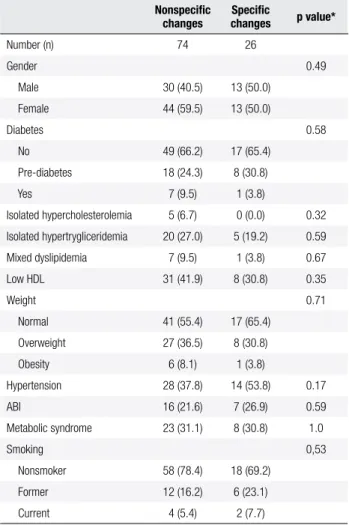
A total of 100 (76.3%) ECG examinations were ana-lyzed by a cardiologist and the indings were divided into two categories: 1) nonspeciic (n = 74): reports that were described as normal, disorders of ventricular re-po-larization, low amplitude QRS interval and changes in conduction inferior parietal were grouped in this cate-gory; 2) speciic changes (n = 26): blockade, ischemia and arrhythmia. The groups were similar (p> 0.05) for any of the variables studied (Table 3). ABI was positive for PAD in 22.3% of the 131 patients evaluated; how-ever, there was no correlation with ECG alterations.
We observed signiicant lower values for HOMA-b
in T2DM (p = 0.03), and increased HOMA-IR in
obese patients (p = 0.01) and in patients with MS di-agnosis (p = 0.02). When data was analyzed according to gender, we observed increased HOMA-IR values in women with MS diagnosis (p= 0.004), while men had lower HOMA-b in patients with higher blood glucose (p= 0.003) or T2DM diagnosis (p= 0.002). We also found increased HOMA-IR in overweight men (p = 0.01) (data not shown). When HOMA values were analyzed in normal glucose (N), PDM and T2DM in-dividuals, we did not observe signiicant differences in the values of the HOMA-IR among the groups, but we did ind lower values of HOMA-b in the PDM and T2DM groups (Table 4).
DISCUSSION
In this study, the majority of the population (68.7%) consisted of adult individuals born in Japan (Issei),
cha-racterizing a community that was formed recently. Wo-men made up most of the group, which can be attribu-ted to the fact that men move out of the community to work or study, and even the recent wave of immigration back to Japan.
Table 3. Clinical characteristics of the population according to
electrocardiogram indings
Nonspeciic changes
Speciic
changes p value*
Number (n) 74 26
Gender 0.49
Male 30 (40.5) 13 (50.0)
Female 44 (59.5) 13 (50.0)
Diabetes 0.58
No 49 (66.2) 17 (65.4)
Pre-diabetes 18 (24.3) 8 (30.8)
Yes 7 (9.5) 1 (3.8)
Isolated hypercholesterolemia 5 (6.7) 0 (0.0) 0.32 Isolated hypertrygliceridemia 20 (27.0) 5 (19.2) 0.59 Mixed dyslipidemia 7 (9.5) 1 (3.8) 0.67
Low HDL 31 (41.9) 8 (30.8) 0.35
Weight 0.71
Normal 41 (55.4) 17 (65.4)
Overweight 27 (36.5) 8 (30.8)
Obesity 6 (8.1) 1 (3.8)
Hypertension 28 (37.8) 14 (53.8) 0.17
ABI 16 (21.6) 7 (26.9) 0.59
Metabolic syndrome 23 (31.1) 8 (30.8) 1.0
Smoking 0,53
Nonsmoker 58 (78.4) 18 (69.2)
Former 12 (16.2) 6 (23.1)
Current 4 (5.4) 2 (7.7)
Data shown as n (%); * p < 0.05 – Fisher’s Exact Test.
Table 4. HOMA-IR and HOMA-b value analysis in normal glycemic,
pre-diabetes (PDM) and type 2 diabetes mellitus (T2DM) groups
HOMA General Normal blood
glucose PDM T2DM
HOMA-IR 9.7 (9.3) 9.1 (8.4) 6.4 (7.6) 12.9 (14.4) HOMA-b 633.7 (703.7) 733.7 (697.5)*,& 661.1 (711.4) 513.3 (1061.0)
Data shown as mean (±SD); *: p < 0.01 compared to T2DM; & : p = 0.04 compared to PDM.
Obesity is not commonly observed among Japanese migrants in the Americas(10), but the absence of a high BMI does not exclude the occurrence of increased visceral fat deposition. Visceral obesity causes a reduc-tion in glucose utilizareduc-tion mediated by insulin, and is clearly related with insulin resistance.
According to the National Nutrition Survey in Japan (2004), the prevalence of obesity (BMI > 25 kg/m2) in
obesi-Cop
yright
© ABE&M t
odos os dir
eit
os r
eser
vados
.
ty were respectively 29.6% and 46.3% among men, and 15.6% and 39% among women. Our data are similar to those reported in the Japanese-Brazilian subjects from Bauru, Sao Paulo/Brazil, where the prevalence of over-weight (BMI > 25 kg/m2) was 40.2%(12).
Similarly, our population showed mean abdomi-nal circumference values close to those found in the Japanese-Brazilian subjects in Bauru (12), but inferior to Asian-American men in Seattle, United States(10). Anyhow, the average values of the abdominal circum-ference in the population of Mombuca were higher than those of Japanese living in Japan, both men and women(13).
Moreover, AO was signiicantly more frequent among men than women. Our results differ from the data on Japanese-Brazilian subjects from Bauru (1). Additionally, in Bauru, the prevalence of AO was greater than the overall prevalence of obesity, while in Mombuca we observed the opposite. In this study, the prevalence of overall obesity (41.9%) was greater than the prevalence of AO (35.7%), supporting other studies that suggest combined analysis of BMI and fat distribu-tion increases the predictive power for CVD and meta-bolic disorders(14).
Mild or moderate elevations of serum triglycerides and lower serum levels of HDL cholesterol are ob-served in overweight and obese individuals. However, serum levels of LDL cholesterol may or may not be increased(15). In Mombuca, averages for total cho-lesterol or fractions and triglycerides were within the normal range.
Several studies have shown that residents of Japa-nese descent outside of Japan are more susceptible to T2DM, dyslipidemia, ischemic heart diseases, and cere-brovascular diseases(12,16,17). The term metabolic syndrome is accepted by the international medical com-munity as a tool capable of identifying individuals with multiple risk factors for CVD and diabetes mellitus(18).
We observed a prevalence of 10.7% of T2DM, 46.6% of hypertension, and 35.8% of metabolic syndrome, showing that there is a signiicant portion of indivi duals exposed to well-established cardiovascular disease risk factors. The highest incidence of these diseases can oc-cur due to greater genetic susceptibility which, along with changes in lifestyle, lead to deterioration of glu-cose metabolism, lipids, increased blood pressure, and obesity(1,19,20).
Epidemiological studies that used ECG showed the importance of left ventricular hypertrophy and
abnor-mal Q waves to predict CVD (21-23). In fact, in the Japanese community of Bauru, Q waves, representing myocardial necrosis, were frequent in the ECGs. Our data showed no speciic differences in age, fasting blood glucose, lipids, changes in blood pressure and BMI ac-cording ECGs indings. This could be explained by the fact that the population is still made up of adults with an average age of less than 60 years, and that the population still retains many of the habits brought from Japan, as well as their work in agriculture. Also, ECG is not very sensitive to predict and diagnose cardiovascular disease.
We did not observe any correlation between ECGs indings and ABI results, either, what may suggest that ABI was not an adequate tool to evaluate atheroscle-rosis in this population. Perhaps this correlation was not possible because of the small number of individuals studied, and because the evaluation of ABI was not per-formed by the same researcher. However, it is known that the prevalence of PAD depends on the age of the population studied and the presence of other risk fac-tors or other atherosclerotic events, such as coronary artery disease or cerebrovascular disease. Anyhow, in accordance with other studies, we found a correlation between HOMA-IR and MS(24), relecting high risk for atherosclerotic disease in this population. We also found that HOMA-b decreases progressively depend-ing on the evolution of glucose abnormalities.
Thus, we conclude that the Japanese-Brazilian sub-jects from Mombuca/Guatapara are exposed to major cardiovascular risk factors, namely high prevalence of MS diagnoses and increased HOMA-IR values. Al-though ECG and ABI were not able to identify indi-viduals at risk for CVD at the time of the study, these tools should still be used. Further studies are necessary to evaluate the use of more sensitive techniques to as-sess cardiovascular disease in this population.
Acknowledgements: we are grateful to patients participating in this study and to Sebastiao L. Brandao Filho for technical as-sistance. This study received no speciic grant from any funding agency in the public, commercial, or not-for-proit sectors.
Disclosure: no potential conlict of interest relevant to this article was reported.
REFERENCES
Cop
yright
© ABE&M t
odos os dir
eit
os r
eser
vados
.
2. Andrade RCG, Figueiredo RC, Foss-Freitas MC, Pace AE, Dal Fa-bbro AL, Franco LJ, et al. Prevalence of diabetes mellitus in the Japanese-Brazilian community of Mombuca, Guatapara, SP. Arq Bras Endocrinol Metab. 2011;55(2):127-33.
3. Fujimoto WY, Leonetti DL, Kinyoun JL, Newell-Morris L, Shuman WP, Stolov WC, et al. Prevalence of diabetes mellitus and impai-red glucose tolerance among second generation Japanese Ame-rican men. Diabetes. 1987;36:721-9.
4. Murabito JM, D’Agostino RB, Silbershatz H, Wilson WF. Intermit-tent claudication. A risk proile from The Framingham Heart Stu-dy. Circulation. 1997;96:44-9.
5. Arai H, Yamamoto A, Matsuzawa Y, Saito Y, Yamada N, Oikawa S, et al. Prevalence of metabolic syndrome in the general Japanese population in 2000. J Atheroscler Thromb. 2006;13(4):202-8. 6. VI Diretrizes Brasileiras sobre Hipertensão. Sociedade Brasileira
de Cardiologia 2010.
7. Alberti KG, Zimmet PZ. Deinition, diagnosis and classiication of diabetes mellitus and its complications. Part 1. Diagnosis and classiication of diabetes mellitus, provisional report of a WHO consultation. Diabet Med. 1998;15:539-53.
8. Geloneze B, Rodovalho-Geloneze S, Parisi C, Pícolo M, Repetto EM, Tambascia MA. Standardization of insulin tolerance test in Brazi-lian population. Diabetes Res Clin Pract. 2000;50(Suppl 1):S102. 9. International Diabetes Federation. Available from: www.idf.org/
metabolic_syndrome.
10. Fujimoto WY, Bergstrom RW, Boyko EJ, Kinyoun JL, Leonetti DL, Newell-Morris LL, et al. Diabetes e diabetes risk factors in second and third-generation Japanese-Americans in Seattle. Diabetes Res Clin Pract. 1994;24(Suppl):S43-52.
11. Ministry of Health, Labor and Welfare, Japan. The National Nutri-tion Survey in Japan. Dai-ichi Shuppan, Tokyo; 2004.
12. Grupo de Estudos do Diabetes na Comunidade Nipo-Brasileira (JBDSG). Diabetes mellitus e doenças associadas em nipo-bra-sileiros. São Paulo: Green Forest do Brasil Editora; 2004. p. 133. 13. Arai H, Yamamoto A, Matsuzawa Y, Saito Y, Yamada N, Pikawa S,
et al. Prevalence of metabolic syndrome in the general Japanese population in 2000. J Atheroscler Thromb. 2006;13(4):202-8.
14. Egger G. The case for using waist to hip ratio measurements in the routine medical checks. Med J Aust. 1992;156:280-5. 15. Katzel LI, Krauss RM, Goldberg AP. Relations of plasma TG and
HDL-c concentrations to body composition and plasma insulin levels are altered in men with small LDL particles. Arterioscler Thromb Vasc Biol. 1994;14:1121-8.
16. Fujimoto WY, Bergstrom RW, Boyko EJ, Chen K, Kahn SE, Leonet-ti DL, et al. Type II diabetes and the metabolic syndrome in Japa-nese-Americans. Diabetes Res Clin Pract. 2000;50(Suppl):S73-6. 17. Fujimoto WY, Bergstrom RW, Boyko EJ, Chen KW, Kahn SE,
Leo-netti DL, et al. Preventing diabetes--applying pathophysiological and epidemiological evidence. Br J Nutr. 2000;84(Suppl):S173-6. 18. World Health Organization. WHO issues new healthy life expec-tancy rankings, Japan number one in new ‘healthy life’ System, 2000. Available from: http://www.who.int/inf-pr-2000/en/pr2000--life-html.
19. Franco LJ. Diabetes in Japanese-Brazilians – Inluence of the ac-culturation process. Diabetes Res Clin Pract. 1996;34:S51-7. 20. Franco LJ, Gimeno SGA, Ferreira SRG, Iunes M. Incremento na
mortalidade associada à presença de diabetes mellitus em nipo -brasileiros. Rev Saúde Pública. 1998;2:118-24.
21. Westerhout CM, Lauer MS, James S, Fu Y, Wallentin L, Armstrong PW; GUSTO IV ACS Investigators. Electrocardiographic left ven-tricular hypertrophy in GUSTO IV ACS: an important risk marker of mortality in women. Eur Heart J. 2007;28:2064-9.
22. Menotti A, Seccareccia F. Electrocardiographic Minnesota code indings predicting short-term mortality in asymptomatic sub-jects. The Italian RIFLE Pooling Project (Risk Factors and Life Ex-pectancy). G Ital Cardiol. 1997;27:40-9.
23. Horibe H, Kasagi F, Kagaya M, Masutani Y, Okayama A, Ueshima H; NIPPON TATA80 Research Group; Working Group of Electrocar-diography Coding for the National Survey of Circulatory Disor-ders, 1980. J Epidemiol. 2005;15:125-34.