ABSTRACT
Sao Paulo Med J. 2007;125(5):297-9.
C
A
SE REPOR
T
Marcelo Lorenzi Marques
Gabriel Salum D’Alessandro
Daher Cezar Chade
Valéria Pereira Lanzoni
Samuel Saiovici
Cláudio José Ramos de Almeida
Primary mucinous adenocarcinoma
of the bladder with signet-ring cells:
case report
Hospital Professor Edmundo Vasconcelos, São Paulo, Brazil
CONTEXT: Primary adenocarcinomas of the bladder are uncommon and usually occur by contiguity with or hematogenic dissemination of other adenocarcinomas such as colorectal, prostate and gynecological tract carcinomas. Mucinous and signet-ring cell histological pat-terns are even rarer and it is often diffi cult to morphologically distinguish them from metastatic colorectal adenocarcinoma.
CASE REPORT: We present and discuss a rare case of primary mucinous adenocarcinoma of the bladder with signet-ring cells in a 57-year-old male patient. Other primary sites for the tumor had been excluded and, in the absence of diges-tive tract tumor and for confi rmation that it was a primary bladder tumor, an immunohistochemistry study was performed.
KEY WORDS: Urinary bladder. Urinary bladder neoplasms. Mucinous adenocarcinoma. Immuno-histochemistry. Signet ring cell carcinoma. INTRODUCTION
Bladder adenocarcinomas account for 0.5 to 2% of all malignant tumors of the bladder. The mucinous and signet-ring cell subtypes are rare and correspond to 20% of these tumors.1,2
Intramural tumor growth causes late symp-toms. This delays the diagnosis and results in a poorer prognosis than for urothelial tumors.1-3
There are a variety of histological types among adenocarcinomas, including papillary, glandular, adenoid cystic, clear cell, muci-nous and signet-ring cell carcinomas. The last of these types is characterized by the presence of intracellular vacuoles fi lled with mucin that displace the hyperchromatic nuclei.2
Because of the rarity of primary muci-nous adenocarcinoma and the difficulties and particularities in diagnosing the primary tumor site, we present here a case of primary mucinous adenocarcinoma of the bladder with signet-ring cells.
CASE REPORT
A 57-year-old male patient presented with a six-month history of painless intermittent macroscopic hematuria without coagula. The patient was a smoker and an alcoholic (Child B
alcoholic liver cirrhosis). He presented weight loss, ascites and lower limb edema. There was no palpable abdominal tumor and digital rectal examination was normal.
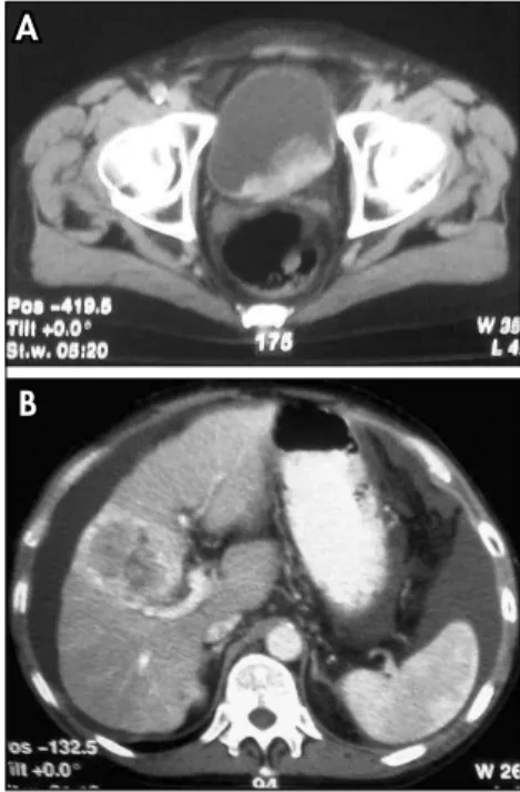
Ultrasound and abdominal tomography revealed ascites, cirrhotic liver with nodules (largest nodule measuring 7.5 cm in diam-eter) and an intravesical mass of 10 cm in diameter that was continuous to the prostate and involved the fl oor and left lateral wall (Figures 1 and 2).
Cystoscopy made it possible to see a ses-sile mass in the left hemitrigone, involving the meatus, left lateral wall, posterior wall and part of the anterior wall, showing rough papillomatous areas and other areas with intact mucosa. Total resection of the intravesical
tu-Figure 1. Abdominal ultrasound showing intravesical mass (10 cm in diameter).
Figure 2. A) Computer tomography show-ing intravesical mass. B) Nodule measur-ing 7.5 cm in diameter in a cirrhotic liver suggesting metastasis.
A
A
298
Sao Paulo Med J. 2007;125(5):297-9. In view of the absence of a digestive tract
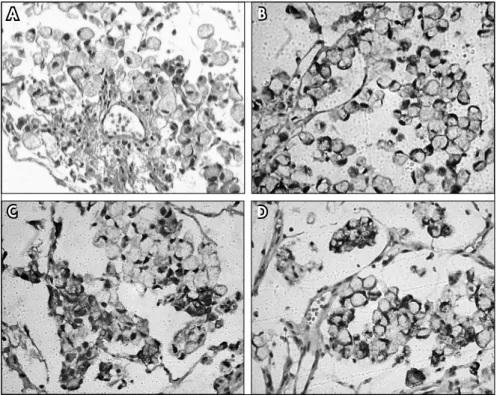
tumor and to confi rm the primary bladder ori-gin of the tumor, immunohistochemical analysis was performed. The tumor was found to be dif-fusely immunopositive for Muc-2 and p63 and focally immunopositive for CK7 and CK20, but negative for TTF-1, PSA, Muc-5AC and CDX-2. The immunohistochemical panel, together with the histological characteristics, characterized a mucus-secreting urothelial carcinoma (Figure 3).
DISCUSSION
Primary bladder adenocarcinomas are uncommon and usually occur by contiguity with or hematogenic dissemination of other adenocarcinomas such as colorectal, prostate and gynecological tract carcinomas.3 The
mucinous and signet-ring cell histological patterns are even rarer and it is often diffi cult to morphologically distinguish them from metastatic colorectal adenocarcinoma.
Immunohistochemical analysis using a panel of specifi c markers is an important al-ternative for etiological differentiation of these tumors. A CDX-2 and CK20-positive and CK7-negative profi le is indicative of digestive tract adenocarcinoma, particularly colorectal carcinoma, and is rare in urothelial tumors, which normally express CK7 alone or together with CK203 and do not express CDX-2.
Few studies on the expression of the cytokeratins CK20 and CK7 in primary bladder adenocarcinoma cases are available in the literature. Torenbeek et al.4 observed
the expression, at least focally, of CK7 in 82% of cases and CK20 in 73%, whereas a CK20-positive and CK7-negative profi le was detected in only 29% of the cases of primary adenocarcinomas of the bladder.3
The expression of markers for mucin-producing tumors such as 2 and Muc-5AC is very heterogeneous and is observed in a wide variety of tumors, particularly those originating from the intestinal tract.5
Immu-nohistochemical evaluation of these specifi c markers to help in diagnosing carcinomas for which the primary origin is uncertain is not usually recommended.
CONCLUSIONS
Primary adenocarcinoma of the bladder is very rare and its precise diagnosis through con-ventional methods is diffi cult to achieve. For this reason, immunohistochemical analysis has major importance in determining the fi nal diagnosis. In this case, the panel was immunopositive for Muc-2, CK7 and CK20 and immunonegative for CDX-2; this, together with the histologi-cal fi ndings, characterized a mucus-secreting urothelial carcinoma. However, the markers for mucin-producing tumors, such as Muc-2 and Muc-5AC, are also observed in a wide variety of other tumors. Therefore, investigation of these markers is not usually recommended.
Figure 3. A) Mucinous adenocarcinoma of the bladder with signet-ring cells (hematoxy-lin-eosin, 200 x). B) Immunohistochemistry positive for Muc-2. C) Immunohistochemistry positive for CK7. D) Immunohistochemistry positive for CK20. (100 x).
mor could not be performed because of its size and infi ltration. On the other hand, the main objective of this procedure was diagnosis.
The histological fi ndings were that this was a case of mucinous adenocarcinoma with signet-ring cells infi ltrating the lamina propria and musculature, with preservation of the mucosa.
Because of the rarity of primary blad-der tumors with these histological features, rigid rectosigmoidoscopy, colonoscopy, upper digestive endoscopy, pelvis resonance and laparoscopy together with liver biopsy were sequentially performed with the aim of detecting possible primary gastrointestinal cancer. Histopathological analysis of the liver fragment revealed mucinous adenocarcinoma associated with cirrhosis.
A
A
BB
C
C
D
D
1. Zaghloul MS, Nouh A, Nazmy M, et al. Long-term results of primary adenocarcinoma of the urinary bladder: a report on 192 patients. Urol Oncol. 2006;24(1):13-20.
2. Fiter L, Gimeno F, Martin L, Gómez Tejeda L. Signet-ring cell adenocarcinoma of bladder. Urology. 1993;41(1):30-3. 3. Wang HL, Lu DW, Yerian LM, et al. Immunohistochemical
distinc-tion between primary adenocarcinoma of the bladder and secondary colorectal adenocarcinoma. Am J Surg Pathol. 2001;25(11):1380-7.
4. Torenbeek R, Lagendijk JH, Van Diest PJ, Bril H, van de Molengraft FJ, Meijer CJ. Value of a panel of antibodies to identify the primary origin of adenocarcinomas presenting as bladder carcinoma. Histopathology. 1998;32(1):20-7. 5. Lau SK, Weiss LM, Chu PG. Differential expression of
MUC1, MUC2, and MUC5AC in carcinomas of various sites: an immunohistochemical study. Am J Clin Pathol. 2004;122(1):61-9.
Sources of funding: None
Confl icts of interest: None
Date of fi rst submission: July 3, 2006
Last received: September 10, 2007
Accepted: Septmeber 12, 2007
299
Sao Paulo Med J. 2007;125(5):297-9.
AUTHOR INFORMATION
Marcelo Lorenzi Marques, MD. Resident, Urology Service,
Hospital Professor Edmundo Vasconcelos, São Paulo, Brazil.
Gabriel Salum D’Alessandro, MD. Resident, General
Surgery, Hospital Professor Edmundo Vasconcelos, São Paulo, Brazil.
Daher Cezar Chade, MD. Resident, Urology Service, Hospital
Professor Edmundo Vasconcelos, São Paulo, Brazil.
Valéria Pereira Lanzoni, MD. Attending physician, Pathology
Service, Hospital Professor Edmundo Vasconcelos, São Paulo, Brazil.
Samuel Saiovici, MD. Attending physician, Urology Service,
Hospital Professor Edmundo Vasconcelos, São Paulo, Brazil.
Cláudio José Ramos de Almeida, MD. Head of the Urology
Service, Hospital Professor Edmundo Vasconcelos, São Paulo, Brazil.
Address for correspondence: Samuel Saiovici
Rua Borges Lagoa, 1450 — Vila Clementino São Paulo (SP) — Brasil — CEP 04038-905 Tel. (+55 11) 5080-4000
E-mail: [email protected]
Copyright © 2007, Associação Paulista de Medicina
RESUMO
Adenocarcinoma mucinoso primário da bexiga com células em anel de sinete: relato de caso
CONTEXTO: Adenocarcinomas vesicais primários são incomuns, o habitual é o comprometimento por con-tigüidade ou via hematogênica de outros adenocarcinomas como colorretal, próstata e trato ginecológico. O padrão histológico correspondente ao mucinoso e com células em anel de sinete é mais raro e, muitas vezes, há difi culdade em distingui-lo morfologicamente do adenocarcinoma colorretal metastático. RELATO DE CASO: Apresentamos e discutimos um caso de adenocarcinama mucinoso com células em anel de sinete primário da bexiga em um paciente masculino, de 57 anos. Foram excluídos outros sítios primários do tumor e, na ausência de tumor do trato digestivo e para confi rmação de tumor vesical primário realizou-se estudo imunoistoquímico.