E
d
it
ör
’e
Mektu
p
L
e
tt
er
s to
E
dit
or
Ilker Sengul1, Demet Sengul2, Gamze Mocan3 1Department of General Surgery, Giresun University Faculty of Medicine, Giresun, 2Department of Pathology, Prof. Dr. A. Ilhan Ozdemir State Hospital, Giresun, 3Division of Cytology, Department of Pathology, Hacettepe University Faculty of Medicine, Ankara, Turkey
Axillary Pilonidal Siınus of a Hirsute Woman
Hirşutizmli Bir Kadında Aksiller Pilonidal Sinüs
DOI: 10.4328/JCAM. 446 Received: 15.10.2010 Accepted: 26.10.2010 Printed: 01.01.2012
Corresponding Author: Ilker Sengul, General Surgery, Vice Dean’s Oice, The Founder Chairman of Department of General Surgery, Deanery of Giresun University Faculty of Medicine, 28100 Giresun / Turkey. Rectorship : +90 454 310 10 00 Deanary: +90 454 214 03 69 GSM: +90 530 885 00 50 GSM-Belgium: +32 484 53 62 61 Fax: +90 454 214 02 47 E-Mail: dr.ilker52@mynet.com, ilker.sengul@giresun.edu.tr To the editor
The terminology of pilonidal sinus was irstly used by Hodges et al [1] in 1880 ater 33 years from Anderson et al who founded a hair in a sacrococcygeal ulcer [2]. Although it is seen mostly in sacrococcygeal area, it may also occur in seldom anatomical localizations such interdigital area, forehead, scalp, umblicus, clitoris, penis, abdomen, neck and axilla [3].
We had presented a hirsute Turkish woman aged 25 in November 2009 with the history and complaint of the intermittent small amount of leakage from her right axilla during the past year. On the physical examination, one small sinus sized 2 mm in diameter in her right axilla was detected. However, it was deined at neither the intergluteal sulcus and nor the other parts of the body. The surgical
treatment was performed via eliptic skin incision. Following the total surgical excision the histopathological evaluations con-irmed the preassumptive diagnosis of axillary pilonidal sinus (Figure 1, 2). Surgical treatment gives to appropriate choice of treatment partially for the disease having one or two sinuses, besides enabling to absolute histopathological diagnoses. Any perioperative or postoperative complication and recurrence has not been detected during the 46 months follow-up [3]. Sion-Vardy et al presented two similar cases of axillary pilonidal sinus one month later, in December 2009. They were a 19-year-old male having the prediagnosis of sebaceous cyst and 22-year-old female having the prediagnosis of lymph node. Both patients had given a history of axillary shaving with razor. Sion-Vardy et al mentioned in their study about that the related previously reported cases were non-hirsute healthy women, aged 17-30 years. However, our presented case in November 2009 is a hirsute 25-year-old woman [4].
So, according to our point of view, the awareness of the probability of a pilonidal disease even in seldom locations like groins of axilla for both hirsute and non-hirsute patients is very important. The malignant degeneration of the disease is very rare, but when it occurs it has much more poor prognosis comparing with squamous cell carcinoma and regular nonmelanoma skin cancer [3].
References
1. Hodges R. M. Boston Med Surg J 1880;103:485. 2. Anderson A. W. Boston Med Surg J 1847;36:74.
3. Sengul I, Sengul D, Mocan G. Axillary pilonidal sinus: A case report of a rare location of pilonidal sinus. North Am J Med Sci 2009;1:316-318.
4. Sion-Vardy N, Osyntsov L, Cagnano E, Osyntsov A, Vardy D, Benharroch D. Unexpected location of pilonidal sinuses. Clin Exp Dermatol 2009;34:e599-601.
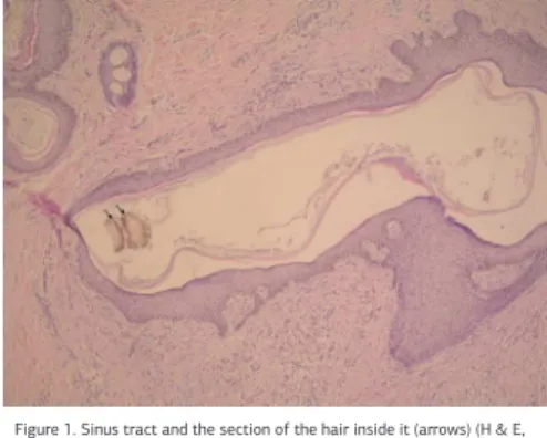
Figure 1. Sinus tract and the section of the hair inside it (arrows) (H & E, original magniication, 10x20).