The Chromone Alkaloid, Rohitukine, Affords
Anti-Cancer Activity via Modulating
Apoptosis Pathways in A549 Cell Line and
Yeast Mitogen Activated Protein Kinase
(MAPK) Pathway
Safia1, Mohd Kamil1, Pooja Jadiya2, Saba Sheikh1, Ejazul Haque1, Aamir Nazir2, Vijai Lakshmi3, Snober S. Mir1*
1Department of Bioengineering & Biosciences, Integral University, Lucknow, Uttar Pradesh, India, 2Laboratory of Functional Genomics and Molecular Toxicology, Division of Toxicology, CSIR-Central Drug Research Institute, Lucknow, Uttar Pradesh, India,3Department of Biochemistry, King George's Medical University, Lucknow, Uttar Pradesh, India
Abstract
The field of cancer research and treatment has made significant progress, yet we are far from having completely safe, efficient and specific therapies that target cancer cells and spare the healthy tissues. Natural compounds may reduce the problems related to cancer treatment. Currently, many plant products are being used to treat cancer. In this study, Rohi-tukine, a natural occurring chromone alkaloid extracted fromDysoxylum binectariferum, was investigated for cytotoxic properties against budding yeast as well as against lung can-cer (A549) cells. We endeavored to specifically study Rohitukine inS.cerevisiaein the con-text of MAPK pathways as yeast probably represents the experimental model where the organization and regulation of MAPK pathways are best understood. MAPK are evolution-arily conserved protein kinases that transfer extracellular signals to the machinery control-ling essential cellular processes like growth, migration, differentiation, cell division and apoptosis. We aimed at carrying out hypothesis driven studies towards targeting the im-portant network of cellular communication, a critical process that gets awry in cancer. Employing mutant strains of genetic model systemSaccharomyces cerevisiae.S.cerevisiae
encodes five MAPKs involved in control of distinct cellular responses such as growth, differ-entiation, migration and apoptosis. Our study involves gene knockouts ofSlt2andHog1
which are functional homologs of human ERK5 and mammalian p38 MAPK, respectively. We performed cytotoxicity assay to evaluate the effect of Rohitukine on cell viability and also determined the effects of drug on generation of reactive oxygen species, induction of apoptosis and expression ofSlt2andHog1gene at mRNA level in the presence of drug. The results of this study show a differential effect in the activity of drug between the WT,
Slt2andHog1gene deletion strain indicating involvement of MAPK pathway. Further, we investigated Rohitukine induced cytotoxic effects in lung cancer cells and stimulated the
OPEN ACCESS
Citation:Safia , Kamil M, Jadiya P, Sheikh S, Haque E, Nazir A, et al. (2015) The Chromone Alkaloid, Rohitukine, Affords Anti-Cancer Activity via Modulating Apoptosis Pathways in A549 Cell Line and Yeast Mitogen Activated Protein Kinase (MAPK) Pathway. PLoS ONE 10(9): e0137991. doi:10.1371/ journal.pone.0137991
Editor:Muzamil Ahmad, Indian Institute of Integrative Medicine, INDIA
Received:April 13, 2015
Accepted:August 24, 2015
Published:September 25, 2015
Copyright:© 2015 Safia et al. This is an open access article distributed under the terms of the
Creative Commons Attribution License, which permits unrestricted use, distribution, and reproduction in any medium, provided the original author and source are credited.
Data Availability Statement:All relevant data are within the paper.
productions of ROS after exposure for 24 hrs. Results from western blotting suggest that Rohitukine triggered apoptosis in A549 cell line through upregulation of p53, caspase9 and down regulation of Bcl-2 protein. The scope of this study is to understand the mechanism of anticancer activity of Rohitukine to increase the repertoire of anticancer drugs, so that prob-lem created by emergence of resistance towards standard anticancer compounds can be alleviated.
Introduction
The ever evolving affliction of cancer is mounting its challenges on researchers and clinicians as the disease continues to impose immense amount of health burden on a devastating global scale. Significant understanding of its mechanistic cues has been achieved through research efforts that have now proven that this ailment finds a strong cause in altered communication between and within cells [1]. Thus far, effective non-surgical remedies against the disease include chemotherapy and radiation based treatment regimens. However, a number of poten-tial anti-cancer therapies, based on molecules from natural origin, have exhibited promise in treating cancer while exerting minimal undesired effects (anemia, nausea and hair loss) and countering the challenge of drug resistance [2]. In addition to side effect and drug resistance, the cost of chemotherapy drug is also very high as compared to the natural compound from the medicinal plants.
Rohitukine (C16H19NO5; 5, 7-dihydroxy- 8-(3-hydroxy-1-methyl-4-piperidinyl)-2-methyl- 4H-chromen-4-one), isolated fromAmoora rohituka,Dysoxylum binectariferum
andSchumanniophyton problematicum, is known to possess anti-inflammatory, anti-implanta-tion, anti-fertility, anti-proliferative and immunomodulatory properties [3]. However antican-cer mechanism of action of Rohitukine is not known and as per our comprehension for the first time it has been evaluated in genetic model system of budding yeast as well as in lung can-cer cells. Hundreds of yeast genes exhibit a link to human disease genes as nearly 30% of noto-rious genes involved in human diseases have yeast orthologs [4]. It is interesting to note that 47% of the yeast genes could be successfully humanized [5].S.cerevisiaeis also helping in revealing important aspects of many diseases such as neurofibromatosis type l, colon cancer [6].
We endeavored to specifically study Rohitukine inS.cerevisiaein the context of MAPK pathways as yeast represents the experimental model where the organization and regulation of MAPK pathways are best understood [7]. MAPK are evolutionarily conserved protein kinases that transfer extracellular signals to the machinery controlling essential cellular processes like growth, migration, differentiation, cell division and apoptosis. Therefore, mutation in any of the kinases of these pathways is directly linked to cancer [8]. It is, hence, prudent to focus fur-ther research efforts towards designing mechanism-based anti-cancer compounds that act on specific molecular targets linked with the etiology of the disease [9]. Hence kinase cascade pres-ents novel opportunities for development of new cancer therapies designed to be less toxic than conventional chemotherapeutic drugs [10]. The studies were conducted employing genetic model systemSaccharomyces cerevisiaeas it has been usefully exploited for elucidating the anticancer therapy in association with exposure to 5-fluorouracil [11]. Yeast is also valued as a striking model for anticancer drug research [12] as it has proven helpful in uncovering the cellular targets of different drugs including precious anti-cancer drug KP1019 [13]. The bud-ding yeast has five types of MAPK inclubud-ding: Fus3, Kss1, Smk1, Hog1 and Slt2. Slt2 is the Competing Interests:The authors have declared
MAPK of the cell wall integrity pathway and functional homolog of human ERK5 that are acti-vated in response to growth factors and stress conditions [14]. Hog1 is functional homolog of mammalian p38 MAPK and is chiefly activated in response to osmotic stress [15].
The studies reported herein, make use of the genetic model systemS.cerevisiaetowards deciphering the effects of Rohitukine on all important process of cellular communication medi-ated by MAP kinase pathway, thereby affecting cellular survival and death via apoptosis. The study also investigates the effect of Rohitukine on apoptosis within human lung cancer cell line and explores the possible mechanisms involved via studies on important modulators of the process.
Materials and Methods
Extraction of Rohitukine
Rohitukine was isolated from stem ofDysoxylum binectariferumas described previously [16]. Briefly, air-dried stem bark of the plant was extracted with 95% ethanol and then concentrated by reduced pressure. It is further fractionated into four fractions (chloroform, soluble n-buta-nol, n-hexane and insoluble n-butanol fraction). From chloroform fraction, a known alkaloid rohitukine {5,7-dihydroxy-2-methyl-8- [4-(3-hydroxy-1-methyl)-piperidinyl]-4H-1-benzo-pyran-4-one)} was isolated by repeated column chromatography over silica gel and further purification by HPLCLC- 20AD using methanol solvent 55:45 v/v, flow rate 1.0 ml/min. The characterization of compound was performed using IR, NMR, mass, derivatization, and com-parison with available literatures. The purity of rohitukine was 99.6% and yield was 1%.
Yeast culture and maintenance
In present study, Wild Type strain BY4741 (MATa his3Δ1 leu2Δ0 met15Δ0 ura3Δ0) and
knockout strain ofSlt2andHog1gene (gift from Dr. A. Chakrabarti and Dr. R. C. Meena from
Defence Institute of Physiology and Allied Sciences, DRDO, India) were employed. The yeast cells were grown in YPD media (1% yeast extract, 2% bactopeptone, 2% glucose) as per the method described earlier [17].
Determination of Minimum inhibitory concentration
Minimum inhibitory concentration (MIC) of drug was determined both spectrophotometri-cally (by measuring O.D. at 600 nm using multiwell microplate reader: Multi Skan, Thermo Scientific) and visually. Rohitukine was dissolved in Dimethyl sulfoxide. The MIC for Rohitu-kine was determined by plotting O.D. at 600 nm versus concentrations of drug (20μg/ml to 100μg/ml) [18]. The concentration at MIC of the drug was used in all experiments.
Evaluation of growth inhibition by spotting assay
After the drug treatment growth inhibition of yeast cells was assessed by spotting assay. Cells were grown on standard yeast extract-peptone-dextrose (YPD) media. For Spotting assays, 5-fold serial dilutions in YPD media were prepared from exponentially growing culture of the different strains. 2μL of each dilution was then spotted onto YPD plate in absence and presence of drug [19].The growth differences were recorded following incubation of the plates for 24hrs at 30°C.
Detection of reactive oxygen species (ROS) in budding yeast
some modification [20]. Briefly, levels of ROS were measured after 24 hrs of drug treatment by adding 0.5μM of H2-DCF-DA to cells for 15 min in dark. Cells were washed thrice with 1X PBS. Fluorescence microscopy was performed using a Zeiss Axioplan-2 microscope using an excitation wavelength of 485 nm and an emission wavelength of 520 nm. ROS production was quantified using image J software (Image J, National Institutes of Health, and Bethesda, MD). A total of 50 cells from each group were quantified for fluorescence intensity and statistical sig-nificance was calculated with respect to untreated control group.
Estimation of mitochondrial content employing MitoTracker Deep Red
staining
To check the effect of drug on mitochondrial content, Mito Tracker Deep Red staining (Cat. no.-22426, Invitrogen) was done as described previously with some modifications [20]. Briefly, 100μl yeast cells were incubated with 100 nM Mito Tracker stain for 50 min at 30°C in dark fol-lowed by three times washing in 1X PBS. Imaging of cells was carried out using fluorescence microscope with an excitation wavelength of 637 nm and an emission wavelength of 660 nm. Fluorescence intensity of mitochondrial content was quantified using Image-J software.
Assay for apoptotic cell death using Acridine Orange (AO) staining
Acridine orange staining was done to check the induction of early stage apoptosis. Acridine orange (Hi-media- 116) was dissolved in PBS (pH = 7). A 100μl volume of yeast cells was stained with 1μl of 2.5 mg/ml of AO to get the working concentration of 25μg/ml. Staining was carried out for 30 minutes in dark and the cells were washed with PBS [21]. Imaging of stained cells was carried out using fluorescence microscope with an excitation wavelength of 502 nm and an emission wavelength of 520 nm. Fluorescence intensity of stained cells was quantified by Image J software.Assay for studying DNA fragmentation
DNA fragmentation was determined by Nuc Blue Live Cell Stain (R37605 Life Technology Corporation) according to the manufacturer’s instructions. Imaging of stained cells was done by fluorescence microscope with an excitation wavelength of 352 nm and an emission wave-length of 460 nm and fluorescence intensity of stained cells was quantified by Image J software.
Semi-Quantitative Reverse transcription PCR for the analysis of mRNA
levels of
Slt2
and
Hog1
gene in the presence of Rohitukine
Total RNA was extracted and reverse transcribed using Revert Aid™First Strand cDNA Synthe-sis Kit (Fermentas Life Sciences, cat- K1622). cDNA was amplified using specific primers listed
inTable 1and PCR products were separated on 1.5% agarose gel and visualized by ethidium
bromide staining.
Protein-ligand interaction employing computational tools
and ERK5 active site were 18.783, 35.698, 30.394 for Hog1 and 15.928, -17.057, 1.101 for ERK5 respectively. Docking simulations were performed using the Lamarckian genetic algorithm (LGA) and the Solis & Wets local search method. Ten different runs were performed for each docking. The final figures were generated with the help of Discovery Studio Visualizer (Accelrys).
Cell Culture
A549 cells (human lung cancer cell line) were obtained from National Centre for Cell Sciences (NCCS) Pune, India, and cultured in DMEM (Dulbecco’s Modified Eagle Media) F-12 (1:1) (HiMedia AL187A) supplemented with 10% fetal bovine serum, 0.2% sodium bicarbonate and 1% antibiotic and antimycotic solution. Cultures were maintained at 37°C and 5% CO2 and 95% humid atmosphere.
MTT assay
MTT (HiMedia-TC191) assay is based on the reduction of MTT by mitochondrial dehydroge-nase to a purple formazan product, gives an indication of mitochondrial integrity, which is interpreted as assessment of percent cell viability [22]. Briefly, cells were seeded in 96–well tis-sue culture plates (104cells/well) in complete DMEM F-12 medium, followed by incubation in
5% CO2, 95% atmosphere for 24hrs at 37°C. After 24hrs exposure of drug (10μM—60μM), MTT (5 mg/ml of stock in PBS) was added (10 ml/ well in 100 ml of cell suspension), and plates were incubated for 4hrs. After incubation, the reaction mixture was carefully taken out and 200μl of dimethyl sulfoxide (DMSO) was added to each well, the contents were mixed well by pipetting up and down several times. The plates were kept on rocker shaker for 10 min at room temperature and then read at 550 nm using multiwell microplate Reader (Multi Skan, Thermo Scientific). Untreated cells were run under identical conditions and served as basal control. Each experiment was repeated thrice and standard deviations were derived from three inde-pendent experiments.
Determination of Reactive Oxygen Species (ROS) in lung cancer cells
ROS generation was estimated by using 2’, 7’-diclorodihydrofluorescein di-acetate(H2-DCF-DA; Cat. no.–D399; Invitrogen) as described previously [23]. Briefly, cells seeded in black 96-well plate at a density of 104cells/well were incubated with 1mM H2-DCF-DA; for
30 min at 37°C followed by incubation with different concentrations of drug for 24hrs. The measurement of ROS was carried out during the course of the treatment period at 485 nm exci-tation and 535 nm emission wavelengths. ROS generation was also confirmed by fluorescence micrograph of cellular ROS. Briefly, cells were plated in 48-well tissue culture plate and treated with 1mM H2-DCF-DA; for 30 min followed by incubation with different concentrations of
Table 1. Sequence of primers used.
Gene Primer (5’-3’) PCR Product Size (bp) Annealing Temperature (°C)
ACT1 GCCATTTTGAGAATCGATTTG (F) 254 56
TTAGAAACACTTGTGGTGAAC (R)
SLT2 AGCAACAGCAGCCTTCAGA (F) 460 60
GAACGCGAGGAAGTATCCAA (R)
HOG1 ATTTGGGTTGGTTTGCTCAG (F) 254 54
TTTCCAAGGGTCTTGTTTGC (R)
Rohitukine for 24 hrs at 37°C. Fluorescence images were captured using a Zeiss Axioplan-2 microscope using FITC filter under 20X objective.
Isolation of Total Cellular Protein from A549 cells
Rohitukine treated and untreated cells were pelleted, washed with cold PBS and lysed in RIPA lysis buffer containing 1 mM EDTA, 50 mM Tris, pH 7.4, 150 mM NaCl, 1% NP-40, 0.25% sodium deoxycholate, 0.1% SDS, 1 mM NaF, 1 mM Na3VO4, 1 mM PMSF and 1μg/mL
leupep-tin [24]. Cell lysate was gently vortexed for 30 sec after 1 h incubation in lysis buffer. Superna-tant was collected by centrifugation at 14,000×g for 15 min and stored in aliquots at -20°C. Protein content was quantified using Bradford protein assay.
Western Blot Analysis Detecting Apoptosis-related proteins
Cell lysates were denatured and twenty microgram of the protein was separated on 12% SDS– polyacrylamide gel electrophoresis. It was electro-transferred to PVDF membrane. The mem-branes were blocked at room temperature with 5% skimmed milk in Tris-buffered saline (TBS) with 0.05% Tween-20 (TBS-T) for 2hrs. After washing with TBS-T membranes were incubated with the primary antibodies against p53 (1:3000), caspase9 (1:3000), Bcl-2 (1:5000) andβ-actin
(1:4000) for overnight at 4°C. After washing, the membrane was incubated with HRP-conju-gated secondary antibody (anti-rabbit or anti-mouse, 1:10000; Invitrogen, USA) at room tem-perature for 1h. Western blot bands were detected using chemiluminescent substrate
(Millipore) using Chemidoc (GE).β-actin was used as internal control for equal loading and
normalization of protein. Protein Ladder (3B BlackBio Biotech-3B75) (3.5–245kDa) was used to determine molecular weight of the protein bands. Densitometry of the bands obtained was done by NIH software Image J version 1.41 (USA).
Statistical analysis
All results are presented as mean ± SEM Statistical significance between various groups was carried out employing Student’s t test by using Graph Pad prism 5 software. Forin vitrostudy data were expressed as mean ±S.D. and statistical significance of the results were determined using one-way ANOVA by Tukey’s multiple comparison test.
Results and Discussion
Δ
Slt2 and
Δ
Hog1 strains are hypersensitive to Rohitukine treatment
In this study, we determined cytotoxicity of Rohitukine in budding yeast and also investigate whether MAP kinase pathways are involved in the Rohitukine induced cell death, so we deter-mined the effect of Rohitukine on the cell viability ofΔSlt2 andΔHog1 strains. Rohitukine
shows cytotoxicity against all type of yeast strains. The MIC50for Rohitukine was determined
by plotting O.D. at 600 nm versus concentrations of drug. MIC50value for WT was found to be
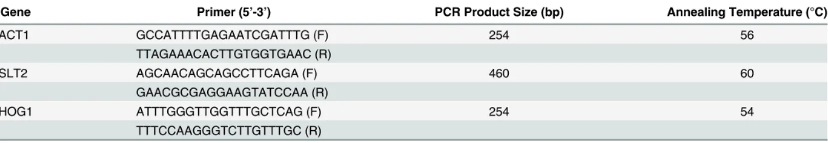
80μg/ml and for both gene knock-out strains was found to be 60μg/ml. All experiments were carried out at dose below the MIC50value (40μg/ml).Fig 1shows that Rohitukine exerts
cyto-toxic effects on yeast. Although the concentration of Rohitukine that kills approximately 50% of yeast cells is higher than the IC50values reported for cancer cells in vitro [25] this result is
The results showed that gene knockout strains were more sensitive to the drug as compared to WT strain at same MIC (40μg/ml) as indicated by density of the spots in spotting assay for cell viability (Fig 1A) and by O.D at 600 nm (Fig 1B) Result of spotting assay confirmed that the yeast cells when treated with Rohitukine lost cell viability in dose dependent manner. In case of WT cells control spot was scored as 0, 40μg/ml of Rohitukine treated cells spot was scored as 1, cells were scored as 2 at 80μg/ml of drug and at 100μg/ml cells spot scored as 3 after 24 hrs of drug treatment. For both types of gene knockouts (Δslt2,Δhog1) strain control
spot was scored as 0, 40μg/ml of drug treated cells scored as 2, 80μg/ml of drug treated cells scored as 3 and 100μg/ml of drug treated cells scored as 4 after 24 hrs of drug treatment.ΔSlt2
strain was hypersensitive to various genotoxic agents having different mode of action including methylmetanosulfonate, UV radiation and phleomycin [28]. Slt2 activation after induction of a single DSB (double-strand break) in the GAL1: HO strain, which has a specific effect on integ-rity of DNA, showing a genuine role for Slt2 in the response to genotoxic stress [29]. Conse-quently, these genes get activated in the presence of drug in WT cells so it could be possible thatΔSlt2 andΔHog1 strains showed hypersensitivity to drug [30].
Rohitukine triggers cell death by inducing oxidative stress and reducing
mitochondrial content in
Δ
Slt2 and
Δ
Hog1 strains
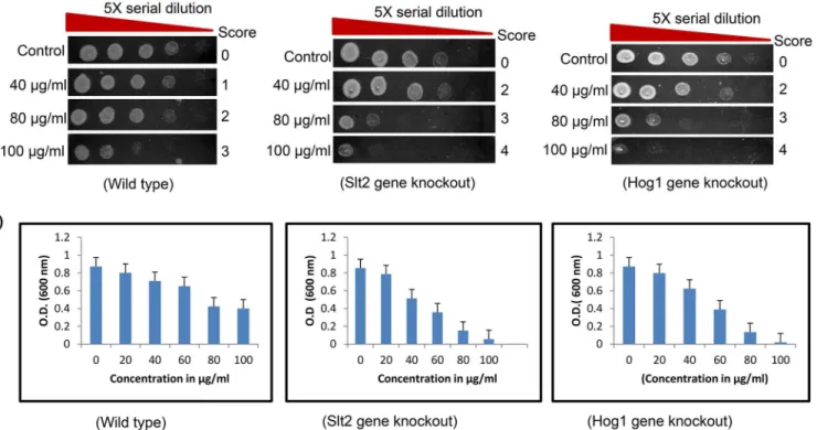
ROS production was measured to analyze the role of ROS in yeast cell death mediated by Rohi-tukine. We found that Rohitukine induced significant amount of ROS after 24 hrs of drug treatment in WT and in gene knockout strains (Fig 2A). Quantification of fluorescence inten-sity of H2-DCF-DA staining(Fig 2B) also showed that Rohitukine treated yeast cells produced
Fig 1. Hypersensitivity ofΔSlt2 andΔHog1 strains to Rohitukine.(a) Yeast cells viability at different concentration of Rohitukine after 24 hrs of drug treatment, 5-fold serial dilutions from exponentially growing cultures of WT,ΔSlt2 andΔHog1 strains were spotted onto YPD medium containing 40μg/ml, 80μg/ml and 100μg/ml of drug. (b)The percentage of surviving cells relative to untreated controls.
comparatively increased levels of ROS as compared to untreated cells, increase being 1.3 (P<0.001), 2.0 (P<0.001) and 1.7 (P<0.001) fold for WT,ΔSlt2 andΔHog1 strains respectively
after Rohitukine treatment as compared to untreated control cells. HoweverΔSlt2 andΔHog1
strains produced more ROS as compared to WT cells after treatment and increase being 1.4 (P<0.001) and 1.2 (P<0.001) fold forΔSlt2 andΔHog1 strains respectively as compared to
WT after drug treatment, which further reinforces the hypersensitivity of both mutant strains to drug. This discrepancy may be because of absence of MAPK which is known to be activated by oxidative stress. Additionally MAPK deficient yeast cells accumulate ROS to a higher extent than WT cells during stationary phase [31]. MAP kinase pathways are influenced not only by receptor ligand interactions, but also by different stressors like oxidative stress induced poten-tial activation of MAPK pathways. Generally, increased ROS production in the cells causes acti-vation of MAPKs but the mechanisms by which ROS can activate these kinases are unclear
Fig 2. Rohitukine promoted ROS production and loss Mitochondrial content in gene knockout strains of yeast.(a) DCFDA staining (b) Graphical representation of Relative formation of reactive oxygen species (ROS) measured by H2DCFDA staining in WT and gene knockout strains of yeast as quantified using Image J software***p<0.001.(c) Mitotracker Deep Red staining (d) Graphical representation for fluorescence intensity of mitochondrial content of the budding yeast as quantified using Image J software***p<0.001.
[32]. These mutants may be impaired in autophagy pathway which is required to prevent excessive ROS accumulation. Inability to increase the expression of respiratory chain compo-nents and ROS scavengers likely leads to the accumulation of ROS in autophagy-defective cells.
S.cerevisiaeHog1 MAPK is activated in response to high osmolarity and is required for cell survival under these conditions [33]. In response to several stresses, Hog1p becomes phosphor-ylated and translocates to the nucleus. Hog1 null mutants were found to be hypersensitive to those stress conditions, which lead to Hog1p activation, in particular to extracellular oxidizing agents [34].
We also checked whether Rohitukine exposure affects mitochondrial content. We observed that mitochondrial content decreased to a greater extent inΔSlt2 andΔHog1 strain after 24 hrs of Rohitukine treatment but WT cells showed increase in mitochondria content after drug treat-ment (Fig 2C). Quantification of fluorescence intensity of mitochondrial content (Fig 2D) showed that Rohitukine treated WT cells showing a 1.6 (P<0.001) fold increase whereas Rohitu-kine treatedΔSlt2 andΔHog1 strains exhibiting 1.2 (P<0.001) and 2.0 (P<0.001) fold reduction respectively as compared to their untreated control. However,ΔSlt2 showed 2.3 (P<0.001) and
ΔHog1 strain exhibited 2.2 (P<0.001) fold reduction as compared to WT in presence of drug. The mitochondria also play a very important role in regulation of many mechanisms con-trolling cell survival and death [35]. Changes in mitochondria are related to aging, decreased synthesis of mitochondrial proteins and reduced activity of oxidative enzymes cause decrease in mitochondrial ATP synthesis [36]. MAP kinase pathways are involved in intrinsic apoptosis in the presence of isoorientin in human hepatoblastoma cancer cells [37]. Flavopiridol (Rohitu-kine derivative) causes cell death by decrease in mitochondrial membrane potential in human leukemia cells [38]. Flavopiridol also causes STI571 (Bcr/Abl kinase inhibitor) induced apopto-sis and damage of mitochondria and apoptoapopto-sis in BCR-ABL-positive human leukemia cells [39]. Consequently MAPK mutants show the increased production of ROS which maybe the likely cause leading to mitochondria dysfunction.
Rohitukine causes induction of early stage apoptosis and DNA damage
in yeast
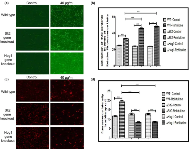
Data of A.O staining showed that Rohitukine causes induction of apoptosis in gene knockout strains as compared to the WT strain after 24 hrs of drug treatment (Fig 3A). Quantification of fluorescence intensity of A.O staining (Fig 3B) showed that WT,ΔSlt2 andΔHog1 stains of yeast exhibiting a 1.3 (p<0.001), 2.0 (p<0.001) and 1.7 (p<0.001) fold increase respectively after drug treatment as compared to their untreated control. However,ΔSlt2 strain showed 1.7 (p<0.001) andΔHog1 strain exhibited 1.2 (p<0.001) fold increase as compared to the WT strain when treated with Rohitukine.
We also observed DNA fragmentation after 24 hrs of drug treatment at MIC by NucBlue Live Cell Stain for DNA. Result of DNA staining (Fig 3C) clearly showed DNA fragmentation in WT as well as in both types of gene knockout strains after drug treatment indicating an apo-ptotic phenotype. We quantified images for fluorescence intensity of DNA staining (Fig 3D). There was 1.5 (p<0.001), 3.0 (p<0.001) and 3.9 (p<0.001) fold increase in Rohitukine treated WT,ΔSlt2 andΔHog1 strain respectively as compared to untreated control. However,ΔSlt2 showed 1.1 (p<0.001) andΔHog1 strain exhibited 1.3 (p<0.001) fold increase as compared to the Rohitukine treated WT cells.
Rohitukine interaction affects expression of
Slt2
and
Hog1
gene in wild
type strain
The mRNA levels ofSlt2andHog1were found to be 3.7 and 2.8 fold increased respectively in WT strain treated with Rohitukine when compared to that of control group (Fig 4A). However mRNA expression ofSlt2gene inΔHog1 strain and expression ofHog1gene inΔSlt2 strain remained un-affected after Rohitukine treatment.Fig 4Bdepicts the fold changes in mRNA levels within different treatment groups normalized against that of control. The selective increase of Slt2 and Hog1 in wild type conditions and not in the knockouts of either gene may be a result of cross-talks between different MAPK pathways which are very common [42]. The over expression ofHog1gene after Rohitukine treatment may be dependent on the presence of
Slt2gene or vice-versa.
Fig 3. Rohitukine causes DNA damage and induction of apoptosis.(a) A.O. staining (b) Graphical representation for fluorescence intensity of apoptotic death of the Yeast cells as quantified using Image J software***p<0.001 (c) DNA damage revealed by Nuc Blue Live Cell Stain (d) Graphical
representation for fluorescence intensity of nucleic acid of the yeast cells as quantified using Image J software***p<0.001.
Zymolyase activates both MAPKs and Slt2 activation depends on the Sho1 branch of the HOG pathway. Both MAPK pathways are essential for cell survival in presence of stress because mutant strains deficient in different components of both pathways are hypersensitive to zymolyase [43]. Thus, a sequential activation of two MAPK pathways may be required for cellular adaptation to stress condition and cell wall damage after the Rohitukine treatment.
From previous studies it is known that Hydroxyurea treatment increases phosphorylation of Slt2 MAP kinase [28]. Slt2 is responsible for cell wall integrity and is activated by cell wall damage, so it might be the possible reason for hypersensitivity of slt2 mutant to drug treatment. Recent studies have implicated the role of Hog1 MAPK in mediating tolerance to a variety of stress conditions including osmotic, oxidative, heat, arsenic, and citric acid stress [44]. A study by Azad et al examined the requirement for a functional HOG pathway to cope with curcumin (100μM) induced stress [45].
Our docking studies revealed that catalytic domain of P38 (Hog1) interacts with Rohitukine through the nine amino acid residues namely VAL89, ARG5, ARG94,VAL345, ILE346, PHE348, PHE8, PHE90, and ASP88, while in case of ERK5 (Slt2) interaction was found with 4 amino acid residues namely TYR245, VAL200, TYR199 and ASN244 (Fig 4C). The free bind-ing energy and estimated inhibition constant (ki) for the‘Rohitukine-P38 domain interaction’ was determined as -6.47 Kcal/mol and 18.18uM respectively; while the same for ‘Rohitukine-ERK5 domain interaction’was found to be -6.31 Kcal/mol, 23.6 u Mol respectively.
Rohitukine induced cytotoxic effects by ROS generation in A549 cell line
To examine the cytotoxicity of Rohitukine, A549 cells were treated with different doses of Rohitukine (10μM to 60μM) for 24 hrs and the viabilities of cells were determined using theFig 4. (a) RT-PCR analysis ofSlt2andHog1gene in budding yeast after drug treatment (i:Untreated control, ii: drug treated) (b) The expression of Slt2 and Hog1 mRNA, expressed as the ratio of densitometric measurement of the sample to the corresponding internal control (β-actin) (i: Untreated control, ii: drug treated) (c) Docking studies of Rohitukine with human two different type of member of MAPK pathway. (i) p38 (Hog1 in
MTT assay. As shown inFig 5A, Rohitukine significantly reduced percentage of viable A549 cells in dose-dependent manner. Among all the tests, cells incubated with 40μM Rohitukine for 24 hrs showed anti-proliferation effect, with cell viability decreased to 50% of the untreated control cells. All experiments were carried out at dose below IC50value.
The crude methanol extract ofF.proliferatumthat is the source of Rohitukine shows cyto-toxicity against HCT-116 and MCF-7 human cancer cell lines (IC50= 10μg/ml for both cancer
cell lines) [25]. Pure Rohitukine from stem barks ofD.binectariferumwas subjected for anti-cancer activity in different lung and ovarian carcinoma cells. IC50value for ovarian carcinoma
cells SKOV3 was found to be 20μM and for breast cancer cells T47D, MDAMB273, MCF7 was found to be 50μM, 3μM, 15μM respectively [46].
Fig 5. Rohitukine affected the percentage of viable and induced ROS in A549 cells.(a) Cell viability was determined using the MTT assay. Cells (1 × 10⁴ cells/well; 96 well plates) were plated in DMEM F12 medium + 10% fetal bovine serum (FBS) with 0, 10, 20, 30, 40, 50 and 60μM for 24 hrs, (b) ROS generation was assessed in terms of relative fluorescence units using 10 mM DCFH-DA in A549 cells after 24 hrs exposure to Rohitukine in black-bottomed 96-well plates and (c) Fluorescence micrographs of ROS generation at 20μM, 30μM, and 40μM Rohitukine concentrations in lung cancer cells obtained at 20X objective after 24 hrs of treatment. The results are represented as means±S.D of three independent experiments. Nonsignificant (ns),*P<0.05,** P<0.01,***P<0.001 versus 0μM.
Flavopiridol, a semi synthetic derivative of Rohitukine exhibited anticancer activity by inhibiting cell cycle dependent kinases (CDKs) [47] Rohitukine possessed anti-estrogenic effect in female Sprague-Dawley rats [48] and compound that show antiestrogenic activity could also have antiproliferative effect on breast cancer cell by cell cycle arrest, including decreased cyclin Dl expression [49].
After determination of cytotoxic effect of Rohitukine in lung cancer cell we checked the effect of drug on ROS generation as in sight of earlier finding that copious chemical stimuli prompt apoptosis via ROS generation [50]. We employed H2-DCF-DA (specific fluorescence probes) staining to examine ROS generation in A549 cells after 24 hrs of Rohitukine (10μM to 40μM) treatment. Significant elevation in ROS levels could be observed at all the tested doses (Fig 5B). Fluorescence micrographs of H2-DCF-DA stained cells further confirmed the above fluorometric findings (Fig 5C). Data from the current study revealed that Rohitukine induces oxidative stress in A549 cells.
Oxidative stress by ROS is a stimulator of numerous cell responses, such as apoptosis in var-ious mammalian cells [50]. Flavopiridol have been shown to alter the redox status of leukemic cells and mediated apoptosis is dependent upon generation of radical oxygen species [51].
This study showed that Rohitukine acts as a ROS generator to trigger cell death in lung can-cer cells, supporting its utility as a cytotoxic therapeutic agent [52].
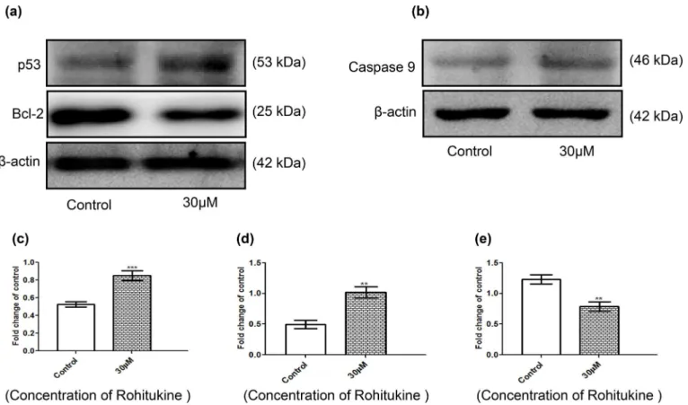
Rohitukine altered the apoptosis-associated protein levels in A549 cells
Cells were exposed to 30μM of Rohitukine for 24 hrs and then the total protein was prepared and western blot analysis was used to detected protein expression of p53, caspase9 and Bcl-2.β-actin was used as an internal loading control. These results are presented inFig 6which indi-cated that expression of p53 (Fig 6A) and caspase9 (Fig 6B) was increased, while the expression of Bcl-2 (Fig 6A) was decreased. The quantitative results showed that Rohitukine increased the protein levels of p53 and caspase9 by 0.40 folds (Fig 6C) and 0.23 (Fig 6D) folds respectively where as decreased the protein levels of Bcl-2 by 0.37 folds (Fig 6E) of the control level at 30μM. Upregulation of proteins like, p53 caspase9 and down regulation of anti-apoptotic pro-teins like Bcl-2 gives insight into the mechanism of action followed by Rohitukine to induce the apoptosis.
Tumor suppressor gene TP53 gets activated during genotoxic stress and promotes cell cycle arrest by the activation of p21 leading to the activation of apoptosis [53]. Caspase-9 activation leads to apoptosis. The majority of cancer therapy initiate apoptosis through the caspase-9 acti-vation, the modulation of caspase-9 expression may be exploited in designing new ways to con-trol apoptosis in neurodegenerative or malignant diseases [54]. Bcl-2, an upstream effector molecule in the apoptotic pathway, has been recognized to be a potent negative regulator of apoptosis, and most cancers generally overexpress Bcl-2 [55]. MAPK pathways also regulate apoptosis and activation of p38 is generally associated with the induction of apoptosis. Berber-ine (a benzylisoquinolBerber-ine alkaloid) significantly inhibited growth and induced cell cycle arrest of NSCLC cells (non small cell lung cancer cells) in a dose-dependent manner. It increased phosphorylation of p38 MAPK in a time-dependent and induced protein expression of tumor suppressor p53. The specific inhibitor of p38 MAPK (SB203580), and silencing of p38αMAPK
(SB203580 and U0126) decrease the cell viability and induced apoptosis in human CNE2 cells (human nasopharyngeal carcinoma cell line) [58].
Induction of apoptosis by flavopiridol in human leukemia cells (U937) proceeds via the intrinsic, cytochrome c-related pathway (caspase9 activation), and is not dependent upon the extrinsic, procaspase-8-associated cascade causing caspase activation and initiation of the apo-ptotic cascade [59]. Further Flavopiridol potently down regulated the levels of several antiapop-totic proteins in B-CLL cells in vitro, However, expression of the pro-apopantiapop-totic proteins Bax and Bak was not significantly influenced by Flavopiridol [60].
The effect of Rohitukine on regulating the expression of apoptosis-related proteins further supported the observation that Rohitukine induced apoptosis in A549 cells.
Dysoxylum binactariferumstem bark as well as its major active constituent Rohitukine pos-sesses diverse biological activities including anti-inflammatory, immunomodulatory, anti leish-manial and cancer activities. However, for the first time mechanism of action of anticancer activity of Rohitukine have been evaluated for budding yeast and lung cancer cell line as well.
Our study also usedS.cerevisiaewhich is a powerful tool for studying the effects of drug on eukaryotic cells. We showed that Rohitukine enhances oxidative stress which leads to induction of apoptosis. The pattern of apoptosis induction is differential in WT and MAP kinase gene knockout strains indicating a critical role of MAPK in induction of cell death after Rohitukine treatment. The results of our study provide first evidence of the role of MAPK pathway in
Fig 6. Rohitukine affected the apoptosis-associated protein levels in A549 cells. Cells were treated with Rohitukine at 30μM for 24hrs, and then the total proteins were prepared and determined as described in methods.(a) The levels of proteins expression of p53 and Bcl-2 (b) proteins expression of caspase9 were estimated by Western blotting. Band intensities were calculated by densitometry and change in protein expression after Rohitukine treatment was calculated with respect to controls and expressed as fold change in graph. (c), (d) & (e) densitometry for p53, Bcl-2, and caspase9 blot respectively. Results were normalized toβ-actin. The data are represented as means±SD of three independent experiments
(**P<0.01 versus control).
mediation of anticancer activity of Rohitukine by triggering an apoptotic phenotype inS. cere-visiae. It is shown to impart its anti-cancer property by induction of ROS which is a key marker of apoptosis, upregulation of proapoptotic protein (p53) and down regulation of antiapoptotic protein (Bcl-2). This proposed mechanism might have broad implications in cancer
therapeutics.
Acknowledgments
We would like to acknowledge Dr. A. Chakrabarti and Mr. R.C.Meena from Defence Institute of Physiology and Allied Sciences, India, for gifting the yeast strains. Ms. Safia is grateful to University Grants Commission (UGC), Government of India for Maulana Azad National Fel-lowship, (F1-17.1/2011/MANF-MUS-UTT-5405).
Author Contributions
Conceived and designed the experiments: S AN VL SSM. Performed the experiments: S MK PJ SS EH. Analyzed the data: S AN VL SSM. Contributed reagents/materials/analysis tools: AN VL SSM. Wrote the paper: S AN SSM.
References
1. Dhillon AS, Hagan S, Rath O, Kolch W. MAP kinase signalling pathways in cancer. Oncogene. 2007; 26: 3279–3290. PMID:17496922
2. Newman DJ, Cragg GM. Natural products as sources of new drugs over the last 25 years. J Nat Prod. 2007; 70: 461–477. PMID:17309302
3. Carlson B, Lahusen T, Singh S, Loaiza-Perez A, Worland PJ, Pestell R, et al. Downregulation of cyclin D1 by transcriptional repression in MCF-7 human breast carcinoma cells induced by Flavopiridol. Can-cer Res. 1999; 59: 4634–4641. PMID:10493518
4. Karathia H, Vilaprinyo E, Sorribas A, Alves R.Saccharomyces cerevisiaeas a model organism: a com-parative study. PLoS One. 2011; 6: 1–10.
5. Kachroo AH, Laurent JM, Yellman CM, Meyer AG, Wilke CO, Marcotte EM. Systematic humanization of yeast genes reveals conserved functions and genetic modularity. Science. 2015; 348: 921–925. doi:
10.1126/science.aaa0769PMID:25999509
6. Matuo R, Sousa FG, Soares DG, Bonatto D, Saffi J, EscarQueil AE, et al.Saccharomyces cerevisiae as a model system to study the response to anticancer agents. Cancer Chemother Pharmacol. 2012; 70: 491–502. PMID:22851206
7. Raymond EC, Jeremy T. Function and regulation in MAPK signaling pathways: Lessons learned from the yeastSaccharomyces cerevisiae. Biochim Biophys Acta. 2007; 1773: 1311–1340. PMID:
17604854
8. Dhillon AS, Hagan S, Rath O, Kolch W. MAP kinase signalling pathways in cancer. Oncogene. 2007; 26: 3279–3290. PMID:17496922
9. Haber DA, Gray NS, Baselga J. The evolving war on cancer. Cell. 2011; 145: 19–24. doi:10.1016/j. cell.2011.03.026PMID:21458664
10. Judith S, Leopold S. Development of anticancer drugs targeting the MAP kinase pathway. Oncogene. 2000; 19: 6594–6599. PMID:11426644
11. Seiple L, Jaruga P, Dizdaroglu M, Stivers TJ. Linking uracil base excision repair and 5-fluorouracil toxic-ity in yeast. Nucleic Acids Res. 2006; 34: 1140–151.
12. Simon AJ, Bedalov A. Yeast as a model system for anticancer drug discovery. Nat Rev Cancer. 2004; 4: 481–487. PMID:15170450
13. Stevens SK, Strehle AP, Miller RL, Gammons SH, Hoffman KJ, McCarty JT, et al. The Anticancer Ruthenium Complex KP1019 Induces DNA Damage, Leading to Cell Cycle Delay and Cell Death in Saccharomyces cerevisiae. Mol Pharmacol. 2013; 83: 225–234. doi:10.1124/mol.112.079657PMID:
23090979
14. Yan C, Luo H, Lee JD, Abe J, Berk BC. Molecular cloning of mouse ERK5/ BMK1 splice variants and characterization of ERK5 functional domains. J Biol Chem. 2001; 276: 10870–10878. PMID:
15. Westfall PJ, Ballon DR, Thorner J. When the stress of your environment makes you go HOG wild. Sci-ence. 2004; 306: 1511–1512. PMID:15567851
16. Lakshmi V, Pandey K, Kapil A, Singh N, Samant M, Dube A. In vitro and in vivo leishmanicidal activity ofDysoxylum binectariferumand its fractions againstLeishmania donovani. Phytomedicine. 2007; 14: 36–42. PMID:17190644
17. Sherman F. Getting started with yeast, Methods Enzymol.1991; 194: 3–21. PMID:2005794
18. Verma M, Sharma A, Naidu S, Bhadra AK, Kukreti R, Taneja V. Curcumin Prevents Formation of Poly-glutamine Aggregates by Inhibiting Vps36, a Component of the ESCRT-II Complex. PloS One. 2013; 7: doi:10.1371-journal.pone.0042923
19. Cui Y, Zhao S, Wu Z, Dai P, Zhou B. Mitochondrial release of the NADH dehydrogenase Ndi1 induces apoptosis in yeast. Mol Biol Cell. 2012; 23: 4373–4381. doi:10.1091/mbc.E12-04-0281PMID:
22993213
20. Jadiya P, Nazir A. Environmental Toxicants as Extrinsic Epigenetic Factors for Parkinsonism: Studies Employing Transgenic C. elegans Model. CNS Neurol Disord Drug Targets. 2012; 11: 976–983. PMID:
23244436
21. Tong N, Zhang J, Chen Y, Li Z, Luo Y, Zuo H, et al. Berberine Sensitizes Mutliple Human Cancer Cells to The Anticancer Effects Of Doxorubicin In Vitro. Oncol Lett. 2012; 3: 1263–1267. PMID:22783430
22. Mosmann T. Rapid colorimetric assay for cellular growth and survival: application to proliferation and cytotoxicity assays. J Immunol Methods.1983; 65: 55–63. PMID:6606682
23. Wan CP, Myung E, Lau BH. An automated microfluorometric assay for monitoring oxidative burst activ-ity of phagocytes. J Immunol Methods. 1993; 159: 131–138. PMID:8445246
24. Takada Y, Sethi G, Sung B, Aggarwal BB. Flavopiridol Suppresses Tumor Necrosis Factor-Induced Activation of Activator Protein-1, c-Jun N-Terminal Kinase, p38 Mitogen-Activated Protein Kinase (MAPK), p44/p42 MAPK, and Akt, Inhibits Expression of Antiapoptotic Gene Products, and Enhances Apoptosis through Cytochrome c Release and Caspase Activation in Human Myeloid Cells. Mol Phar-macol. 2008; 73: 1549–1557. doi:10.1124/mol.107.041350PMID:18287248
25. Kumara PM, Sreejayan N, Priti V, Ramesha BT, Ravikanth G, Vasudeva R, et al.Dysoxylum binectari-ferumHook.f (Meliaceae), a rich source of rohitukine. Fitoterapia. 2010; 81; 145–148. doi:10.1016/j. fitote.2009.08.010PMID:19686817
26. Stepanov A, Nitiss KC, Neale G, Nitiss JL. Enhancing drug accumulation inSaccharomyces cerevisiae by repression of pleiotropic drug resistance genes with chimeric transcription repressors. Mol Pharma-col. 2008; 74: 423–431. doi:10.1124/mol.107.044651PMID:18469141
27. Moye Rowley WS. Transcriptional control of multidrug resistance in the yeast Saccharomyces. Prog Nucleic Acid Res Mol Biol. 2003; 73: 251–279. PMID:12882520
28. Carot MS, Bano MC, Igual JC. The yeast mitogen-activated protein kinase Slt2 is involved in the cellular response to genotoxic stress. Cell Div. 2012; doi:10.1186/1747-1028-7-1
29. Bandyopadhyay S, Mehta M, Kuo D, Sung MK, Chuang R, Jaehnig EJ, et al. Rewiring of genetic net-works in response to DNA damage. Science. 2010; 303: 1385–1389.
30. Harrison JC, Zyla TR, Bardes ES, Lew DJ. Stress-specific activation mechanisms for the“cell integrity”
MAPK pathway. J Biol Chem. 2004; 4: 2616–2622.
31. Suzuki WS, Onodera J, Ohsumi Y. Starvation Induced Cell Death in Autophagy-Defective Yeast Mutants Is Caused by Mitochondria Dysfunction. PLoS One. 2011; doi:10.1371/journal.pone.0017412
32. Son Y, Cheong YK, Kim NH, Chung HT, Kang DG, Pae H. Mitogen-Activated Protein Kinases and Reactive Oxygen Species: How Can ROS Activate MAPK Pathways? J Signal Transduct. 2011; doi:
10.1155/2011/792639
33. Jiang SY, Lee J, Roper CW, Wong W. Role of Hog-1 Mitogen Activated Protein Kinase in the Saccharo-myces CerevisiaeUV Response. J Exp Microbiol Immunol. 2007; 11: 112–119.
34. Kaba H, Nimtz M, Muller P, Bilitewski U. Involvement of the mitogen activated protein kinase Hog1p in the response of Candida albicans to iron availability. BMC Microbiol. 2013; doi: 10.1186/1471-2180-13-16
35. Lane N. Mitochondrial disease: powerhouse of disease. Nature. 2006; 440: 600–602. PMID:
16572142
36. Petersen KF, Befroy D, Dufour S, et al. Mitochondrial dysfunction in the elderly: possible role in insulin resistance. Science. 2003; 300: 1140–1142. PMID:12750520
38. Rosato R, Grant S. Synergistic induction of mitochondrial damage and apoptosis in human leukemia cells by flavopiridol and the histone deacetylase inhibitor suberoylanilide hydroxamic acid (SAHA). Leu-kemia. 2002; 16:1331–1343. PMID:12094258
39. Yu C, Krystal G, Dent P, Grant S. Flavopiridol Potentiates STI571-induced Mitochondrial Damage and Apoptosis in BCR-ABL-positive Human Leukemia Cells. Clin Cancer Res. 2002; 8: 2976–2984. PMID:
12231544
40. Madeo F, Frohlich E, Frohlich KU. A yeast mutant showing diagnostic markers of early and late apopto-sis. J Cell Biol.1997; 139: 729–734. PMID:9348289
41. Mitsui K, Nakagawa D, Nakamura M, Okamoto T, Tsurugi K. Valproic acid induces apoptosis depen-dent of Yca1p at concentrations that mildly affect the proliferation of yeast. FEBS Lett. 2005; 579: 723–
727. PMID:15670835
42. Queralt E, Igual JC. Functional connection between the Clb5 cyclin, the protein kinase C pathway and the Swi4 transcription factor inSaccharomyces cerevisiae. Genetics. 2005; 171: 1485–1498. PMID:
16118191
43. Garcia R, Rodriguez-Pena JM, Bermejo C, Nombela C, Arroyo J. The high osmotic response and cell wall integrity pathways cooperate to regulate transcriptional responses to zymolyase-induced cell wall stress inSaccharomyces cerevisiae. J Biol Chem. 2009; 284: 10901–10911. doi:10.1074/jbc. M808693200PMID:19234305
44. Lawrence CL, Botting CH, Antrobus R, Coote PJ. Evidence of a new role for the high-osmolarity glyc-erol mitogen-activated protein kinase pathway in yeast: regulating adaptation to citric acid stress. Mol Biol Cell. 2004; 24: 3307–3323.
45. Azad GK, Singh V, Thakare MJ, Baranwal S, Tomar RS. Mitogen-activated protein kinase Hog1 is acti-vated in response to curcumin exposure in the budding yeastSaccharomyces cerevisiae. BMC Micro-biol. 2014; doi:10.1186/s12866-014-0317-0
46. Kumara PM, Zuehlke S, Priti V, Ramesha BT, Shweta S, Ravikanth G, et al. Fusarium proliferatum, an endophytic fungus fromDysoxylum binectariferumHook.f, produces Rohitukine, a chromane alkaloid possessing anti-cancer activity. Antonie Van Leeuwenhoek. 2013; 101: 323–329.
47. Sedlacek HH, Czech J, Naik R, Kaur G, Worland P, Losiewicz M, et al. Flavopiridol (L86 8275; NSC 649890), a new kinase inhibitor for tumor therapy. Int J Oncol. 1996; 9: 1143–1168. PMID:21541623
48. Keshri G, Oberoi RM, Lakshmi V, Pandey K, Singh MM. Contraceptive and hormonal properties of the stem bark ofDysoxylum binectariferumin rat and docking analysis of rohitukine, the alkaloid isolated from active chloroform soluble fraction. Contraception. 2007; 76: 400–407. PMID:17963866
49. Watts C, Brady A, Sarcevic B, Defazio A, Musgrove E, Sutherland R. Antiestrogen inhibition of cell cycle progression in breast cancer cells in associated with inhibition of cyclin-dependent kinase activity and decreased retinoblastoma protein phosphorylation. Mol Endocrinol. 2009; 9: 1804–1813. 50. Hsuuw YD, Chang CK, Chan WH, Yu JS. Curcumin prevents methylglyoxal-induced oxidative stress
and apoptosis in mouse embryonic stem cells and blastocysts. J Cell Physiol. 2005; 205: 379–386. PMID:15887245
51. Decker RH, Dai Y, Grant S. The cyclin-dependent kinase inhibitor flavopiridol induces apoptosis in human leukemia cells (U937) through the mitochondrial rather than the receptor-mediated pathway. Cell Death Differ. 2001; 8: 715–724. PMID:11464216
52. Su YT, Chang HL, Shyue SK, Hsu SL. Emodin induces apoptosis in human lung adenocarcinoma cells through a reactive oxygen species-dependent mitochondrial signaling pathway. Biochem Pharmacol. 2005; 70: 229–241. PMID:15941563
53. Shen Y, White E. p53-dependent apoptosis pathways. Adv Cancer Res. 2001; 82: 55–84. PMID:
11447765
54. Kuida K, Haydar TF, Kuan CY, Gu Y, Taya C, Karasuyama H, et al. Reduced apoptosis and cyto-chrome c-mediated caspase activation in mice lacking caspase 9. Cell. 1998; 94: 325–37. PMID:
9708735
55. Hockenbery DM, Oltvai ZN, Yin XM, Milliman CL, Korsmeyer SJ. Bcl-2 functions in an antioxidant path-way to prevent apoptosis. Cell. 1993; 75: 241–251. PMID:7503812
56. Zheng F, Tang Q, Wu JJ, Zhao SY, Liang ZY, Li L, et al. p38αMAPK-mediated induction and interaction of FOXO3a and p53 contribute to the inhibited growth and induced-apoptosis of human lung adenocar-cinoma cells by berberine. J Exp Clin Cancer Res. 2014; doi:10.1186/1756-9966-33-36
57. Tsuchiya T, Tsuno NH, Asakage M, Yamada J, Yoneyama S, Okaji Y, et al. Apoptosis induction by p38 MAPK inhibitor in human colon cancer cells. Hepatogastroenterology. 2008; 55: 930–935. PMID:
58. Wen J, Wang XC, Zhang YW, Nie YL, Talbot SG, Li GC, et al. Mitogen activated protein kinase inhibitor induced apoptosis and enhance the diallyle disulfide induced apoptotic effects in human CNE2 cells. J Health Sci. 2008; 54: 129–136.
59. Decker RH, Dai Y, Grant S. The cyclin-dependent kinase inhibitor flavopiridol induces apoptosis in human leukemia cells (U937) through the mitochondrial rather than the receptor-mediated pathway. Cell Death Differ. 2001; 8: 715–724. PMID:11464216