Subjective facial analysis
and its correlation with dental relationships
Gustavo Silva Siécola1, Leopoldino Capelozza Filho2, Diego Coelho Lorenzoni1, Guilherme Janson3, José Fernando Castanha Henriques3
Introduction: Subjective facial analysis is a diagnostic method that provides morphological analysis of the face. Thus, the aim of the present study was to compare the facial and dental diagnoses and investigate their relationship. Methods: This sample consisted of 151 children (7 to 13 years old), without previous orthodontic treatment, analyzed by an orthodontist. Standardized extraoral and intraoral photographs were taken for the subjective facial classification according to Facial Pattern classification and occlusal analyses. It has been researched the occurrence of different Facial Patterns, the relationship between Facial Pattern classification in frontal and profile views, the relationship between Facial Patterns and Angle classification, and between anterior open bite and Long Face Pattern. Results: Facial Pattern I was verified in 64.24% of the children, Pattern II in 21.29%, Pattern III in 6.62%, Long Face Pattern in 5.96% and Short Face Pattern in 1.99%. A substantial strength of agreement of approximately 84% between frontal and profile classification of Facial Pattern was observed (Kappa = 0.69). Agreement between the Angle classification and the Facial Pattern was seen in approximately 63% of the cases (Kappa = 0.27). Long Face Pattern did not present more open bite prevalence. Conclusion: Facial Patterns I and II were the most prevalent in children and the less prevalent was the Short Face Pattern. A significant concordance was observed between profile and frontal subjective facial analysis. There was slight concordance between the Facial Pattern and the sagittal dental relationships. The anterior open bite (AOB) was not significantly prevalent in any Facial Pattern.
Keywords: Malocclusion. Diagnosis. Orthodontics.
1 Doctoral program in Orthodontics, Bauru School of Dentistry, Universidade de São Paulo (Bauru/SP, Brazil).
2 Professor of Orthodontics, Department of Oral Biology, Universidade do Sagrado Coração (Bauru/SP, Brazil).
3 Professor, Department of Orthodontics, Bauru School of Dentistry, Universidade de São Paulo (Bauru/SP, Brazil).
Submitted: September 21, 2016 - Revised and accepted: November 17, 2016
» The authors report no commercial, proprietary or financial interest in the products or companies described in this article.
DOI: http://dx.doi.org/10.1590/2177-6709.22.2.087-094.oar
How to cite this article: Siécola GS, Capelozza Filho L, Lorenzoni DC, Jan-son G, Henriques JFC. Subjective facial analysis and its correlation with dental relationships. Dental Press J Orthod. 2017 Mar-Apr;22(2):87-94.
DOI: http://dx.doi.org/10.1590/2177-6709.22.2.087-094.oar
» Patients displayed in this article previously approved the use of their facial and in-traoral photographs.
Contact address: Gustavo Silva Siécola
Alameda Octávio Pinheiro Brisolla 9-75 - Bauru/SP (Brasil) - CEP: 17.012-901 E-mail: [email protected]
Introdução: a análise facial subjetiva é um método diagnóstico que privilegia a avaliação morfológica da face; assim, o principal objetivo do presente trabalho foi comparar os diagnósticos faciais e dentários e investigar a correlação entre eles. Métodos: a amostra consistiu de 151 crianças (7 a 13 anos de idade), sem nenhum tratamento ortodôntico prévio, analisadas por um orto-dontista. Foram realizadas fotografias padronizadas extrabucais e intrabucais, para a classificação subjetiva dos Padrões Faciais e das relações dentárias segundo a classificação de Angle. Investigou-se a ocorrência de diferentes tipos de Padrões Faciais, em vistas frontal e de perfil; a relação entre os Padrões Faciais e as relações dentárias de Classe e, também, entre a má oclusão de mordida aberta anterior e o Padrão Face Longa. Resultados: o Padrão Facial I (PF I) esteve presente em 64,24% das crianças; o PF II, em 21,29%; o PF III, em 6,62%; o PF Face Longa, em 5,96% e o PF Face Curta, em 1,99%. Observou-se concor-dância substancial entre a avaliação do PF na vista frontal e na de perfil, igual a 84% (Kappa = 0,69). Houve concorconcor-dância entre a avaliação da relação dentária de Classe e do PF em 63% da amostra (Kappa = 0,27). O PF Face Longa não demonstrou maior prevalência da má oclusão de mordida aberta. Conclusão: os Padrões Faciais I e II foram os mais prevalentes em crianças, enquanto o menos prevalente foi o Padrão Face Curta. Verificou-se concordância significativa entre as análises faciais frontal e de perfil. Existe uma ligeira concordância entre o Padrão Facial e a relação sagital dentária. A mordida aberta anterior não se apresentou mais prevalente em nenhum tipo de Padrão Facial.
INTRODUCTION
According to the classiication of malocclusion pro-posed by Angle1 in 1885,occlusal problems could be
grouped regarding their similar origin and treatment, which would be very clinically useful. However, limita-tions were recognized with their routine use, especial-ly because it is based onespecial-ly on dental relations without incorporating skeletal or facial features. The advent of cephalometric radiographs solved some shortcomings of Angle classiication, incorporating information on hard tissue relationships, but still, little attention was placed on the sot tissues and facial analysis. Cephalo-metric analysis of sot proile was the most inluential method in building awareness of the sot tissue as a di-agnostic instrument.2,3,4 Although it was emphasized
long time ago that the sot-tissue facial proile may be of equal importance to the craniofacial skeleton in the assessment of orthodontic patients,5,6 only more
re-cently did orthodontists recognize the functional and esthetic aspects of the sot tissue as determinants in di-agnosis and treatment planning.7
Several studies have shown the relationship be-tween facial features and the skeletal and occlusal changes in malocclusions, concluding that informa-tion about skeletal and dental abnormalities can be derived from sot tissue analysis. Therefore, facial analysis should be used as a diagnostic tool. Signiicant correlations have been observed between skeletal and sot tissues in the anteroposterior discrepancy.8-11 A
re-cent study has demonstrated that sagittal and vertical occlusal deviations can be related to sot tissue mor-phology in children at the age of 4-6.12 With this new
concept about orthodontic facial diagnosis, variation is accepted as natural, and the average cephalometric measurements do not usually relect the facial features of the patient. The orthodontist’s task is to achieve the occlusal and facial outcomes that would mostly beneit an individual. Since the sot tissues largely determine the limitations of orthodontic treatment, from the perspectives of function and stability, as well as esthet-ics, the orthodontist must plan treatment within the patient’s limits of sot tissue adaptation and sot tissue contours.7 Driven by the same concern, and aiming to
organize a diagnostic method supported by protocols and capable of providing speciic predictions, in 2004, Capelozza Filho13 developed a classiication system for
orthodontic problems based on facial morphology.
According to this classiication, the morphological analysis of the face is the main diagnostic tool for Facial Pattern determination14 and the face is classiied into
ive clinically subjective Patterns regardless of numerical averages: Facial Patterns I, II, III; Long and Short Face Patterns. This analysis has been frequently used with acceptable level of reproducibility.13,15
As described, previous studies have analyzed the re-lationship between the facial features and skeletal and occlusal characteristics. Such studies typically used fa-cial measurements taken in photographs. Evaluations of the relationship between Angle classiication and sub-jective facial analysis, which were the aim of this study, are scarce. Yet, it has been researched the occurrence of diferent Facial Patterns, the relationship between Facial Pattern classiication in frontal and proile views, and between anterior open bite and Long Face Pattern.
MATERIAL AND METHODS Subjects
The sample included a total of 151 students (82 male; 69 female; 7 to 13 years old) from two primary schools from the city of Bauru, São Paulo, Brazil. These students were at diferent stages of dentition, including mixed and permanent dentitions, and could not be or have undergone orthodontic treatment. The ethical approval was obtained from Bauru School of Dentistry, Universidade de São Paulo
(University of São Paulo, Bauru/SP, Brazil).
Facial photographs
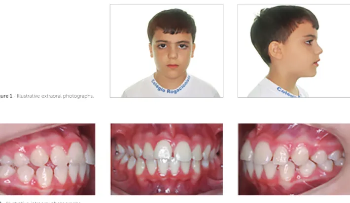
Standardized extraoral and intraoral photographs were taken for the subjective facial and occlusal analysis. The extraoral photographs consisted in the frontal and proile views, and the intraoral set consisted of a frontal, right and let side views (Figs 1 and 2).
Figure 1 - Illustrative extraoral photographs.
Figure 2 - Illustrative intraoral photographs.
Facial and occlusal classifications
The subjective facial evaluation was performed by a calibrated evaluator who classiied the subjects into ive groups according to their facial features: Pattern I, II, III; Long or Short Face facial patterns. This classii-cation is subjective and based on the analysis of various facial features, which are described in detail in related works.13,15,16,17 In brief, Facial Pattern I has harmonious
facial growth in the sagittal and vertical direction, good relationship between maxilla and mandible and propor-tionality between the facial thirds. This Pattern show as particularities: facial symmetry, good zygomatic projec-tion, pleasant nasolabial angle, passive lip sealing or dis-crete interlabial space, well deined chin-neck line and angle. Facial Pattern II has a convex proile with either maxillary excess or mandibular deiciency, or even a combination of both. Facial Pattern III presents a lat to concave facial proile, resulting from maxillary deicien-cy, mandibular excess or a combination of both. Verti-cally, the Long Face Pattern shows a vertical excess of the lower facial third combined or not with a decreased middle third and an active lip contact. Contrarily, the Short Face Pattern shows a decreased lower facial third, with lip compression at rest. In this classiication, the vertical Pattern (Long or Short Face) is always leading
on the sagittal. For example, if the patient has features of Pattern III and also Long or Short Face, it is classiied as a Long or Short Face. The Facial Pattern classiica-tion was separately performed on the frontal and proile views, which ensures characteristics of diferent
sever-ity.13,14,15 An Asymmetry Pattern was also incorporated
in the classiication, qualitatively and subjectively evalu-ated on frontal view, for the presence of visible man-dibular laterognathism.
The subjects’ anteroposterior occlusion was also classiied into Classes I, II or III according to Angle’s classiication.1 One researcher performed this
and characterized by the presence of negative overbite observed in intraoral frontal view photograph — except for cases in which permanent incisors were in active eruption process.
Intraexaminer agreement
The examiner was submitted to a Kappa test in two different periods, after a 30-day interval. In the first evaluation, 28 patients were examined in both frontal and profile views, and repeated after 30 days.
Statistical analyses
Concordance between the facial subjective classi-fication in the frontal and profile views, as well as be-tween the facial profile classification and sagittal den-tal relationship was investigated with Kappa statistics. Association between anterior open bite and Long Face Pattern was evaluated with Fisher’s exact test and the level of statistical signiicance was set at 5%. All statistical analy-ses were performed with Statistical Package for Social Sci-ences (SPSS), version 5.0 (SPSS Inc., Surrey, UK).
RESULTS
The intra-examiner concordance was analyzed by the Kappa test, showing to be moderate and sub-stantial depending on the view (0.60 for the frontal view and 0.74 for the profile view).
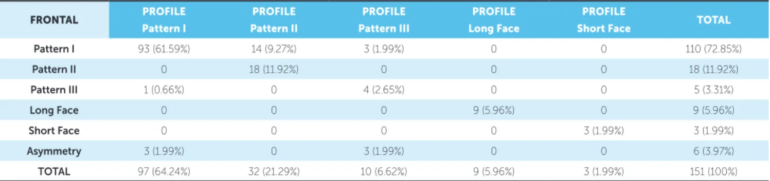
Table 1 shows the occurrence of different Facial Patterns according to the frontal and profile analy-ses, respectively. Pattern I individuals were the ma-jority in both classifications (frontal = 72.85% and profile = 64.24%). In the profile evaluation, although Facial Pattern I was also prevalent, the percentage of Facial Pattern II showed an increase when compared with the frontal view, with a reduction in the per-centage of Pattern I. Moreover, Kappa test showed a substantial strength of agreement (0.69) between frontal and profile analyses. When analyzing the rela-tion between the frontal and profile views, there is a tendency of the score between them being the same, i.e., 127 out of 145 patients presented the same Facial Pattern classification. This tendency cannot be deter-mined in the Asymmetry Facial Pattern because it is not possible to be evaluated in the profile view.
In the sagittal analysis, an agreement of 63.31% be-tween the classiication of Facial Pattern (I, II and III) and teeth relationship (Class I, II and III) was veriied, i.e., agreement occurred in most of the cases, but it was not mandatory (Table 2). Besides, Kappa test showed only a slight strength of agreement (0.27).
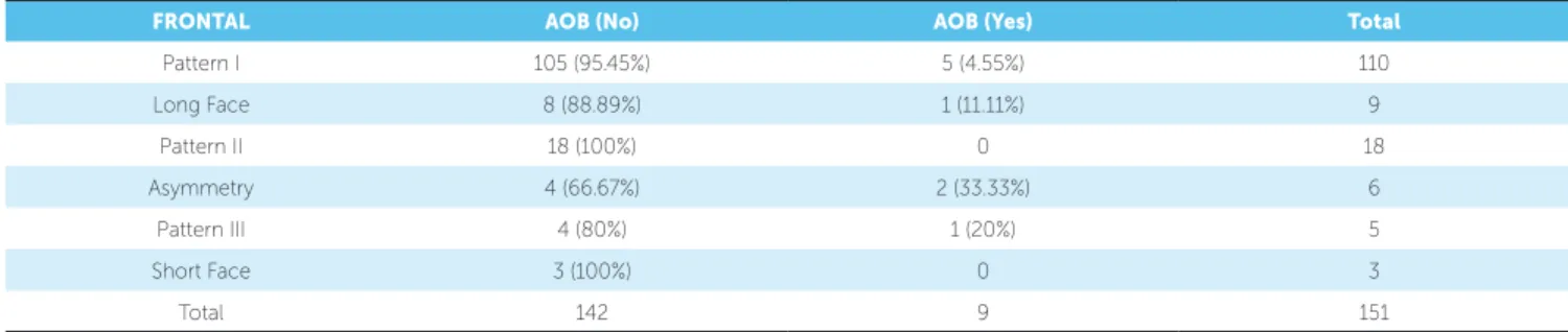
Table 3 shows the prevalence of anterior open bite between diferent Facial Patterns on the frontal analysis, demonstrating that the Long Face Pattern showed no more anterior open bite than the others (p = 0.501).
FRONTAL PROFILE
Pattern I
PROFILE Pattern II
PROFILE Pattern III
PROFILE Long Face
PROFILE
Short Face TOTAL
Pattern I 93 (61.59%) 14 (9.27%) 3 (1.99%) 0 0 110 (72.85%)
Pattern II 0 18 (11.92%) 0 0 0 18 (11.92%)
Pattern III 1 (0.66%) 0 4 (2.65%) 0 0 5 (3.31%)
Long Face 0 0 0 9 (5.96%) 0 9 (5.96%)
Short Face 0 0 0 0 3 (1.99%) 3 (1.99%)
Asymmetry 3 (1.99%) 0 3 (1.99%) 0 0 6 (3.97%)
Table 2 - Agreement percentage between sagittal dental relationship (Angle classification) and the Facial Pattern.
Table 3 - Prevalence of anterior open bite (AOB) among different Facial Patterns in the frontal analysis.
PROFILE Class I Class II Class III Total
Pattern I 66 (47.48%) 24 (17.27%) 7 (5.04%) 97 (69.78%)
Pattern II 12 (8.63%) 18 (12.95%) 2 (1.44%) 32 (23.02%)
Pattern III 6 (4.32%) 0 4 (2.88%) 10 (7.19%)
Total 90 (64.75%) 36 (25.9%) 13 (9.35%) 139 (100%)
FRONTAL AOB (No) AOB (Yes) Total
Pattern I 105 (95.45%) 5 (4.55%) 110
Long Face 8 (88.89%) 1 (11.11%) 9
Pattern II 18 (100%) 0 18
Asymmetry 4 (66.67%) 2 (33.33%) 6
Pattern III 4 (80%) 1 (20%) 5
Short Face 3 (100%) 0 3
Total 142 9 151
DISCUSSION
Facial analysis performed unconsciously and dai-ly by ordinary people, directdai-ly influences perception of the characteristics of people we interact with, and is notoriously influenced by the occlusal character-istics and vice versa.17 It is known that the ratings
of attractiveness, intelligence, conscientiousness, pleasantness and acceptance differed significantly depending on the occlusal status depicted. Persons with normal occlusion were rated as most attractive, intelligent, agreeable and extraverted, whereas per-sons with prognathism were rated as least attractive, intelligent, and extraverted.18 Furthermore, persons
with ideal smiles are considered more intelligent and have a greater chance of inding a job when compared with persons with non-ideal smiles.19 These points
highlight the important of facial appearance in orth-odontic diagnosis and planning, showing that obtaining good occlusal outcomes, regardless of the facial damage, is not the best way today. The search for the appropriate diagnosis and treatment for the patient’s Facial Pattern seems to be the best choice, specially when the patient’s complaint is the face. Therefore, subjective facial analy-sis is a diagnostic tool which has had its importance in-creased for being the parameter by which patients and
the people they live with will evaluate the treatment results.17 Besides, organizing orthodontic diagnosis
ac-cording to the Facial Patterns allows orthodontists to treat malocclusions based on the location of skeletal dis-crepancies—if they are present—, or the etiology of the malocclusion, establishing treatment protocols that are tailored speciically to each Pattern in each age group, with short-term protocols and predictable long-term prospects by taking into account discrepancy severity.14
Facial analysis can be performed in several ways, in-cluding the use of angular or linear measurements,20 but
these methods cause signiicant errors when trying to tailor each individual to population average standards.7
It is known that diagnoses performed from Angle clas-siication and cephalometric references lead to results which are not always compatible with the patient’s es-thetic expectations.21 Combined with the establishment
of ideal occlusal relationship, the best esthetic as possible must be pursued.22 For that, diagnosis must be primarily
based on the frontal and proile facial morphology, on the smile assessment and complemented by occlusal evalua-tion, whose discrepancy commonly is a consequence of skeletal error.14 Radiographs are also an important tool,
To show that subjective facial analyses are reproduc-ible, a previous publication applied statistical tests to eval-uate the agreement, evidencing that agreement between raters and a gold standard was moderate, with raters ex-hibiting greater agreement between them (Kappa = 0.85) than with the gold standard (0.48). So, with training and experience, the subjective and qualitative facial analysis can be efectively and individually used for each patient.14
In this research, intra-examiner agreement was also ana-lyzed by the Kappa test, showing to be moderate and sub-stantial depending on the view (0.60 for the frontal view and 0.74 for the proile view), similarly to the literature. It is important to point out that the evaluator that classiied Facial Patterns in this research was the same called as gold standard in previous publication, and who was the creator of Facial Pattern classiication.14
A substantial agreement of approximately 84% be-tween frontal and proile classiication of Facial Pattern was observed (Table 1). Among discordant cases, most were considered Pattern I in the frontal analysis and Pat-tern II in proile assessment. This fact occurs because changes in Pattern II are especially sagittal and essentially viewed in proile view. So, if these changes are subtle, they will probably not be identiied in the frontal analysis. This explains the diferences between the proportions of Patterns I and II found in the frontal and proile
analy-ses.13,15 When an individual is classiied as Pattern I in the
frontal view and as Pattern II or III in the proile view, this means that the discrepancy was not severe enough to compromise the front view, thus, treatment prognosis may be better. If an individual is diagnosed as Pattern II in the proile analysis and Pattern I in the frontal analysis, this individual should be approached as a Pattern II.13,15
The Facial Patterns frequencies in children in de-ciduous dentition has already been demonstrated for Pattern I (63.22%), Pattern II (33.10%) and Pat-tern III (3.68%).24 In this investigation, in mixed and
initial permanent dentitions, Pattern I has been ob-served in 64.24%, Pattern II in 21.29%, Pattern III in 6.62%, Long Face Pattern in 5.96% and Short Facial Pattern in 1.99%. The Short and Long Face Patterns have not been evaluated by researchers in the deciduous dentition because, in this phase of craniofacial growth and development, they are not well deined. Compar-ing the results, only small diferences on Facial Patterns distributions between deciduous and mixed/permanent dentitions can be observed. These data reinforce the fact
that facial morphology is early deined and kept along the growth.25 The literature conirms that, by showing
that Pattern II is present from deciduous dentition and mandibular growth and does not improve facial skel-etal relationship along the evolution of the deciduous, mixed and permanent dentitions.26-29
Agreement between Angle classiication and Facial Pattern was seen in approximately 63% of cases (Ta-ble 2). The percentage of concordance was relevant, however the kappa test showed only slight force. Al-though less marked, natural dentoalveolar compensa-tion also exists in the sagittal plane, where individuals with facial Patterns II or III may present Class I den-tal relationships consequent to denden-tal compensations. Moreover, occlusal problems with dentoalveolar origin may lead to Class II or III dental relationships in patients with facial Pattern I. In these, the treatment prognosis is better because there are no skeletal discrepancies.13
In general, dental positioning is a consequence of skeletal error which characterizes the malocclusion. This correlation foresees the dental problems which dif-ferent types of Patterns will tend to present. To make this evaluation precociously, understanding how the growth is going to occur implies in deining real and coherent possibilities of treatment, with a more realis-tic and stable prognosis. In general, Angle classiication tends to relect sagittal behavior of the facial skeleton in all Facial Patterns. The only available research about this relation reveals a tendency of Class following the Facial Pattern from the deciduous dentition, which was more evident in Pattern II. In Pattern I, Class I prevailed (62.99%), followed by Class II (35.82%) and Class III (1.18%). In Pattern II, Class II prevailed (81.35%) fol-lowed by a low incidence of Class I (18.64%). In Pat-tern III, Class III was present in 50% of the children, followed by Class I in 48.64%, and Class II in 1.35%.30
observed in Pattern II, where the occurrence of dental relation of Class II decreased in the mixed and perma-nent dentitions in relation to what was described for the deciduous dentition. Possible explanations for these small diferences among the researches are the refer-ence for the Class classiication (in deciduous dentition, authors used the deciduous canine and, in mixed and permanent dentition, the permanent molars are used), the adjustment of molar’s relation with the major in-ferior Leeway space and the diferential mandibular growth in adolescence.31 An interesting suggestion for
future research is to investigate the Facial Pattern and its relationship with Angle classiication separately in the mixed and permanent dentitions, in order to point out possible diferences, which cannot be performed in this sample due to the restrict number of participants.
The anterior open bite (AOB) was not signiicantly prevalent in any Facial Pattern (Table 3). A possible ex-planation is that the children evaluated in this research were mostly in the mixed dentition, and the main etio-logical factors of anterior open bite at this stage are the oral habits, causing dentoalveolar changes, regardless of the Facial Pattern. Oral habits have a high frequency in children and deleterious habits most frequently as-sociated are paciier and thumb sucking, and tongue thrust.32,33 Other explanation is the high potential for
dentoalveolar vertical compensation, evidenced by ex-cessive gingival tissue exposure without anterior open bite, characterizing the gingival smile.34
CONCLUSIONS
1. Facial Patterns I (64.24%) and II (21.29%) were the most prevalent in children, followed by Pattern III (6.62%), Long Face Pattern (5.96%) and Short Face Pattern (1.99%).
2. Agreement between proile and frontal subjective facial analysis was substantial. The divergences seem to be related to patients with slight sagittal skeletal error that are preferably identiied in proile rather than in frontal analysis.
3. There was slight concordance between Facial Pat-tern and sagittal dental relationships.
4. The anterior open bite was not signiicantly prev-alent in any Facial Pattern.
Authors contribution
1. Angle EH. Classification of malocclusion. Dent Cosmos. 1899;41(3):248-64.
2. Holdaway RA. A soft-tissue cephalometric analysis and its use in orthodontic treatment planning. Part I. Am J Orthod. 1983 July;84(1):1-28.
3. Holdaway RA. A soft-tissue cephalometric analysis and its use
in orthodontic treatment planning. Part II. Am J Orthod. 1984 Apr;85(4):279-93.
4. Alvarez AT. The A line: a new guide for diagnosis and treatment planning. J Clin Orthod. 2001 Sept;35(9):556-69.
5. Lavelle CL. Facial proiles in diferent occlusal categories. J Dent. 1973 Feb;1(3):141-6.
6. Bass NM. The aesthetic analysis of the face. Eur J Orthod. 1991 Oct;13(5):343-50.
7. Ackerman JL, Proit WR, Sarver DM. The emerging soft tissue paradigm
in orthodontic diagnosis and treatment planning. Clin Orthod Res. 1999 May;2(2):49-52.
8. Bittner C, Pancherz H. Facial morphology and malocclusions. Am J
Orthod Dentofacial Orthop. 1990 Apr;97(4):308-15.
9. Ferrario VF, Sforza C, Puleo A, Poggio CE, Schmitz JH. Three-dimensional
facial morphometry and conventional cephalometrics: a correlation study. Int J Adult Orthodon Orthognath Surg. 1996;11(4):329-38. 10. Dimaggio FR, Ciusa V, Sforza C, Ferrario VF. Photographic soft-tissue
proile analysis in children at 6 years of age. Am J Orthod Dentofacial Orthop. 2007 Oct;132(4):475-80.
11. Kasai K. Soft tissue adaptability to hard tissues in facial proiles. Am J Orthod Dentofacial Orthop. 1998 June;113(6):674-84.
12. Godt A, Bechtold TE, Schaupp E, Zeyher C, Koos B, Baas E et al. Correlation between occlusal abnormalities and parameters investigated by three-dimensional facial photography. Angle Orthod. 2013
Sept;83(5):782-9.
13. Capelozza Filho L. Diagnóstico en Ortodoncia. Maringá: Dental Press; 2005.
14. Reis SAB, Abrão J, Claro CAA, Fornazari RF, Capelozza Filho L. Agreement among orthodontists regarding facial pattern diagnosis. Dental Press J Orthod. 2011 July-Aug;16(4):60-72.
15. Reis SAB, Abrão J, Capelozza Filho L, Claro CAA. Estudo comparativo do peril facial de indivíduos Padrões I, II e III portadores de selamento labial passivo. Rev Dental Press Ortod Ortop Facial. 2006 Jul-Ago;11(4):36-45. 16. Cardoso MA, Bertoz FA, Capelozza Filho L, Reis SA. Características
cefalométricas do padrão face longa. Rev Dental Press Ortod Ortop Facial. 2005 Mar-Apr;10(2):29-43.
17. Reis SAB, Abrão J, Capelozza Filho L, Claro CAA. Análise facial subjetiva. Rev Dental Press Ortod Ortop Facial. 2006 Set-Out;11(5):159-72. 18. Olsen JA, Inglehart MR. Malocclusions and perceptions of attractiveness,
intelligence, and personality, and behavioral intentions. Am J Orthod Dentofacial Orthop. 2011 Nov;140(5):669-79.
REFERENCES
19. Pithon MM, Nascimento CC, Barbosa GC, Coqueiro Rda S. Do dental esthetics have any inluence on inding a job? Am J Orthod Dentofacial Orthop. 2014 Oct;146(4):423-9.
20. Wen YF, Wong HM, McGrath CP. Longitudinal photogrammetric analysis of soft tissue facial changes: a systematic review of the literature and a summary of recommendations. J Craniofac Surg. 2015 July;26(6):1830-4. 21. Michiels G, Sather AH. Validity and reliability of facial proile evaluation in
vertical and horizontal dimensions from lateral cephalograms and lateral photographs. Int J Adult Orthodon Orthognath Surg. 1994;9(1):43-54. 22. Sarver DM, Ackerman JL. Orthodontics about face: the re-emergence
of the esthetic paradigm. Am J Orthod Dentofacial Orthop. 2000 May;117(5):575-6.
23. Atchison KA, Luke LS, White SC. Contribution of pretreatment radiographs to orthodontists’ decision making. Oral Surg Oral Med Oral Pathol. 1991 Feb;71(2):238-45.
24. Silva Filho OG, Herkrath FJ, Queiroz APC, Aiello CA. Padrão facial na dentadura decídua: estudo epidemiológico. R Dental Press Ortodon Ortop Facial. 2008 Jul-Ago;13(4):45-59.
25. Silva Filho OG, Bertoz FA, Capelozza Filho L, Almada EC. Crescimento facial espontâneo Padrão II: estudo cefalométrico longitudinal. Rev Dental Press Ortod Ortop Facial. 2009 Jan-Feb;14(1):40-60. 26. Antonini A, Marinelli A, Baroni G, Franchi L, Defraia E. Class II
malocclusion with maxillary protrusion from the deciduous through the mixed dentition: a longitudinal study. Angle Orthod. 2005 Nov;75(6):980-6.
27. Baccetti T, Franchi L, McNamara JA Jr, Tollaro I. Early dentofacial features of Class II malocclusion: a longitudinal study from the deciduous through the mixed dentition. Am J Orthod Dentofacial Orthop. 1997 May;111(5):502-9.
28. Bishara SE. Mandibular changes in persons with untreated and treated Class II division 1 malocclusion. Am J Orthod Dentofacial Orthop. 1998 June;113(6):661-73.
29. Kerr WJ, Hirst D. Craniofacial characteristics of subjects with normal and postnormal occlusions—a longitudinal study. Am J Orthod Dentofacial Orthop. 1987 Sept;92(3):207-12.
30. Silva Filho OG, Queiroz APC, Herkrath FJ, Silva GFB. Correlation between facial pattern and sagittal relationship between dental arches in deciduous dentition: epidemiological considerations. Rev Dental Press Ortod Ortop Facial. 2008 Jan-Feb;13(1):101-12.
31. Bishara SE, Jamison JE, Peterson LC, DeKock WH. Longitudinal changes in standing height and mandibular parameters between the ages of 8 and 17 years. Am J Orthod. 1981 Aug;80(2):115-35.