BASIC RESEARCH
Thermographic evaluation of hind paw skin
temperature and functional recovery of locomotion
after sciatic nerve crush in rats
Viviane Z. Sacharuk,I,IIGisele A. Lovatel,I,IIJocemar Ilha,I,IISimone Marcuzzo,I,IIAlexandre Severo do Pinho,II Le´der L. Xavier,III Milton A. Zaro,I,IVMatilde AchavalI,II
IPrograma de Po´s-Graduac¸a˜o em Neurocieˆncias, Instituto de Cieˆncias Ba´sicas da Sau´de, Universidade Federal do Rio Grande do Sul, Porto Alegre, RS, Brazil.IILaborato´rio de Histofisiologia Comparada, Departamento de Cieˆncias Morfolo´gicas, Instituto de Cieˆncias Ba´sicas da Sau´de, Universidade Federal do Rio Grande do Sul, Porto Alegre, RS, Brazil.IIILaborato´rio de Biologia Celular e Tecidual, Faculdade de Biocieˆncias, Pontifı´cia Universidade Cato´lica do Rio Grande do Sul, Porto Alegre, RS, Brazil.IVInstituto Brasileiro de Tecnologia em Calc¸ado (IBTEC), Novo Hamburgo, Rio Grande do Sul, RS, Brazil.
INTRODUCTION: Peripheral nerves are often damaged by direct mechanical injury, diseases, and tumors. The peripheral nerve injuries that result from these conditions can lead to a partial or complete loss of motor, sensory, and autonomic functions, which in turn are related to changes in skin temperature, in the involved segments of the body. The aim of this study was to evaluate the changes in hind paw skin temperature after sciatic nerve crush in rats in an attempt to determine whether changes in skin temperature correlate with the functional recovery of locomotion.
METHODS:Wistarrats were divided into three groups: control (n = 7), sham (n = 25), and crush (n = 25). All groups were subjected to thermographic, functional, and histological assessments.
RESULTS:DT in the crush group was different from the control and sham groups at the 1st, 3rdand 7rdpostoperative days (p,0.05). The functional recovery from the crush group returned to normal values between the 3rdand 4th week post-injury, and morphological analysis of the nerve revealed incomplete regeneration at the 4thweek after
injury.
DISCUSSION:This study is the first demonstration that sciatic nerve crush in rats induces an increase in hind paw skin temperature and that skin temperature changes do not correlate closely with functional recovery
KEYWORDS: Thermography; Skin temperature; Nerve crush; Functional recovery.
Sacharuk V, Lovatel G, Ilha J, Marcuzzo S, Xavier L, Zaro M, Achaval M. Thermographic evaluation of hind paw skin temperature and functional recovery of locomotion after sciatic nerve crush in rats. Clinics. 2011;66(7):1259-1266.
Received for publication onApril 6, 2011;First review completed onApril 12, 2011;Accepted for publication onApril 12, 2011
E-mail: [email protected]
Tel.: 55 51 33083624
INTRODUCTION
Peripheral nerves are often damaged by direct mechanical injury, diseases, tumors or iatrogenic lesions.1 The most common etiologies of acute traumatic peripheral nerve injury include penetrating injury, crush, stretch, and ischemia.2The incidence of traumatic injuries in the USA is estimated at .500,000 new patients annually.1 Nerve injury can have a profound impact on patient life, and the cost to society can be felt not only in direct health sector expenses but also in the loss of production.3Many human and animal studies have shown that peripheral nerve injuries can be accompanied by skin temperature variations in the region innervated by the injured nerve.4-12 Skin
temperature is determined by the diameter of skin blood vessels, which is controlled neurologically by two opposing mechanisms: the sympathetic system, which exerts vaso-constrictor effects, and antidromic stimulation, which is responsible for vasodilator effects. Thus, under pathological circumstances, variations in skin temperature may occur either due to changes in the activity of the sympathetic nervous system or due to altered antidromic stimulation resulting from neurogenic inflammation.10,13
Although animal models using sciatic nerve chronic constriction have shown variations in skin temperature after nerve injury,9,10,14 there are no reports in the literature regarding the effects of nerve crush injury on skin tempera-ture. Thermography is a diagnostic tool that can assess normal and abnormal functioning of the sensory and sympathetic nervous system. Variations in skin temperature are easily documented using thermography, a highly sensitive non-invasive technique capable of detecting alterations in skin surface temperature.4-6,9-12,15-19,21,22 Measurement of skin Copyrightß2011CLINICS– This is an Open Access article distributed under
temperature using thermography could be of use in following the temporal evolution of nerve injuries because it allows clinical quantification of peripheral nerve injuries,5,6 facilitat-ing evaluation, diagnosis, and follow-up treatment.
This study investigated the effects of sciatic nerve injury on hind paw skin temperature by temporal analysis of skin temperature. Temporal analyses of hind paw skin tempera-ture were made using infrared thermography on different days after lesion formation; the findings correlated with functional recovery indices (sciatic functional index and toe spread index). To verify whether the recovery was only functional or also morphological, histological analyses were also performed.
MATERIALS AND METHODS
Subjects
The experiment was performed on 57 3-month-old male Wistar rats, weighing 265–400 g (initial age and weight), obtained from a local breeding colony (ICBS, Universidade Federal do Rio Grande do Sul, Brazil). The rats were housed in standard plexiglass boxes, with a 12512 h light/dark cycle,
at a controlled temperature (23¡1
˚
C). Food and water wereavailable ad libitum. All animals were cared for in accordance with Brazilian law and in compliance with the National Institute of Health’s Guidelines for Care and Use of Laboratory Animals (publication no. 85-23, revised 1985). This study was approved by the Ethics Committee of the
Universidade Federal do Rio Grande do Sul, RS – Brazil.
Experimental Protocol
The animals were randomly divided into three groups: (1) control rats that did not undergo a surgical procedure (control, n = 7), (2) rats subjected to a sham surgical procedure without sciatic nerve crush (SHAM, n = 25), and (3) rats subjected to a surgical procedure with sciatic nerve crush (CRUSH, n = 25). For surgical procedures, animals were anesthetized using ketamine and xylazine (90 and 15 mg/Kg, respectively, i.p.), and the right sciatic nerve was exposed through a skin incision extending from the greater trochanter to the mid-thigh, followed by splitting the overlying gluteal muscle. The nerve crush injury was performed with 1-mm hemostatic forceps for 30 seconds, as previously described by Bridge et al.56 The muscle and skin were then closed with 4-0 nylon sutures, and the animals were returned to their cages.
Assessment of Skin Temperature
Hind paw plantar surface skin temperature measure-ments were obtained using an infrared video camera (ELECTROPHYSICS - PV320T, USA) placed at an angle of approximately 90
˚
from the surface, 25 cm from the plantar region of the hindlimb and 75 cm above the floor. This camera has a thermal sensitivity of approximately 0.08˚
C, with an error of 2˚
C. The images were digitally recorded at 30-second intervals. The thermographic procedures were conducted in a room at a constant temperature of 23˚
C (¡1˚
C) and humidity between 30 and 50% (Thermo/hydro/clock, MT-230, Minipa, Brazil). Surface temperature was measured from 9:00 to 12:00 a.m., after allowing each animal at least one hour to acclimate to the ambient temperature. Animals were lightly anesthetized with sodium pentobarbi-tal (30-40 mg/kg, i.p., Crista´lia, Brazil) to suppress the righting reflex while preserving the withdrawal reflex and
were returned to their Plexiglass boxes. Anesthetized rats were gently placed in a prone position using a body-shaped depression, and the dorsal surface of the hind paw was fixed with adhesive tape. The hind paws were positioned, and the heat emitted from the plantar region was measured using an infrared detector (Figure 1).
Temperature maps were obtained before the surgical procedure and 1, 3, 7, 14, 21, and 28 days following the nerve crush injury. Body core temperature was measured using a digital thermometer inserted 2 cm into the rectum prior to and immediately after infrared thermographic measurements. The animals were then returned to their Plexiglass boxes. Infrared images were evaluated using analytical software (Velocity, USA). The area of interest was the plantar region of the hindlimb that corresponds to the dermatomes of the sciatic nerve.16
region of the contralateral side (left). Differences greater than 0.6
˚
C from the mean temperature within the analyzed area (spots) were considered abnormal or asymmetrical, while differences less than 0.6˚
C were considered normal or symmetrical. TheDT is shown in˚
C. If the value ofDT was positive, the experimental side was warmer than the contralateral side. IfDT was negative, the experimental side was colder than the contralateral side.Assessment of Functional Recovery
All animals were subjected to a series of motor activity assessments 7, 14, 21, and 28 days after lesion. Recovery of locomotor activity, as determined by the sciatic functional index (SFI)47 and the toe spread index (TSI),48 was considered to be proof of adequate re-innervation of the right hindlimb.
Both indices were based on measurements of the footprints of walking rats, which provide a reliable and easily quantifiable method for evaluating the functional condition of the sciatic nerve. For this test, rats were trained to walk on a white sheet of paper covering the bottom of a 100-cm-long, 8.5-cm-wide track ending in a dark box. Afterwards, the animals had the ventral portion of their hind feet painted with dark dye and were placed back on the track to walk.
The rats’ footprints were used to determine the following measurements: (1) the distance from the heel to the third toe, the print length (PL); (2) the distance from the first to the fifth toe, the toe spread (TS); and (3) the distance from the second to the fourth toe, the intermediary toe spread (ITS). For TSI, only parameter (2), TS, was measured. All measurements were obtained for both the experimental (E) and normal (N) sides.47Several prints from each foot were obtained on each track, but only three prints were used to determine the mean measurements in the E and N sides.
The SFI and TSI were calculated according to the equations below:
1) SFI = -38.3 (EPL – NPL)/NPL+109.5 (ETS – NTS)/NTS +13.3 (EIT – NIT)/NIT – 8.8.43
2) TSI = (ETS – NTS)/NTS46
In the SFI, an score of zero (¡11) represents normal
function, and an index of approximately –100 indicates total impairment.49In the TSI, normal function is represented by an index of zero.
Histological Assessment
After 28 days post-lesion, animals were anesthetized with sodium thiopental (50 mg/kg, i.p.; Crista´lia, Brazil) and transcardially perfused using a peristaltic pump (Milan, Brazil) with 300 ml of saline solution, followed by 400 ml of
0.5% glutaraldehyde (Merck, Germany) and 4% parafor-maldehyde (Reagen, Brazil) in 0.1 M phosphate buffer (PB, pH 7.4) at room temperature. Afterwards, for nerve regeneration analysis, short segments (,3 mm) of the right sciatic nerve were rapidly excised 5 mm under the distal portion of the nerve. The specimens were maintained in the fixative solution described above for one hour at room temperature and at 4
˚
C until processed. They were then post-fixed in 1% OsO4(Sigma Chemicals Co., USA) in PB, dehydrated in a graded series of alcohol and propylene oxide (Electron Microscopy Science, USA), embedded in resin (Durcupan, ACM-Fluka, Switzerland), and polymer-ized at 60˚
C (Ilha et al., 2006). Transverse semi-thin sections (1mm) were obtained using an ultramicrotome (MT 6000-XL, RMC, Tucson, USA) and stained with 1% toluidine blue (Merck, Germany) in 1% sodium tetraborate (Ecibra, Brazil). Afterwards, images of the nerve were captured and digitized (100% in oil immersion) using a Nikon Eclipse E-600 microscope (Tokyo, Japan) coupled to a digital camera for morphological analysis.Statistical analysis
The DT data were examined using the Kruskal-Wallis Test, and the temporal evolution of DT in each group was analyzed using the Friedman Test. The SFI and TSI were analyzed using analysis of variance (ANOVA) for repeated measures followed by the Bonferroni test. Statistical analysis of the data was performed using SPSSH 11.5 (Statistical Package for the Social Sciences, Inc., Chicago, Illinois) with significance set atp#0.05. All data are shown as mean¡
standard error of the mean (SEM).
RESULTS
Skin Temperature
The baseline and post-injury measurements obtained using infrared thermography are shown in Table 1. Given that in our study, only temperature differences between the paws equal to or in excess of 0.6
˚
C (DT$0.6˚
C) were considered asymmetric, thermography showed there was no significantDT in the hind paws between the groups prior to the surgical procedure. In this evaluation, the DT was less than 0.6
˚
C (calculated as the skin temperature on the right side minus that on the left side) (p,0.05; Table 1). However, theDT in the crush group was significantly different from those in the control and sham groups on the 1st, 3rd, and 7thpostoperative days after sciatic nerve crush (p,0.05; Table 1). TheDT of the crush group was greater than 0.6˚
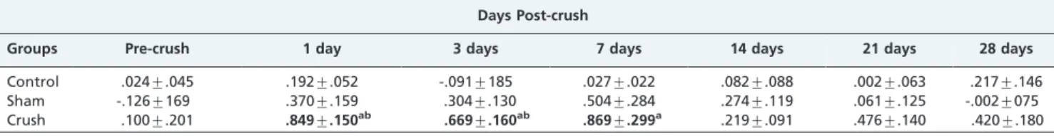
C (p,0.05; Table 1), and the experimental side was abnormally warmer than the control side (Figure 1). It is noteworthy that no differences wereTable 1 -Skin temperature differences (DT) between the experimental (right) and normal (left) sides at various time points following injury. TheDT is shown in
˚
C.Days Post-crush
Groups Pre-crush 1 day 3 days 7 days 14 days 21 days 28 days
Control .024¡.045 .192¡.052 -.091¡185 .027¡.022 .082¡.088 .002¡.063 .217¡.146
Sham -.126¡169 .370¡.159 .304¡.130 .504¡.284 .274¡.119 .061¡.125 -.002¡075
Crush .100¡.201 .849¡.150ab .669
¡.160ab .869
¡.299a .219
¡.091 .476¡.140 .420¡.180
found between the control and sham groups throughout the postoperative period.
When analyzing the temporal evolution ofDT in the crush group, we found a significant temperature difference between hind paws through the 7thpostoperative day after the crush procedure (p,0.05 for Friedman test). This difference was not observed in the control or sham groups (p.0.05 for Friedman test).
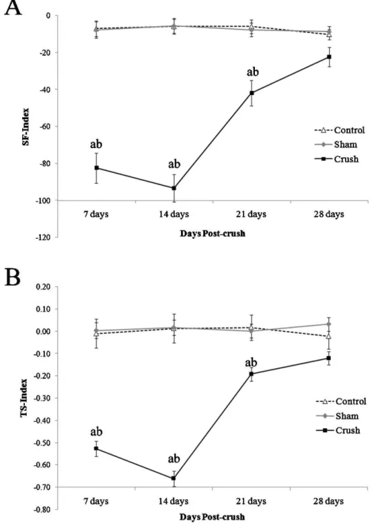
Functional Recovery
The SFI and TSI were measured on postoperative days 7, 14, 21, and 28, and the data are presented in Figure 2 as the means¡SEM.
In the control and sham groups, the SFI and TSI were considered normal throughout the experiment, with the SFI being approximately -10 and the TSI approximately zero. No differences were found between the non-operated
control and sham-operated groups following the survival period after surgery.
In the crush group, the SFI and TSI values on post-operative-days 7, 14, and 21 were significantly lower than those observed in the control and sham groups (p,0.05; Figure 2). This result indicated a loss of motor function in the right hindlimb in all animals that received sciatic crush, a result that confirms that the sciatic nerve crush was fully effective.
On postoperative day 28, no statistical differences were found between the groups in either the SFI or the TSI, indicating a nearly complete restoration of hind limb motor function.
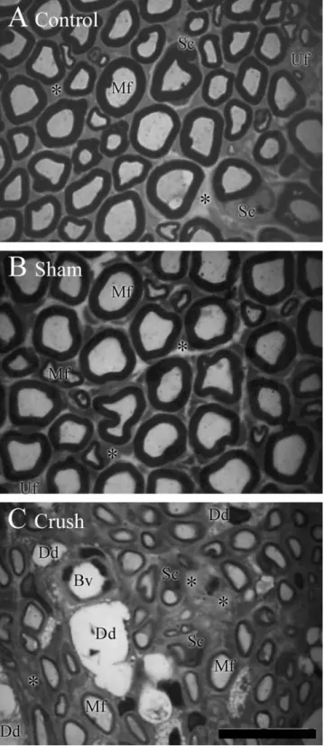
Histology
Histological sections were qualitatively analyzed at the distal portion of the sciatic nerve. The histological appear-ance of the sciatic nerve was essentially normal for the sham and control groups. These groups exhibited a similar distribution of small- and large-diameter myelinated and unmyelinated nerve fibers and Schwann cells, each sur-rounded by a well-defined endoneurium, and regular proportions between myelin sheath thickness and fiber diameter (Figure 3). However, in the crush group, histolo-gical analysis reveals an apparent unimodal fiber spectrum with the predominance of small-diameter thin myelin sheath fibers, a reduction in size, and an alteration in the shape of nerve fibers, an increase of endoneurium between the nerve fibers and the presence of degeneration debris (Figure 3).
DISCUSSION
This study investigated the effects of sciatic nerve crush on hind paw skin surface temperature. The findings were compared with functional recovery indices (SFI and TST). To verify whether the recovery was only functional or also morphological, histological analyses were also performed. We used thermography to examine skin temperature in three groups of animals: 1) the control group, 2) the sham group, and 3) the crush group.
In a classical study from Ochoa and Bennet,9 in normal animals, the observedDT values were distributed symme-trically around a mean of approximately zero (0.03¡0.34
˚
C),and a DT equal to or greater than 0.8
˚
C was classified as asymmetrical. In our study, a DT was considered signifi-cantly asymmetrical when it was equal to or greater than 0.6˚
C. This choice of cutoff point is based on the fact that this value represents 1.99 standard deviations from the mean found in our control group (0.06¡0.27˚
C). The Ochoa studyused a similar principle to define the temperature limits of normal and abnormalDT values.
In our results, we observed that the control and sham groups exhibited DT values less than 0.6
˚
C with no statistical differences between the groups. Moreover, our findings show that after sciatic nerve crush, the DT of plantar surface skin was greater than 0.6˚
C on postoperative days 1, 3, and 7. The affected side was warmer on the 7th postoperative day when compared with the contralateral Figure 3 - Digitized images of transverse semi-thin sections(1mm) obtained from regenerating sciatic nerves in control (A), sham (B) and crush (C) groups. Note the similarity and apparent bimodal fiber spectrum, the large and small myelinated fibers and the scanty endoneurial space between the myelinated fibers when clusters of unmyelinated fibers are present in the control and sham groups. Moreover, note that the crush group shows an apparent unimodal fiber spectrum with a predomi-nance of small-diameter thin myelin sheath fibers and an increased size of the endoneurium between the nerve fibers and the presence of degeneration debris in this group. Mf indicates myelinated nerve fiber; Uf, unmyelinated nerve fiber;
side. Thermography demonstrated that sciatic nerve crush is sufficient to change hind paw skin temperature.
Several experimental studies of peripheral nerve injuries have identified variations in skin temperature after nerve injury, but this study is one of the first detailed reports demonstrating that sciatic nerve crush induces a variation in the skin temperature of the plantar region of the hind paw.4-12,23
Thermographic studies have described two thermal patterns in which the affected side was either warmer or colder than the contralateral side. Similar to our results, previous studies, using chronic constriction9,10 and com-plete transection11 of the sciatic nerve and thermography, reported that the skin temperature on the experimental side was warmer during the first week. One study showed that immediately following sciatic transection, there was a considerable increase (5.1
˚
C) in plantar hind paw tempera-ture in relation to the contralateral side, which was maintained for 42 days.11In contrast to our results obtained using sciatic nerve crush, in the chronic constriction model,9,10 the skin temperature of the experimental side became progressively colder than the contralateral side after the first week following nerve injury. This difference between the findings presented in our study and previous studies may be due to the different experimental models used. The chronic constriction model is a more aggressive approach, with several important differences when compared to the crush model. While the crush model is characterized by increased nervous fiber regeneration,24both models exhibit decreased release of catecholamines, mainly adrenaline and noradre-naline, from postganglionic sympathetic vasoconstric-tor fibers10,12,25-28 in the first few days following injury.25,26,12,30,31 These changes in catecholamine release can explain the warming in the experimental side during the first week with both the crush and chronic constriction of the sciatic nerve.
Thus, our results are similar to and may be explained, at least in part, by those reported by Werdehausen and cols (2007), who showed that sciatic nerve blockage in humans is responsible for increases in skin temperature within the course of superficial veins in the big toe and above the ankles. This study also showed that skin warming is not related to the segmental or neural innervation pattern.29
This nerve blockade, and consequent, sympathetic blockade are responsible for a reduced adrenaline and noradrenaline release in the sympathetic vasoconstrictor fibers,10,12,25-29 corroborating our hypothesis. In our study, increases in temperature were observed throughout the plantar region of the hind paw after sciatic nerve crush. While there may have been a more pronounced temperature increase in the big toe or in the toes, unfortunately, the analytical spots used in our study were insufficiently small to focus on a single toe.
Another mechanism that may explain the warming of the skin on the experimental side observed in this study could be the antidromic activation of type-C cutaneous nociceptor nerve terminals. This activation causes a release of vasoactive neuropeptides, such as calcitonin gene-related peptide (CGRP) and substance P (SP). These peptides reach arterioles by diffusion and trigger a chemical cascade that acts directly on vascular smooth muscle, culminating in the dilatation of arterioles9,10,13,15,17,35-41and cutaneous vasodi-latation, inducing an increase in local temperature.39,40
Nevertheless, the rat foot receives cutaneous sensory innervation from both the saphenous and sciatic nerves43. In our study, the saphenous nerve was left intact. After sciatic nerve injury, collateral sprouting of the saphenous nerve into the sciatic nerve territory occurs frequently,43,44which permits fibers containing SP and CGRP to extend anatomi-cally into adjacent glabrous skin.45,46 This may have influenced the skin temperature readings obtained in this study.
Functional recovery was evaluated by walking track analysis using the SFI47and TSI.48The SFI and TSI methods are commonly used to evaluate peripheral nerve regenera-tion51-58 and show sensory input, motor response and cortical integration, indicating that reduced cutaneous information of the plantar aspect of the hind paw leads to a significant functional deficit.51-53,56The SFI and TSI in the present study indicated significant hind paw impairment after sciatic crush which persisted until postoperative day 21. At postoperative day 28, both SFI and TSI showed no statistical differences from the baseline value, indicating almost complete restoration of paw function. These results are in accordance with a recent study by our group54,55and with previous authors48,56-60 that reported an almost complete restoration of hindlimb function by the 3rdor 4th week after injury.
Peripheral nerve recovery was evaluated by qualitative histological examinations of the distal portion of the sciatic nerve. Histological analysis is commonly used to evaluate recovery from nerve injuries.47,54-56,61In this study, analysis of histological sections from the distal portion of the sciatic nerve indicated that sciatic nerve crush produced morpho-logical changes characterized by an apparent predominance of small-diameter thin myelin sheath fibers and an increase in endoneurial space. The patterns observed in our histological qualitative analysis are very similar to those observed in other studies performed in our laboratory,54,55 in which detailed histological qualitative analysis showed there was no recovery in the morphological patterns of sciatic nerve in crush-injured animals when compared to control or sham groups until five weeks54 and 13 weeks post-injury.55 Additionally, other studies have shown similar changes in nerve morphology after sciatic nerve crush.56,61,62 In their histological studies, Bridge and cols56 reported that an increase in myelination and a decrease in endoneurial space were only observed at eight weeks after nerve injury. As the period of the present experiment was four weeks, this may explain why we still found signs of nerve degeneration between the regenerated fibers in our study.
Our results from the histological study and regarding functional recovery show that almost complete functional recovery can occur without complete nerve regeneration. This discrepancy between the results can be explained by the fact that histological analysis only provides a picture of the trophic conditions of the nerve, but it is unable to measure the proportion of regenerated fibers that reach an appropriate peripheral target or the conduction capacity of the nerve.47,48
post-injury. However, motor performance only returns to normal values between three and four weeks post-injury. Notably, even though skin temperature and motor function of the experimental hind limb had returned to normal by four weeks after the crush injury, morphological analysis of the distal portion of the sciatic nerve still revealed incomplete regeneration at this time.
This study showed that there is no clear correlation between the scores obtained in the functional recovery indices (SFI and TSI) and the changes in hind paw skin temperature. This may be explained by the fact that changes in skin paw temperature were only observed during the first week after sciatic nerve crush, whereas functional recovery was only observed 28 days after the crush. Therefore, changes in skin temperature probably cannot be used as a nerve regeneration ‘‘marker,’’ because such changes are probably more closely related to changes in sympathetic vasomotor control9,10,25-28 and neurogenic inflamma-tion9,10,13,15,35,36,38-41than nerve regeneration.
The results of this research are clinically relevant because they provide the basis for performing further studies on the application of thermography for the diagnosis and evolu-tion of peripheral nerve injury in humans. This knowledge could potentially help clinicians develop more efficient therapeutic strategies for this pathological condition.
ACKNOWLEDGMENTS
This research was supported by UFRGS, IBTEC and the Brazilian funding agencies CNPq and CAPES. Matilde Achaval and Le´der Xavier are CNPq investigators.
REFERENCES
1. Rodrı´guez FJ, Valero-Cabre´ A, Navarro X. Regeneration and functional recovery following peripheral nerve injury. Drug Dis Today: Disease Models. 2004;1:177-85, doi: 10.1016/j.ddmod.2004.09.008.
2. Campbell WW. Evaluation and management of peripheral nerve injury. Clinical Neurophysiology. 2008;119:1951-65, doi: 10.1016/j.clinph.2008. 03.018.
3. Rosberg HE, Carlsson KS, Dahlin LB. Prospective study of patients with injuries to the hand and forearm: costs, function and general health. Scand J. Plast. Reconstr. Surg. Hand. 2005;39:360-9, doi: 10.1080/ 02844310500340046.
4. Pulst M, Haller P. Thermographic assessment of impaired sympathetic function in peripheral nerve injuries. J Neurol. 1981;226:35-42, doi: 10. 1007/BF00313316.
5. Brelsford KV, Uematsu S. Thermographic presentation of cutaneou sensory and vasomotor activity in the injured peripheral nerve. J Neurosurg. 1985;62:711-5, doi: 10.3171/jns.1985.62.5.0711.
6. Uemtasu S. Thermographic imaging of cutaneous sensory segment in patients with peripheral nerve injury. J. Neurosurg. 1985;62:716-20, doi: 10.3171/jns.1985.62.5.0716.
7. Uematsu S, Edwin DH, Jankel WR, Kozikowski J, Trattner M. Quantification of thermal asymmetry- Part 1: Normal values and reproducility. J Neurosurg. 1988;69:552-5, doi: 10.3171/jns.1988.69.4. 0552.
8. Uematsu S, Jankel WR, Edwin DH, Kim W, Kozikowski J, Rosenbaum A, et al. Quantification of thermal asymmetry- Part 2: Applicattion in low-back pain and sciatica. J Neurosurg. 1988;69:556-661, doi: 10.3171/jns. 1988.69.4.0556.
9. Bennet JG, Ochoa JL. Thermographic observations on rat with experi-mental neuropatic pain. Pain. 1991;45:61-7, doi: 10.1016/0304-3959(91)90165-T.
10. Wakisaka S, Kajander K, Bennet G. Abnormal skin temperature and abnormal sympathetic vasomotor innervation in an experimental painful peripheral neuropathy. Pain. 1991;46:299-313, doi: 10.1016/0304-3959(91)90113-C.
11. Gerow G, Callton M, Meyer JJ, Demchak JJ, Christiansen J. Thermographic evaluation of rats with complete sciatic nerve transac-tion. J Manipulative Physiol Ther. 1990;13:257-61.
12. Ziproudina N, Ming Z, Ha¨nninen OOP. Plantar infrared thermography measuments and low back pain intensity. J Manipulative Physiol Ther. 2006;29:219-23, doi: 10.1016/j.jmpt.2006.01.003.
13. Ochoa JL, Yarnitsky D, Marchettini P, Dotson R, Cline M. Interactions between sympathetic vasoconstrictor outflow and C nociceptor-induced antidromic vasodilatation. Pain. 1993;54:191-6, doi: 10.1016/0304-3959(93)90208-7.
14. Hord AH, Denson DD, Huerkamp MJ, Seiler JG. Changes in rat paw perfusion after experimental mononeuropathy: assessment by Laser Dopller Fluxmetry. Anesth Analg. 1999;88:103-8.
15. Cline M, Ochoa J, Torebjo¨rk E. Chronic hyperalgesia and skin warming caused by sensitized C nociceptors. Brain. 1989;112:621-47, doi: 10.1093/ brain/112.3.621.
16. Takahashi Y, Murata A, Nakajima Y. Dilatation of subcutaneous perforating blood vessels associated with capsaicin-induced cutaneous axon reflex: demonstration with subtraction thermography. J Autonomic Nerv Syst. 1999;75:87-92, doi: 10.1016/S0165-1838(98)00172-6.
17. Koyama N, Hirata K, Hori K, Dan K, Yokota T. Computer-assisted infrared termographic study of axon reflex induced by intradermal melittin. Pain. 2000;84:133-9, doi: 10.1016/S0304-3959(99)00192-X. 18. Herry C, Frize M. Quantitative assessement of pain-related thermal
dysfunction through clinical digital infrared thermal imagin. BioMedical Engineering OnLine.2004;19:1-14.
19. Viana DML, Carrive P. Changes in cutaneous and body temperature during and after conditioned fear to context in the rat. Eur J Neurosci. 2005;21:2505-12, doi: 10.1111/j.1460-9568.2005.04073.x.
20. Su¨mbera R, Zelova´ J, Kunc P, Knı´kova´ I, Burda H. Patterns of surface temperatures in two mole-ratas (Bathyergidae) with different social systems as revealed by IR-thermography. Physiol Behav. 2007;92:526-32, doi: 10.1016/j.physbeh.2007.04.029.
21. Ng EYK. A review of thermography as promising non-invasive detection modality for breast tumor. International Journal of Thermal Sciences. 2009;48:849-59, doi: 10.1016/j.ijthermalsci.2008.06.015.
22. Zhang Y, Ge H, Yue S, Kimura Y, Nielsen LA. Attenuated skin blood flow response to nociceptive stimulation of latent myofascial trigger points. Arch Phys Med Rehabil. 2009;90:325-32, doi: 10.1016/j.apmr.2008. 06.037.
23. Ha¨bler HJ, Stegmann JU, Timmermann L. Funcional evidence for the differential control of superficial and deep blood vessels by sympathetic vasoconstrictor and primay afferent vasodilator fibers in rat hairless skin. Exp Brain Res. 1998;118:230-4, doi: 10.1007/s002210050276.
24. Lancelotta MP, Sheth RN, Meyer RA, Belzberg AJ, Griffin JW, Campbell JN. Severity and duration of hyperalgesia in rat varies with type of nerve lesion. Neurosurgery. 2003;53:1200-9, doi: 10.1227/01.NEU.0000089482. 80879.9A.
25. Eranko O, Ha¨rko¨nen M. Effect of axon division on the distribution of noradrenaline and acetylcholinesterase in sympathetic neurons of the rat. Acta Physiol Scand. 1965;63:411-2, doi: 10.1111/j.1748-1716.1965. tb04082.x.
26. Boyle FC, Gillespie JS. Accumulation and loss of noradrenaline central to a constriction on adrenergic nerves. Eur J Pharmacol. 1970;12:77-84, doi: 10.1016/0014-2999(70)90031-2.
27. Wasner G, Heckmann K, Maier C, Baron R. Vascular abnormalities in acute reflex sympathetic dystrophy (CRPS I). Arch Neurol. 1999;56:613-20, doi: 10.1001/archneur.56.5.613.
28. Wasner G, Schattschneider, Heckmann K, Maier C, Baron R. Vascular abnormalities in reflex sympathetic dystrophy (CRPS I): mechanism and diagnostic value. Brain. 2001;124:587-99, doi: 10.1093/brain/124.3.587. 29. Werdehausen R, Braun S, Hermannas H, Freynhagen R, Lipfert P, Steves
MF. Uniform distribution of skin-temperature invrease after different regional-anesthesia techniques of the lower extremity. Regional Anesthesia and Pain Medicine. 2007;32:73-8.
30. Koitstinaho J, Wadhwani KC, Balbo A, Rapoport SI. Regeneration of perivascular adrenergic innervations in rat tibial nerve after nerve crush. Acta Neuropathol. 1991;81:486-90, doi: 10.1007/BF00310127.
31. Desmeules JA, Kayser V, Weil-Fuggaza J, Bertrand A, Guilbaud G. Influence of the sympathetic nervous system in the development of abnormal pain-related behaviours in a rat model of neuropathic pain. Neuroscience. 1995;67:941-51, doi: 10.1016/0306-4522(95)00098-4. 32. Kurves H, Daemen M, Slaaf D, Stassen F, Van Den Wildenberg F,
Kitslaar P, et al. Partial peripheral neuropathy and denervation induced adrenoceptor supersenstivity. Acta Orthop Bel. 1998;64:64-70. 33. Sato J, Perl ER. Adrenergic excitation of cutaneous pain receptors
induced by peripheral nerve injury. Science. 1991;251:1608-10, doi: 10. 1126/science.2011742.
34. Tracey DJ, Cunningham JE, Romm MA. Peripheral hyperalgesia in experimental neuropathy:mediation by a2-adrenoreceptors on post-ganglionic sympathetic terminals. Pain. 1995;60:317-27, doi: 10.1016/ 0304-3959(94)00141-Z.
35. Lembeck F, Holzer P. Substance P as neurogenic mediator of antidromic vasodilation and neurogenic plasma extravasation. Naunyn-Schmiedeberg’s Arch Pharmacol. 1979;310:175-83, doi: 10.1007/ BF00500282.
37. Hornyak MF, Naver HK, Rydenhag B, Wallin BG. Sympathetic activity influences the vascular axon reflex in the skin. Acta Physiol Scand. 1990;139:77-84, doi: 10.1111/j.1748-1716.1990.tb08899.x.
38. Birklein F, Schmels M. Neuropeptides, neurogenic inflammation and complex regional pain syndrome (CRPS). Neurosci Lett. 2008;437:199-202, doi: 10.1016/j.neulet.2008.03.081.
39. Bayliss WM. On the origin from the spinal cord of the vasodilator fibers of the hind limb and on the nature of these fibers. J Physiol. 1901;26:173-209.
40. Steinhoff M, Sta¨nder S, Seeliger S, Ansel JC, Schmelz M, Luger T. Modern aspects of cutaneous neurogenic inflammation. Arch Dermatol. 2003;139:1479-87, doi: 10.1001/archderm.139.11.1479.
41. Kurvers HAJM, Tangelder GJ, De Mey JGR, Reneman RS, Slaaf DW, Rouwet EV, et al. Influence of partial nerve injury in the rat on efferent function of sympathetic and antidromically acting sensory nerve fibers. The Journal of Trauma: Injury, Infection, and Critical Care. 1996;41:981-7, doi: 10.1097/00005373-199612000-00007.
42. Kurvers HA, Tangelder GJ, De Mey JG, Slaaf DW, Beuk RJ, van den Wildenberg FA, et al. Skin blood flow abnormalities in a rat model of neurophatic pain: result of decreased symphathetic vasoconstrictor outflow? Journal of the Autonomic Nervous System. 1997;63:19-29, doi: 10.1016/S0165-1838(96)00127-0.
43. Devor M, Schonfeld D, Seltzer Z, Wall PD. Two modes of cutaneous reinnervation following peripheral nerve injury. J Comp Neur. 1979;185:211-20, doi: 10.1002/cne.901850113.
44. Attal N, Filliatreau G, Perrot S, Jazat F, Di Giamberardino L, Guilbauld G. Behavioural pain-related disorders and contribution of the saphenous nerve in crush and chronic constriction injury of the rat sciatic nerve. Pain. 1994;59:301-12, doi: 10.1016/0304-3959(94)90083-3.
45. Kinnman E, Aldskogius H. Collateral sprounting of sensory axons in the glabrous skin of the hind paw after chronic sciatic nerve lesion in adult and neonatal rats: a morphological study. Brain Research. 1986;377:73-82, doi: 10.1016/0006-8993(86)91192-3.
46. Kinnman E, Aldskogius H, Johansson O, Wiesenfeld-Hallin Z. Collateral reinnervation and expansive regenerative reinnervation by sensory axons into ‘‘foreign’’denervated skin: an immunohistochemical study in the rat. Exp Brain Research. 1992;91:61-72.
47. De Medinaceli. Interpreting nerve morphometry data after experimental traumatic lesions. J Neurosci Methods. 1995;58:29-37, doi: 10.1016/0165-0270(94)00156-B.
48. Smit X, De Kool BS, Blok JH, Visser GH, Hovuis SER, Neck JWV. Recovery of neurophysiological features with time after rat sciatic nerve repair: a magneto neurographic study. Journal of the Peripheral Nervous System. 2006;11:126-34, doi: 10.1111/j.1085-9489.2006.00077.x.
49. Vareja˜o ASP, Meek MF, Ferreira AJA, Patrı´cio JAB, Cabrita AMS. Functional evaluation of peripheral nerve regeneration in the rat: walking track analysis. J Neurosci Methods. 2001;108:1-9, doi: 10.1016/ S0165-0270(01)00378-8.
50. Vareja˜o ASP, Cabrita AMS, Geuna S, Melo-Pinto P, Filipe VM, Gramsbergen A, et al. Toe out angle: a functional index for the evaluation of sciatic nerve recovery in the rat model. Exp Neurol. 2003;183:695-9, doi: 10.1016/S0014-4886(03)00208-5.
51. Vareja˜o ASP, Filipe VM. Contribuition of cutaneous inputs from the hind paw to the control of locomotion in rats. Behav Brain Res. 2007;176:193-201, doi: 10.1016/j.bbr.2006.09.018.
52. Martins RS, Siqueira MG, Da Silva CF, Plese JPP. Correlation between parameters of electrophysiological, histomorphometric and sciatic functional index evaluations after rat sciatic nerve repair. Arq Neuropsiquiatr. 2006;64:750-6, doi: 10.1590/S0004-282X2006000500010. 53. Luı´s AL, Amado S, Geuna S, Rodrigues JM, Simo˜es MJ, Santos JD, et al.
Long-term functional ADN morphological assessments of standardized rat sciatic nerve crush injury with a non-serrated clamp. J Neurosci Methods. 2007;163:92-104, doi: 10.1016/j.jneumeth.2007.02.017. 54. Ilha J, Araujo R, Talysz T, Hermel ES, Rigon P, Xavier LL, et al.
Endurance and resistance exercise training programs elicits specific effects on sciatic nerve regeneration after experimental truamatic lesion in rats. Neurorehabilitation and Neural Repair. 2008;22:355-366, doi: 10. 1177/1545968307313502.
55. Malysz T, Ilha J, Nascimento PS, De Angelis K, Schaan BA, Achaval M. Beneficial effects of treadmill training in experimental diabetic nerve regeneration. Clinics. 2010;65:1329-37, doi: 10.1590/S1807-59322010001200017. 56. Bridge PM, Ball DJ, Mackinnon SE, Nakao Y, Brandt K, Hunter DA, Hertl C. Nerve crush injuries- A model for axonotmesis. Exp Neurol. 1994;127:284-90, doi: 10.1006/exnr.1994.1104.
57. Dijkstra JR, Meek MF, Robinson PH, Gramsbergen A. Methods to evaluate functional nerve recovery in adult rats: Walking track analysis, video analysis and withdrawal reflex. J Neurosci. 2000;96:89-96. 58. Van Meeteren NLU, Brakkee JH, Helders PJM, Croiset G, Gispen WH,
Wiegant VM. Recovery of function after sciatic nerve crush lesion in rats selected for diverging locomotor activity in the open field. Neurosci Lett. 1997;238:131-4, doi: 10.1016/S0304-3940(97)00870-7.
59. Van Meeteren NLU, Brakkee JH, Helders PJM, Gispen WH. The effects of exercise training on functional recovery after sciatic nerve crush in the rat. J Peripheral Nerve System. 1998;3:277-82.
60. Vogelaar CF, Vrinten DH, Hoekman MFM, Brakkee JH, Burbach JPH, Hamers FPT. Sciatic nerve regeneration in mice and rats: recovery of sensory innervations is followed by a slowly retrating neuropathic pain-like syndrome. Brain Res. 2001;1027:67-72, doi: 10.1016/j.brainres.2004. 08.036.
61. Ro¨yatta¨ M, Wei H, Pertovaara A. Spinal nerve ligation-induced neuropathy in yhe rat: sensory disordes and correlation between histology of the peripheral nerves. Pain. 1999;80:161-70, doi: 10.1016/ S0304-3959(98)00199-7.