BASIC RESEARCH
Exercise alleviates hypoalgesia and increases the level
of calcitonin gene-related peptide in the dorsal horn
of the spinal cord of diabetic rats
Patrı´cia Severo do Nascimento,I,IIGisele Agustini Lovatel,IJocemar Ilha,I,II,III Le´der L. Xavier,IVBeatriz D’Agord Schaan,I,VMatilde AchavalI,II
IUniversidade Federal do Rio Grande do Sul (UFRGS), Instituto de Cieˆncias Ba´sicas da Sau´de, Programa de Po´s-Graduac¸a˜o em Neurocieˆncias, Porto Alegre/
RS, Brazil.IIUniversidade Federal do Rio Grande do Sul (UFRGS), Instituto de Cieˆncias Ba´sicas da Sau´de, Departamento de Cieˆncias Morfolo´gicas, Laborato´rio de Histofisiologia Comparada, Porto Alegre/RS, Brazil.IIIUniversidade do Estado de Santa Catarina (UDESC), Departamento de Fisioterapia, Floriano´polis/SC, Brazil. IVPontifı´cia Universidade Cato´lica do Rio Grande do Sul (PUCRS), Faculdade de Biocieˆncias, Departamento de Cieˆncias Morfofisiolo´gicas, Laborato´rio de Biologia Celular e Tecidual, Porto Alegre/RS, Brazil.VUniversidade Federal do Rio Grande do Sul e Hospital de Clı´nicas de Porto Alegre, Faculdade de Medicina, Departamento de Medicina Interna, Porto Alegre/RS, Brazil.
OBJECTIVE: The aim of this study was to evaluate the effects of treadmill training on nociceptive sensitivity and immunoreactivity to calcitonin gene-related peptide in the dorsal horn of the spinal cord of diabetic rats.
METHODS: Male Wistar rats were divided into three groups: control, diabetic and trained diabetic. Treadmill training was performed for 8 weeks. The blood glucose concentrations and body weight were evaluated 48 h after diabetes induction and every 30 days thereafter. The nociceptive sensitivity was evaluated using the tail-flick apparatus. The animals were then transcardially perfused, and the spinal cords were post-fixed, cryoprotected and sectioned in a cryostat. Immunohistochemistry for calcitonin gene-related peptide analysis was performed on the dorsal horn of the spinal cord.
RESULTS:The nociceptive sensitivity analysis revealed that, compared with the control and trained diabetic animals, the latency to tail deflection on the apparatus was longer for the diabetic animals. Optical densitometry demonstrated decreased calcitonin gene-related peptide immunoreactivity in the dorsal horn of the spinal cord in diabetic animals, which was reversed by treadmill training.
CONCLUSION: We concluded that treadmill training can alleviate nociceptive hypoalgesia and reverse decreased calcitonin gene-related peptide immunoreactivity in the dorsal horn of the spinal cord of diabetic animals without pharmacological treatment.
KEYWORDS: Hypoalgesia; Tail-Flick Test; Calcitonin Gene-Related Peptide Content; Dorsal Horn of Spinal Cord; Diabetic Neuropathy.
do Nascimento PS, Lovatel GA, Ilha J, Xavier LL, Schaan BD, Achaval M. Exercise alleviates hypoalgesia and increases the level of calcitonin gene-related peptide in the dorsal horn of the spinal cord of diabetic rats. Clinics. 2012;67(9):1087-1091.
Received for publication onApril 8, 2012;First review completed onApril 20, 2012;Accepted for publication onMay 17, 2012
E-mail: [email protected]
Tel.: 55 51 3308-3624
INTRODUCTION
One of the most common complications of diabetes mellitus is chronic sensorimotor distal symmetric polyneuro-pathy, with sequelae including painless foot ulceration and occasionally, lower limb amputations (1). Sensory neurons, located in the dorsal root ganglia, are also affected by chronic hyperglycemia, resulting in a decrease in the production (2-4) of the most abundant peptide involved in processing
nociceptive information, the calcitonin gene-related peptide (CGRP) (5,6). Consequently, there is an alteration in sensory behavior, contributing to sensory loss and its consequences.
Recently, several studies have demonstrated the beneficial effects of physical exercise in patients (7) and rats with diabetes (8-11). Physical exercise has been indicated for people with diabetes, yet little is known about the effects of exercise on the processing of nociceptive information; thus, the aim of this study was to investigate the effects of treadmill training on nociceptive sensitivity and the CGRP levels in the dorsal horn of the spinal cord of male diabetic rats.
METHODS
Animals
Thirty male Wistar rats (12 weeks old) from a local breeding colony (ICBS, UFRGS) were housed under standard Copyrightß2012CLINICS– This is an Open Access article distributed under
the terms of the Creative Commons Attribution Non-Commercial License (http:// creativecommons.org/licenses/by-nc/3.0/) which permits unrestricted non-commercial use, distribution, and reproduction in any medium, provided the original work is properly cited.
laboratory conditions with food and water available ad libitumand a 12:12 h light/dark cycle. All efforts were made to minimize the number of animals studied and their suffering. The animals were cared for in accordance with Arouca Brazilian law (11794/2008) and the recommendations of the Brazilian Society for Neurosciences, the Review Committee of the School of Veterinary Surgery, University of Buenos Aires, and the International Brain Research Organization. Moreover, the study methods were in com-pliance with the National Institute of Health’s Guidelines for the Care and Use of Laboratory Animals (publication no. 85-23, revised 1985), and the Ethical Committee of the UFRGS approved the study under protocol number 2008-062.
Experimental design
The rats were divided into three groups: non-diabetic rats (C), diabetic rats (D), and diabetic rats subjected to treadmill training (TD) for eight weeks. For the nociceptive analysis using the tail-flick test, ten animals were used per group. For the immunohistochemistry studies, six animals were randomly selected per group.
Diabetes induction
After an overnight fasting period (6 h), the rats from the D and TD groups received a single intravenous injection of streptozotocin (STZ; 50 mg/kg of body weight; Sigma Chemicals Co., St Louis, MO, USA) diluted in 10 mM of citrate buffer (pH 4.5), whereas the C group received only the citrate buffer (9,17). Blood collected from the rats’ tails was used to measure the blood glucose concentrations using test strips (Performa, Roche, USA). Diabetes was defined as fasting glucose.300 mg/dL in the tail vein blood 48 h after the STZ injection (12). The body weights were measured before diabetes induction and every 30 days thereafter, and the blood glucose concentrations were measured 48 h after the induction of diabetes and every 30 days thereafter.
Maximal exercise test and treadmill training
During the 4th week after diabetes induction, all of the
animals underwent adaptation to a treadmill that was originally designed for human use (Runner, Sa˜o Paulo, SP, Brazil) and was modified for use by rats by walking for 10 min at 5 m/min for four days. On the 5th day, the rats were submitted to a maximal exercise test (MET) consisting of a graded exercise on the treadmill with speed increments of 5 m/min every 3 min, starting at 5 m/min and continuing up to the maximal intensity attained by each rat. The exercise was stopped when each animal remained for more than 50% of the time without run or walk. (13,14). The values obtained in the MET were used to plan the treadmill training program, which began during the 5thweek after diabetes induction. To correct the exercise intensity, a second MET was performed during the 5thtraining week.
Exercise was performed on a treadmill twice per day with an interval of 4 h between each session, five days per week. The training intensity was increased gradually according to the MET results. During the first week, the running sessions lasted 10 min, with increases in duration each week, reaching 60 min in the 7thweek, which was maintained until the 8th week. Each training session consisted of a warm-up period, a main period and a cool-off period. During the warm-up period, the rats ran 15% of the session time at 30% of the maximum velocity determined by the MET; during the main period, the rats ran 70% of the session time at 60% of the maximum velocity; and during the cool-off period, the rats ran 15% of the session time at 30% of the maximum MET values (10).
Nociceptive sensitivity analysis
Nociceptive sensitivity was assessed using the tail-flick apparatus (Insight, Ribeira˜o Preto, SP, Brazil). The rats were wrapped in a towel and placed on the apparatus. A light source positioned below the tail was focused on a point 2– 3 cm rostral to the tip of the tail (15). The deflection of the tail activated a photocell and automatically terminated the test. The animals were exposed to the tail-flick apparatus every week to familiarize them with the procedure, as the novelty of the apparatus can itself induce antinociception (16). The tail-flick latency represented the period of time from the beginning of the test to the tail deflection. The final data are presented as a percentage of the control group, which was assigned a value of 100%, and the values are expressed as the means¡standard error of the mean (SEM).
Immunohistochemical procedure
One day after the nociception analysis, rats were anesthe-tized with sodium thiopental (i.p.; 50 mg/kg; Cristalia, Brazil). Heparin (1000 IU; Cristalia, Itapira, SP, Brazil) was injected into the left cardiac ventricle, and the animals were then transcardially perfused through the left ventricle using a peristaltic pump (Control Company, Friendswood, TX, USA, 20 mL/min) with 400 mL of 0.9% saline solution followed by 400 mL of a fixative solution of 4% paraformaldehyde (Synth, Diadema, SP, Brazil) in 0.1 M phosphate buffer, pH 7.4 (PB). The lumbar spinal cords were removed, post-fixed in the same solution at room temperature for 4 h and cryoprotected by immersion in 15% and 30% sucrose (Synth, Diadema, SP, Brazil) solutions in PB at 4
˚
C until they sank. After these procedures, the spinal cords were quickly frozen in isopentane (Merck, Darmstadt, Germany) cooled in liquid nitrogen and kept in a freezer (-70˚
C) for further analyses.Transverse sections (35mm) from the lumbar spinal cords
were obtained using a cryostat (CM1850, Leica, Wetzlar, Germany) at -20
˚
C and collected in PB saline (PBS), pH 7.4. The free-floating sections were pre-treated with 3% hydro-gen peroxide for 30 min, carefully washed, treated with 2%Table 1 -Body weight and blood glucose in the studied groups.
Before induction
48 h after
induction 30 days 60 days 90 days
Group Weight (g)
Glycemia (mg/dL)
Weight (g)
Glycemia (mg/dL)
Weight (g)
Glycemia (mg/dL)
Weight (g)
Glycemia (mg/dL)
C 298¡5 86¡5 351¡4 90¡2 384¡3 89¡2 406¡3 94¡3
D 307¡16 389¡21* 269¡10* 525¡25* 256¡11* 534¡7* 254¡11* 527¡26*
bovine serum albumin (Inlab, Sa˜o Paulo, SP, Brazil) in PBS containing 0.4% Triton X-100 (PBS-Tx; Sigma Chemical Co, St Louis, MO, USA) for 30 min and incubated for 48 h with polyclonal CGRP-antibody (1:2250; courtesy of Dr. Rodrigo, Cajal Institute, Spain) and gentle stirring at 4
˚
C. The primary antibody was then removed, and the sections were washed in PBS-Tx for 30 min. The sections were then immersed in a secondary antibody (goat anti-rabbit lgG; Sigma Chemical Co., St Louis, MO, USA) diluted 1:50 in PBS-Tx for 2 h at room temperature with gentle stirring. After washing with PBS-Tx three times for 15 min, a soluble complex of horseradish peroxidase and rabbit anti-horseradish perox-idase (Sigma Chemical Co., St Louis, MO, USA) diluted 1:500 was applied for 1 h30 min at room temperature. The reaction was revealed in a medium containing 0.06% 3,39 -diaminobenzidine (DAB, Sigma Chemical Co., St Louis, MO, USA) dissolved in PBS for 10 min, and 1 mL of 3%H2O2/mL was then added to the DAB medium for an
additional 10 min. Finally, the sections were rinsed in PBS, mounted on glass slides, dehydrated in ethanol, cleared with xylene and covered with Entellan (Merck, Darmstadt, Germany) and coverslips. Control sections were prepared by omitting the primary antibody and replacing it with PBS.
Optical Densitometry
Semi-quantitative densitometric analysis was used to measure the intensity of the CGRP immunoreaction using a Nikon Optiphot-2 microscope (100x, Tokyo, Japan) coupled to a Micrometrics camera (Accu Scope, Commack, NY, USA) and Image Pro Plus Software 6.0 (Media Cybernetics, Rockville, MD, USA). The digitized images obtained from the dorsal horn of the spinal cords were converted to an 8-bit gray scale (0–255 gray levels). All of the lighting conditions and magnifications were held constant. The picture elements (pixels) employed to measure the optical density were obtained from squares measuring 1160mm2(area of interest;
AOI) overlaid on the gray scale image. Both the left and right sides of each spinal cord were used. For each rat, 10 measurements were taken from the dorsal horn of the spinal cord. The results for the spinal cords are presented as the total mean value from the three studied regions.
The background staining subtraction and correction were performed in accordance with our previously published protocol (17). The optical density (OD) was calculated using the following formula:
OD x,yð Þ~{log INT x,y½ð ð Þ{BLÞ=ðINC{BLÞ
where ‘‘OD(x,y)’’ is the optical density at pixel(x,y); ‘‘INT(x,y)’’ is the intensity at pixel(x,y); ‘‘BL’’, or black, is the intensity generated when no light penetrates the material; and ‘‘INC’’ is the intensity of the incidental light.
Statistical analysis
The blood glucose and body weight data were analyzed using repeated measures analysis of variance (ANOVA), and the differences between the groups were assessed using the Bonferroni post hoc test. The data obtained from the nociceptive test and the optical densitometry of CGRP-ir were analyzed using a one-way ANOVA and the Bonferroni
post hoctest. Statistical significance was set at p,0.05. The data were run on the Statistica 6.0 software package (StatSoft,
Inc., Tulsa, OK, USA). All of the data are represented as the means¡standard error of mean (SEM).
RESULTS
Body weight and blood glucose concentrations
Before diabetes induction, there were no differences in the body weight between the C (298¡5 g), D (307¡16 g)
and TD (297¡9 g) groups (p.0.05). However, 30, 60, and 90 days after diabetes induction, the rats from the D (269¡10 g; 256¡11 g; 254¡11 g, respectively) and TD
(281¡11 g; 270¡8 g; 290¡17 g, respectively) groups pre-sented lower body weights than the C group (351¡4 g;
384¡3 g; 406¡3 g, respectively;p,0.001; Table 1).
Two days after diabetes induction, the blood glucose concentrations were higher in the D (389¡21 mg/dL) and
TD (352¡20 mg/dL) groups than in the C (86¡5 mg/dL) group (p,0.001). The blood glucose concentrations were still higher at 30, 60 and 90 days in the D (525¡25 mg/dL;
534¡7 mg/dL; 527¡26 mg/dL, respectively) and TD (438¡46 mg/dL; 525¡22 mg/dL; 535¡27 mg/dL,
respec-tively) groups after diabetes induction compared with the C group (90¡2 mg/dL; 89¡2 mg/dL; 94¡3 mg/dL, respec-tively; p,0.001). There were no differences between the D and TD groups at any time (Table 1).
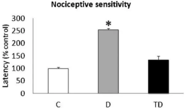
Nociceptive sensitivity
The latency to the tail deflection of the C group was expressed as 100%. Group D required 154.7% more time to deflect the tail than the C group (p,0.001) and 120% more time to deflect the tail than the TD group (p,0.001). The TD Figure 1 -Nociceptive sensitivity: the latency to tail deflection on the tail-flick apparatus for the C, D and TD groups. ANOVA followed by Bonferroni’spost hoctest. *corresponds top,0.001 compared with the C and TD groups.
group took only 34.7% more time to deflect the tail than the C group (p.0.05; Figure 1).
Optical densitometry of CGRP-ir
The OD analysis of the dorsal horn of the spinal cord revealed that CGRP-ir was lower in the D (0.056¡0.004)
group than in the C (0.098¡0.003; p,0.001) and TD (0.096¡0.01; p,0.05) groups. There was no difference between the C and TD groups (p.0.05; Figure 2). Representative images are presented in Figure 3.
DISCUSSION
In the present study, we have demonstrated that treadmill training alone, with no pharmacological intervention, can prevent the nociceptive loss caused by the hyperglycemic state in rats with STZ-induced diabetes. This beneficial effect was associated with changes in the CGRP-ir in the dorsal horn of the spinal cord.
As expected, diabetic rats display lower body weights and higher blood glucose levels than non-diabetic rats (8,10). Treadmill training did not alter the body weight or blood glucose levels in rats (8,10,18) or in type 1 diabetic patients (19). STZ induces an insulin-deficient state similar to type 1 diabetes in humans, which is not typically improved by exercise interventions (20), in contrast to type 2 diabetes, which is an insulin-resistant state (21).
In the analysis of nociceptive sensitivity, measured using the tail-flick test, the diabetic rats demonstrated an increase in the latency to deflect the tail from the apparatus, which indicates an STZ-induced loss of sensitivity. These data are in accordance with those of previous studies, which found deficits in motor and sensory nerve conduction (22,23) and thermal (23,24) and nociceptive hypoalgesia (24) in animals. Our findings are also in agreement with clinical data indicating that diabetes in humans can cause nociceptive hypoalgesia (25-27), which is an important determinant of lower limb ulcers and amputations.
However, diabetic rats that were subjected to treadmill training displayed a latency to deflect the tail from the apparatus similar to that of the non-diabetic animals, indicating that training is able to prevent alterations in the sensitivity behavior of diabetic animals. These data have not previously been reported in the literature. We also examined the effects of training on the CGRP-ir in the dorsal horn of the spinal cord, and the data reveal that physical exercise can maintain the content of this peptide in the spinal cord at levels similar to those of control animals. Diabetes is well known to cause a reduction in the content of CGRP in peripheral nerves (2,3,28), the neurons of the dorsal root ganglia and the dorsal horn of the spinal cord (29). Although physical training has been considered in the treatment of diabetic patients for some time, it was not
known, until now, whether training was able to prevent alterations in nociceptive signaling and CGRP content in the spinal cord. The benefits of treadmill training in these parameters could arise from the effects of nerve growth factor (NGF) and neurotrophin-3 (NT-3). These neuro-trophic factors promote neuronal survival and differentia-tion by activating phosphatidylinositol 3-kinase signaling (PI3-K). Moreover, there are various studies demonstrating that treadmill training increases the level of NGF in the sensory neurons (30) and in the hippocampus (31,32) of NT-3 in the spinal cord (NT-3NT-3) and of NGF and NT-NT-3 in the soleus muscle (34,35).
In conclusion, treadmill training is able to alleviate the nociceptive hypoalgesia caused by diabetes and this improvement is related to the content of CGRP in the dorsal horn of the spinal cord.
ACKNOWLEDGMENTS
We thank Silvia Barbosa for her technical assistance. This study was supported by grants from CNPq and CAPES. do Nascimento PS was supported by a Ph.D. scholarship from CNPq. Achaval M, Schaan BD and Xavier LL are CNPq investigators. We are indebted to Roche, which donated the test strips.
AUTHOR CONTRIBUTIONS
do Nascimento PS is the primary author of this manuscript, who organized the research protocol and participated in the research and article preparation. Lovatel GA, Ilha J and Xavier LL participated in the research protocol. Schaan BD participated in the article preparation. Achaval M is responsible for the research and participated in the article preparation.
REFERENCES
1. Boulton AJ. Lowering the risk of neuropathy, foot ulcers and amputa-tions. Diabet Med. 1998;15(Suppl 4):S57-9, http://dx.doi.org/10.1002/ (SICI)1096-9136(1998120)15:4+,S57::AID-DIA741.3.3.CO;2-4. 2. Diemel LT, Stevens EJ, Willars GB, Tomlinson DR. Depletion of
substance P and calcitonin gene-related peptide in sciatic nerve of rats with experimental diabetes; effects of insulin and aldose reductase inhibition. Neurosci Lett. 1992;137(2):253-6, http://dx.doi.org/10.1016/ 0304-3940(92)90416-5.
3. Brewster WJ, Diemel LT, Leach RM, Tomlinson DR. Reduced sciatic nerve substance P and calcitonin gene-related peptide in rats with short-term diabetes or central hypoxaemia co-exist with normal messenger RNA levels in the lumbar dorsal root ganglia. Neuroscience. 1994;58(2):323-30, http://dx.doi.org/10.1016/0306-4522(94)90038-8. 4. Schmidt Y, Unger JW, Bartke I, Reiter R. Effect of nerve growth factor on
peptide neurons in dorsal root ganglia after taxol or cisplatin treatment and in diabetic (db/db) mice. Exp Neurol. 1995;132(1):16-23, http:// dx.doi.org/10.1016/0014-4886(95)90054-3.
5. Fu¨rst S. Transmitters involved in antinociception. Brain Res Bull. 1999;15;48(2):129-41, http://dx.doi.org/10.1016/S0361-9230(98)00159-2. 6. Costentin J. Pain and its main transmitters. Ann Pharm Fr. 2000; 58:77-83. 7. van Dijk JW, Manders RJ, Tummers K, Bonomi AG, Stehouwer CD, Hartgens F, et al. Both resistance- and endurance-type exercise reduce the prevalence of hyperglycaemia in individuals with impaired glucose tolerance and in insulin-treated and non-insulin-treated type 2 diabetic
patients. Diabetologia. 2012;55(5):1273-82, http://dx.doi.org/10.1007/ s00125-011-2380-5.
8. Do Nascimento PS, Malysz T, Ilha J, Araujo RT, Hermel EE, Kalil-Gaspar PI, et al. Treadmill training increases the size of A cells from the L5 dorsal root ganglia in diabetic rats. Histol Histopathol. 2010;25(6):719-32. 9. Harthmann AD, De Angelis K, Costa LP, Senador D, Schaan BD, Krieger EM, et al. Exercise training improves arterial baro- and chemoreflex in control and diabetic rats. Auton Neurosci. 2007;133(2):115-20, http:// dx.doi.org/10.1016/j.autneu.2006.10.004.
10. Do Nascimento PS, Lovatel GA, Barbosa S, Ilha J, Centenaro LA, Malysz T, et al. Treadmill training improves motor skills and increases tyrosine hydroxylase immunoreactivity in the substantia nigra pars compacta in diabetic rats. Brain Res. 2011;1382:173-80, http://dx.doi.org/10.1016/j. brainres.2011.01.063.
11. Rossi DM, Valenti VE, Navega MT. Exercise training attenuates acute hyperalgesia in streptozotocin-induced diabetic female rats. Clinics. 2011;66(9):1615-9, http://dx.doi.org/10.1590/S1807-59322011000900019. 12. Junod A, Lambert AE, Stauffacher W, Renold AE. Diabetogenic action of
streptozotocin: relationship of dose to metabolic response. J Clin Invest. 1969;48(11):2129-39, http://dx.doi.org/10.1172/JCI106180.
13. Ilha J, Araujo RT, Malysz T, Hermel EE, Rigon P, Xavier LL, et al. Endurance and resistance exercise training programs elicit specific effects on sciatic nerve regeneration after experimental traumatic lesion in rats. Neurorehabil Neural Repair. 2008;22(4):355-66.
14. Rodrigues B, Figueroa DM, Mostarda CT, Heeren MV, Irigoyen MC, De Angelis K. Maximal exercise test is a useful method for physical capacity and oxygen consumption determination in streptozotocin-diabetic rats. Cardiovasc Diabetol. 2007;6:38, http://dx.doi.org/10.1186/1475-2840-6-38. 15. Fontella FU, Bruno AN, Balk RS, Ru¨cker B, Crema LM, Correˆa MD, et al. Repeated stress effects on nociception and on ectonucleotidase activities in spinal cord synaptosomes of female rats. Physiol Behav. 2005;85(2):213-9, http://dx.doi.org/10.1016/j.physbeh.2005.04.010. 16. Netto CA,Siegfried B,Izquierdo I. Analgesia induced by exposure to a
novel environment in rats: effect of concurrent and post-training stressful stimulation. Behav Neural Biol. 1987;48(2):304-9, http://dx.doi.org/ 10.1016/S0163-1047(87)90850-8.
17. Xavier LL, Viola GG, Ferraz AC, Da Cunha C, Deonizio JM, Netto CA, et al. A simple and fast densitometric method for the analysis of tyrosine hydroxylase immunoreactivity in the substantia nigra pars compacta and in the ventral tegmental area. Brain Res Brain Res Protoc. 2005;16(1-3): 58-64, http://dx.doi.org/10.1016/j.brainresprot.2005.10.002.
18. Midaoui AE, Chiasson JL, Tancre`de G, Nadeau A. Physical training reverses the increased activity of the hepatic ketone body synthesis pathway in chronically diabetic rats. Am J Physiol Endocrinol Metab. 2006;290:E207-E12, http://dx.doi.org/10.1152/ajpendo.00608.2004. 19. Hollingsworth DR, Moore TR. Postprandial walking exercise in pregnant
insulin-dependent (type I) diabetic women: reduction of plasma lipid levels but absence of a significant effect on glycemic control. Am J Obstet Gynecol. 1987;157(6):1359-63.
20. Huttunen NP, La¨nkela¨ SL, Knip M, Lautala P, Ka¨a¨r ML, Laasonen K, et al. Effect of once-a-week training program on physical fitness and metabolic control in children with IDDM. Diabetes Care. 1989;12(10):737-40, http://dx.doi.org/10.2337/diacare.12.10.737.
21. Umpierre D, Ribeiro PA, Kramer CK, Leita˜o CB, Zucatti AT, Azevedo MJ, et al. Physical activity advice only or structured exercise training and association with HbA1c levels in type 2 diabetes: a systematic review and meta-analysis. JAMA. 2011;305(17):1790-9, http://dx.doi.org/10.1001/ jama.2011.576.
22. Cameron NE, Cotter MA, Robertson S. The effect of aldose reductase inhibition on the pattern of nerve conduction deficits in diabetic rats. Q J Exp Physiol. 1989;74(6):917-26.
23. Lupachyk S, Shevalye H, Maksimchyk Y, Drel VR, Obrosova IG. PARP inhibition alleviates diabetes-induced systemic oxidative stress and neural tissue 4-hydroxynonenal adduct accumulation: correlation with peripheral nerve function. Free Radic Biol Med. 2011;50(10):1400-9, http://dx.doi.org/10.1016/j.freeradbiomed.2011.01.037.
24. Drel VR, Pacher P, Vareniuk I, Pavlov I, Ilnytska O, Lyzogubov VV, et al. A peroxynitrite decomposition catalyst counteracts sensory neuropathy in streptozotocin-diabetic mice. Eur J Pharmacol. 2007; 569(1-2):48-58. 25. Dyck PJ, Dyck PJ, Larson TS, O’Brien PC, Velosa JA. Patterns of
quantitative sensation testing of hypoesthesia and hyperalgesia are predictive of diabetic polyneuropathy: a study of three cohorts. Nerve growth factor study group. Diabetes Care. 2000;23(4):510-7.
26. Bierhaus A, Haslbeck KM, Humpert PM, Liliensiek B, Dehmer T, Morcos M, et al. Loss of pain perception in diabetes is dependent on a receptor of the immunoglobulin superfamily. J Clin Invest. 2004; 114(112):1741-51. 27. Doupis J, Lyons TE, Wu S, Gnardellis C, Dinh T, Veves A. Microvascular
reactivity and inflammatory cytokines in painful and painless peripheral diabetic neuropathy. J Clin Endocrinol Metab. 2009;94(6):2157-63, http:// dx.doi.org/10.1210/jc.2008-2385.
28. Jiang Y, Nyengaard JR, Zhang JS, Jakobsen J. Selective loss of calcitonin gene-related peptide-expressing primary sensory neurons of the A-cell phenotype in early experimental diabetes. Diabetes. 2004;53(10):2669-75, http://dx.doi.org/10.2337/diabetes.53.10.2669.
29. Unger JW, Klitzsch T, Pera S, Reiter R. Nerve growth factor (NGF) and diabetic neuropathy in the rat: morphological investigations of the sural nerve, dorsal root ganglion, and spinal cord. Exp Neurol. 1998;15391):23-34, http://dx.doi.org/10.1006/exnr.1998.6856.
30. Molteni R, Zheng JQ, Ying Z, Go´mez-Pinilla F, Twiss JL. Voluntary exercise increases axonal regeneration from sensory neurons. Proc Natl Acad Sci USA. 2004;101(22):8473-8, http://dx.doi.org/10.1073/ pnas.0401443101.
31. Chae CH, Jung SL, An SH, Park BY, Wang SW, Cho IH, et al. Treadmill exercise improves cognitive function and facilitates nerve growth factor signaling by activating mitogen-activated protein kinase/extracellular signal-regulated kinase1/2 in the streptozotocin-induced diabetic rat hippocampus. Neuroscience. 2009;164(4):1665-73, http://dx.doi.org/ 10.1016/j.neuroscience.2009.09.075.
32. Um HS, Kang EB, Koo JH, Kim HT, Jin-Lee, Kim EJ, et al. Treadmill exercise represses neuronal cell death in an aged transgenic mouse model of Alzheimer’s disease. Neurosci Res. 2011;69(2):161-73, http:// dx.doi.org/10.1016/j.neures.2010.10.004.
33. Coˆte´ MP, Azzam GA, Lemay MA, Zhukareva V, Houle´ JD. Activity-dependent increase in neurotrophic factors is associated with an enhanced modulation of spinal reflexes after spinal cord injury. J Neurotrauma. 2011;28(2):299-309, http://dx.doi.org/10.1089/ neu.2010.1594.
34. Go´mez-Pinilla F, Ying Z, Opazo P, Roy RR, Edgerton VR. Differential regulation by exercise of BDNF and NT-3 in rat spinal cord and skeletal muscle. Eur J Neurosci. 2001;13(6):1078-84.