CLINICAL SCIENCE
Trapezoidal osteochondral autologous plug
single-block graft for treating chondral lesions of the knee:
clinical and functional medium-term results in an
observational study
Cezar Teruyuki Kawano, Ma´rcio Moura Rocha dos Santos, Marcus Guilherme de Oliveira, Tadeu Colens Ourivio
CEMKA (Centro Me´dico Kawano), Sa˜o Paulo/SP, Brazil.
OBJECTIVE:To evaluate the clinical and functional results of autologous trapezoidal plug single-block grafts fixed with absorbable chondral darts in patients with osteochondral knee lesions of varying sizes.
METHODS:Twenty-five patients underwent surgery from February 2000 to June 2008. Seventy-two percent of the patients were male, and the mean age was 29 years.
RESULTS:The right side (56%) and the medial condyle (92%) were most affected. The lesions had an average area of 5.28 cm2, and the mean follow-up was 59 months. All of the variables other than instability showed significant improvements (p,0.05), as shown by the increase in the mean Lysholm score from 55 points preoperatively to 92 points (p,0.001) postoperatively. There was no loosening or collapse of the osteochondral graft. All of the patients had patellofemoral crepitation and pain for an average of six months.
CONCLUSION:Autologous trapezoidal plug single-block grafts are a therapeutic option for defects of varying sizes and provide good clinical results and low morbidity at the donor site in the medium term.
KEYWORDS: Cartilage Diseases; Bone Transplantation; Knee; Subchondral Arthroplasty; Lesions.
Kawano CT, Santos MM, Oliveira MG, Ourivio TC. Trapezoidal osteochondral autologous plug single-block graft for treating chondral lesions of the knee: clinical and functional medium-term results in an observational study. Clinics. 2012;67(10):1191-1195.
Received for publication onJune 19, 2012;First review completed onJune 19, 2012;Accepted for publication onJune 20, 2012 E-mail: [email protected]
Tel.: 55 11 5579-3272
INTRODUCTION
Treating chondral and osteochondral lesions, particularly those located on the weight-bearing surface of the knee, still presents a challenge for orthopedists. Pain, joint effusion, instability and joint locking are common complaints, leading to limitations and a decrease in the physical activity level (1) of the patient. Physical therapy is an important treatment component (2); however, the joint cartilage has limited regeneration potential (3), and its intrinsic repair mechanism is incapable of reestablishing the biomechanical integrity of normal chondral tissue (4). Because of the absence of vascular, neural and lymphatic tissue, the chondrocytes around the lesions are unable to migrate, proliferate, or produce cartilaginous matrices to cover the lesions (5). Therefore, these lesions have a high risk of
leading to permanent complications such as osteoarthrosis. The purpose of any surgical cartilage repair is to reestablish joint congruence while maintaining sufficient biomechanical properties to support body weight and painlessly perform a full range of motion (6).
Surgical techniques, such as debridement to remove cartilaginous tissue and wash the joint, remove proteolytic enzymes and inflammatory mediators; however, these processes only relieve symptoms temporarily (7). Abrasion arthroplasty and microfractures cause the migration of mesenchymal cells and induce metaplasia that forms fibrocartilage in the lesion area (8).
Over the last decade, many studies have reported cover-ing the osteochondral lesion with hyaline cartilage. Various procedures have been used, such as autologous, periosteal, perichondral, and osteochondral transplant, homologous grafts, chondrocyte culture, and genetic therapy.
Autologous osteochondral transplant, a commonly used procedure because of the positive results achieved (3,9-11), consists of removing a cartilaginous fragment and its underlying subchondral bone from a donor area outside the weight-bearing region. The fragment is then transferred to the defective zone, and an attempt is made to improve the Copyrightß2012CLINICS– This is an Open Access article distributed under
the terms of the Creative Commons Attribution Non-Commercial License (http:// creativecommons.org/licenses/by-nc/3.0/) which permits unrestricted non-commercial use, distribution, and reproduction in any medium, provided the original work is properly cited.
surgical indications for chondral lesions in the weight-bearing area of their femoral condyles (described in Table 1) underwent surgery using trapezoidal autologous osteo-chondral grafts fixed with absorbable osteo-chondral darts. The patients were followed up in the outpatient clinic of the Kawano Medical Center (CEMKA). The treatment was approved by the ethics committee of the Hospital Nossa Senhora do Rosa´rio, and each patient signed an informed consent form.
The following inclusion criteria were established: 1) the presentation of osteochondral lesions due to complications from grade III and IV unstable osteochondritis as deter-mined by the magnetic resonance classification developed by Nelson et al. (12) and by arthroscopy according to Clanton and DeLee (13); and 2) grade IV idiopathic chondral lesions according to the Outerbridge (14) classification. Patients who presented mild or degenerative concomitant meniscal lesions and patients with degenerative arthropathy diseases were excluded.
The patients were aged 15 to 50 years (average 29.32¡10.01 years). As shown in Table 1, 72% of the
patients were male. The right knee was affected in 56% of the cases and the left knee in 44% of the cases. The medial
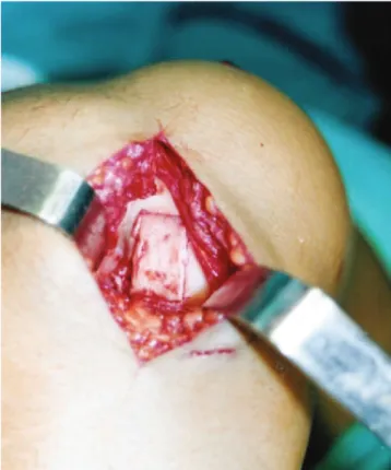
Afterwards, with the knee at 90
˚
of flexion, we made an ipsilateral parapatellar longitudinal incision (Figure 1). With visualization of the lesion (Figure 2), we performed a trapezoid-shaped resection using a millimeter-graded sur-gical chisel and leaving the edges of healthy cartilage and bleeding spongy bone tissue (Figure 3).With the knee extended, we began to remove the anterior-superior ipsilateral osteochondral graft from the affected femoral condyle in the peripheral area of the trochlea. Two central holes were drilled in the graft as the fixation site for the chondral darts, and the graft was transferred to the host area by impaction. The graft was produced in a single trapezoidal block.
To allow the graft to settle without any height differences between it and the surrounding cartilage, the knee was moved through its full range of motion by flexion and extension. Next the graft was fixed with absorbable darts measuring 18 mm long and 1.3 mm wide (Figure 4).
Suction drainage was inserted and maintained for 24 hours, and suturing was performed in layers (medial or lateral retinacular suture, subcutaneous with absorbable sutures and skin with nylon suture).
Postoperative rehabilitation
In the first three weeks after surgery, the knee was immobilized with an inguinal-malleolar orthosis; the patient was instructed not to place weight on it and to perform only isometric exercises. From weeks three to six, active and Table 1 -Demographic and clinical data of the 25 patients
included in the study.
Patient Age Sex Side Condilus Disease Area (cm2)
1 28 F Left Medial Osteochondritis 8 2 29 M Left Medial Osteochondritis 6 3 26 F Right Medial Idiopathic lesion 2 4 32 M Right Medial Osteochondritis 6 5 15 M Left Medial Osteochondritis 8 6 16 M Right Medial Osteochondritis 6 7 22 M Left Medial Osteochondritis 6 8 22 M Right Medial Osteochondritis 4 9 48 M Right Medial Osteochondritis 4 10 36 F Left Medial Osteochondritis 4 11 30 M Right Medial Osteochondritis 9 12 19 F Right Lateral Idiopathic lesion 6 13 19 M Right Medial Osteochondritis 6 14 44 F Left Medial Idiopathic lesion 2 15 25 M Right Medial Idiopathic lesion 4 16 16 M Right Medial Osteochondritis 6 17 21 M Left Lateral Osteochondritis 6 18 44 M Right Medial Idiopathic lesion 6 19 32 M Right Medial Idiopathic lesion 3 20 40 F Right Medial Idiopathic lesion 2 21 29 M Left Medial Idiopathic lesion 4 22 25 M Left Medial Osteochondritis 6 23 50 M Left Medial Osteochondritis 6 24 31 F Right Medial Osteochondritis 6
passive exercises were initiated to increase the range of motion. From the sixth week, the patient was permitted to place partial weight on the knee, and full weight bearing was permitted after eight weeks.
Statistical analysis
The data were analyzed using descriptive and inferential statistics. The quantitative variables are described as the mean, standard deviation, median, maximum and minimum. The qualitative variables were presented as absolute and relative frequencies. The normality of the data was evaluated by the Kolmogorov-Smirnov test. After rejecting this premise (p#0.05), the non-parametric Wilcoxon test was applied to compare the groups (16).
A significance level of 5% was adopted. For the statistical analysis, the Statistical Package for the Social Sciences (SPSS for Windows version 11.0) was used.
RESULTS
The patients completed a minimum follow-up period of four months, and there were no losses. The results of the pre- and postoperative clinical and functional evaluation for
the 25 patients (by the modified Lysholm knee scale) are shown in Tables 2 and 3 and Figure 5. All the patients, with the exception of four, had lesions that measured a minimum of 4 cm2(84%). Each patient showed statistically significant improvement in all of the evaluations, with the exception of instability, which was not present before the surgery in any of the patients.
None of the patients developed collapse or loosening of the osteochondral graft, thrombophlebitis, or infection. The patients presented pain and patellofemoral crepitations for an average of six months, with one patient presenting for ten months.
DISCUSSION
Hyaline cartilage has superior biomechanical properties and attaches to fibrocartilage to mechanically support weight-bearing joints (17). According to the arthroscopic classification that Clanton and DeLee developed for osteochondritis dissecans of the knee, osteochondral detach-ment and the formation of a fibrin layer at the base of the defect occurs in grade III and IV lesions, which hinders the angiogenic, osteoinductor and osteoconductor stimulation for bone incorporation (13).
In an attempt to promote vascular stimulation of the base of the defect and restore the congruence and integrity of the joint, various surgical methods have been described to treat osteochondral lesions. The use of osteochondral autologous graft is a treatment that is now supported by various authors (3,9-11) who believe it has the following advantages. 1) Osteochondral autologous graft is a method that reestablishes over 80% of the lesion surface with hyaline Figure 2 - Osteochondral lesion in a weight-bearing area of the
medial femoral condyle.
Figure 3 -Host area preparation with exposed viable subchon-dral spongy tissue.
cartilage with little interference on its composition, which benefits the biomechanical structure of the knee (3).
2) Because the osteochondral graft consists of spongy bone, it has osteogenic properties that favor its incorpora-tion into the bone, and because it is composed of an osteochondral fragment, the osteochondral graft promotes regular surface maintenance without forming different levels due to the settlement of the affected area (3).
3) Compared with homologous grafts, autologous grafts prevent contamination by transmittable diseases and the possibility of immunological reactions; autologous grafts also lower the cost compared with homological osteochon-dral, periosteal, perichondral and chondrocyte grafts (8).
The main disadvantage of the autologous graft treatment relates to complications in the donor area that are caused by the size of the graft removal. For this reason, many authors
restrict the use of autologous grafts to lesions smaller than 3 to 4 cm2(3,10). Sgaglione et al. observed that the donor bases of grafts larger than 6.5 cm2in diameter are not completely filled by fibrocartilage, which increases morbidity at the site and creates a higher risk of arthritic alterations (9).
In a 10-year follow-up study of 1,000 patients submitted to mosaicoplasty, Hangody and Fu¨les, obtained successful results in 92% of cases; in just 3% of cases, complications occurred at the donor site and there was an improvement one year after surgery. These authors affirmed that the cylindrical format of differently sized grafts leads to approximately 80% to 90% filling of the lesion base, while the remainder (filled by fibrocartilage) does not cause alterations in either the clinical evaluation or the comple-mentary and arthroscopic exams (3).
Pallazzi et al. demonstrated the trapezoidal autologous single-block osteochondral graft technique in 32 knees with solid lesions in the condylar weight-bearing region. Pallazzi et al. obtained an average 87.5% success rate during a 10-year follow-up period. The authors affirmed that the cylindrical resection of the graft results in a greater diameter in the donor area when totaling the multiple fragments of the mosaicoplasty with 80% filled with hyaline cartilage and the remainder filled with fibrocartilage, which causes greater instability of the host area graft (10).
Using the technique of Pallazzi, Camargo et al. reported that fixation of the graft provides bone incorporation, which helps stabilize rotational movements and prevents extrusion or sinking of the osteochondral graft. Furthermore, Camargo et al. affirmed that it is important to preserve 5 mm (on average) at the lateral or medial edges of the condyles to maintain the stability of the patellofemoral track (11).
Sgaglione et al. affirmed that the advantage of using a single-block graft is that it provides a surface of hyaline cartilage and fewer interfaces between the native and transplanted tissue (9).
In our work, we opted to reproduce the technique of Pallazzi, except for the method of graft fixation. We used absorbable darts with press-fit grafts (as originally described); the depth of the base was reduced from 15-20 mm to 14-16 mm because we found viable spongy tissue at this level.
Twenty-five patients underwent surgery; 72% of the patients were male with an average age of 29¡10 years. The medial
Table 3 -Size of the lesion and the Lysholm knee scale results before and after surgery.
Patient Area (cm2) Lysholm (pre) Lysholm (post)
1 8 40 95
2 6 48 95
3 2 59 90
4 6 61 90
5 8 46 100
6 6 51 95
7 6 67 90
8 4 59 95
9 4 74 95
10 4 52 90
11 9 38 90
12 6 71 95
13 6 55 90
14 2 63 90
15 4 46 90
16 6 46 100
17 6 55 90
18 6 45 90
19 3 69 90
20 2 46 84
21 4 60 90
22 6 55 90
23 6 43 84
24 6 63 90
25 6 74 90
Post 25¡0 25 25 25
Block Pre 5.36¡3.4 6 2 10 0.000
Post 15¡0 15 15 15
Total Pre 55.44¡10.59 55 38 74 0.000
condyle was affected in 92% of the knees, and in 68% the main case was osteochondritis dissecans, which was similar to the epidemiological data found in the majority of studies in the literature (1,3,4,6,8,10). However, unlike the other works (9,10), which limited the use of the autologous graft to lesions up to 4 cm2, we used this technique in lesions with areas ranging from 2 to 9 cm2with an average area of 5.28¡1.84 cm2. The
lesions larger than 4 cm2presented clinical results similar to
the lesions with smaller areas, as demonstrated by the results of the Lysholm knee scale (Table 3).
In our sample, we managed to improve the results observed through the modified Lysholm knee scale by an average of 36%. Given that there was no patient with an associated ligament lesion, there was a significant increase in the results for all the variables, except for instability. In the preoperative period, we obtained an average Lysholm score of 55.44¡10.59, and in the postoperative period, we
obtained a final average of 91.52¡3.9.
All the patients presented with patellofemoral crepitation in the early postoperative period over an average of six months. All the patients showed improvement up to one year following surgery.
The use of the trapezoidal plug autologous single-block graft for osteochondral lesions in the weight-bearing areas of the femoral condyles is a therapeutic option for defects of various sizes and provides good clinical results and low morbidity in the base of the donor site in the short and medium term.
AUTHOR CONTRIBUTIONS
Kawano CT was responsible for the study design, data collection and interpretation, manuscript writing and final revision of the manuscript. Oliveira MG was responsible for the data collection and interpretation, bibliographical review, manuscript writing and final revision of the manuscript. Santos MM was responsible for the data collection and interpretation, bibliographical review, manuscript writing and final revision. Ourivio TC was responsible for the data collection and interpretation, bibliographical review, manuscript writing and final manu-script revision.
REFERENCES
1. Jakob RP, Franz T, Gautier E, Mainil-Varlet P. Autologous osteochondral grafting in the knee: Indication, results and reflections. Clin Orthop Relat
Res. 2002;(401):170-84, http://dx.doi.org/10.1097/00003086-200208000-00020.
2. Carvalho NA, Bittar ST, Pinto FR, Ferreira M, Sitta RR. Manual for guided home exercises for osteoarthritis of the knee. Clinics. 2010;65(8):775-80, http://dx.doi.org/10.1590/S1807-59322010000800006. 3. Hangody L, Fu¨les P. Autologous osteochondral mosaicoplasty for the
treatment of full-thickness defects of weight-bearing joints: ten years of experimental and clinical experience. J Bone Joint Surg Am. 2003;85-A Suppl 2):25-32.
4. Horas U, Pelinkovic D, Herr G, Aigner T, Schnettler R. Autologous chondrocyte implantaion and osteochondral cylinder transplantation in cartilage repair of the knee joint. A prospective, comparative trial. J Bone Joint Surg Am. 2003;85-A(2):185-92.
5. Gudas R, Kalesinskas RJ, Kimtys V, Stankevicius E, Toliusis V, Bernotavicius G, et al. A prospective randomized clinical study of mosaic osteochondral autologous transplantation versus microfracture for the treatment of osteochondral defects in the knee joint in young athletes. Arthroscopy. 2005;21(9):1066-75.
6. Kellett CF, Boscainos PJ, Gross AE. Surgical options for articular defects of the knee. Expert Rev Med Devices. 2006;3(5):585-93, http:// dx.doi.org/10.1586/17434440.3.5.585.
7. Karataglis D, Green MA, Learmonth DJ. Autologous osteochondral transplantation for the treatment of chondral defects of the knee. Knee. 2006;13(1):32-5, http://dx.doi.org/10.1016/j.knee.2005.05.006. 8. Alleyne KR, Galloway MT. Management of osteochondral injuries of the
knee. Clin Sports Med. 2001;20(2):343-64, http://dx.doi.org/10.1016/ S0278-5919(05)70310-0.
9. Sgaglione NA, Miniaci A, Gillogly SD, Carter TR. Update on advanced surgical techniques in the treatment of traumatic focal articular cartilage lesions in the knee. Arthroscopy. 2002;18(2 Suppl 1):9-32.
10. Palazzi Coll S, Palazzi Coll C, Palazzi Duarte S. Osteocartilaginous autograft of the knee. International Orthopaedics. 1977;1(1):48-52, http://dx.doi.org/10.1007/BF00269759.
11. Camargo OPA, Severino NR, Aihara T, Cury RPL, Souza DG, Palomino EM, et al. Tratamento da osteonecrose do joelho atrave´s do uso do auto-enxerto osteocartilaginoso [Treatment of knee osteonecrosis using autogenous osteochondral graft]. Rev Bras Ortop. 1995;30(5):298-304. 12. Nelson DW, DiPaola J, Colville M, Schmidgall J. Osteochondritis
dissecans of the talus and knee: prospective comparison of MR and arthroscopic classifications. J. Comput Assist Tomogr. 1990;14(5):804-8, http://dx.doi.org/10.1097/00004728-199009000-00026.
13. Clanton TO, DeLee JC. Osteochondritis dissecans. History, pathophy-siology and current treatment concepts. Clin Orthop Relat Res. 1982;167:50-64.
14. Outerbrigde RE. Osteochondritis dissecans of the posterior femoral condyle. Clin Orthop Relat Res. 1983;(175):121-9.
15. Lysholm J, Gillquist J. Evaluation of knee ligament surgery results with special emphasis on use of a scoring scale. Am J Spots Med. 1982;10(3):150-4, http://dx.doi.org/10.1177/036354658201000306. 16. Rosner B. Fundamentals of biostatistics. 2nd ed. Boston: PWS Publishers;
1986.
17. Farmer JM, Martin DF, Boles CA, Curl WW. Chondral and osteochondral injuries. Diagnosis and management. Clin Sports Med. 2001;20(2):299-320, http://dx.doi.org/10.1016/S0278-5919(05)70308-2.