Hypovitaminosis D in patients undergoing kidney
transplant: the importance of sunlight exposure
Cristiane F. Vilarta,IMarianna D. Unger,ILuciene M. dos Reis,IWagner V. Dominguez,IElias David-Neto,II Rosa M. Moyse´s,I,IIISilvia Titan,IMelani R. Custodio,IMariel J. Hernandez,IVVanda JorgettiI,*
IDivisao de Nefrologia, Faculdade de Medicina FMUSP, Universidade de Sao Paulo, Sao Paulo, SP, BR.IIDivisao de Urologia, Faculdade de Medicina FMUSP,
Universidade de Sao Paulo, Sao Paulo, SP, BR.IIIMestrado em Medicina, Universidade Nove de Julho (UNINOVE), Sao Paulo, SP, BR.IVServicio de Nefrologı´a y Trasplante Renal, Hospital Universitario de Caracas, Universidad Central de Venezuela, Caracas, Venezuela.
OBJECTIVES: Recent studies have shown a high prevalence of hypovitaminosis D, defined as a serum 25-hydroxyvitamin D level less than 30 ng/ml, in both healthy populations and patients with chronic kidney disease. Patients undergoing kidney transplant are at an increased risk of skin cancer and are advised to avoid sunlight exposure. Therefore, these patients might share two major risk factors for hypovitaminosis D: chronic kidney disease and low sunlight exposure. This paper describes the prevalence and clinical characteristics of hypovitaminosis D among patients undergoing kidney transplant.
METHODS:We evaluated 25-hydroxyvitamin D serum levels in a representative sample of patients undergoing kidney transplant. We sought to determine the prevalence of hypovitaminosis D, compare these patients with a control group, and identify factors associated with hypovitaminosis D (e.g., sunlight exposure and dietary habits). RESULTS:Hypovitaminosis D was found in 79% of patients undergoing kidney transplant, and the major asso-ciated factor was low sunlight exposure. These patients had higher creatinine and intact parathyroid hormone serum levels, with 25-hydroxyvitamin D being inversely correlated with intact parathyroid hormone serum levels. Compared with the control group, patients undergoing kidney transplant presented a higher prevalence of 25-hydroxyvitamin D deficiency and lower serum calcium, phosphate and albumin but higher creatinine and intact parathyroid hormone levels.
CONCLUSIONS:Our results confirmed the high prevalence of hypovitaminosis D in patients undergoing kidney transplant. Therapeutic strategies such as moderate sunlight exposure and vitamin D supplementation should be seriously considered for this population.
KEYWORDS: Vitamin D; Kidney Transplantation; Parathyroid Hormone; Hypovitaminosis D; Chronic Kidney Disease.
Vilarta CF, Unger MD, dos Reis LM, Dominguez WV, David-Neto E, Moyse´s RM, et al. Hypovitaminosis D in patients undergoing kidney transplant: the importance of sunlight exposure. Clinics. 2017;72(7):415-421
Received for publication onNovember 24, 2016;First review completed onJanuary 24, 2017;Accepted for publication onMarch 14, 2017
*Corresponding author. E-mail: [email protected]
’ INTRODUCTION
Several studies have demonstrated a high prevalence of hypovitaminosis D (hypo D) in different regions of the world including Latin America, even though these places receive the necessary ultraviolet radiation (UVR) required to produce adequate levels of 25-hydroxyvitamin D [25(OH)D] (1-3).
Recently, vitamin D and its metabolites have been exten-sively studied because of the role they play in functions such as the regulation of calcium (Ca) and phosphorus (P) levels, parathyroid hormone suppression and bone mineralization in adults (4). In addition, there are associations between
vitamin D deficit and the development of cardiovascular and immunological disease, cancer, diabetes mellitus and arterial hypertension (2).
Vitamin D deficiency in patients with chronic kidney disease (CKD) can lead to alterations in bone mineral meta-bolism, resulting in frequent fractures and increased cardio-vascular mortality (4,5). In this context, increases in the serum levels of 25(OH)D at 10 ng/ml are associated with a 14% decreased mortality risk in this population (5).
Kidney transplant (KT) resolves some of these disorders and leads to improvements in the prognosis and quality of life of patients with end-stage renal disease. However, studies have also shown that hypo D is a persistent event in during both early and late transplant periods, requiring supplementation in most cases (4-10). The major causes of hypo D in patients undergoing KT include: the increased catabolism of vitamin D secondary to the use of glucocorti-coids (11), restricted sunlight exposure, and sunscreen use by medical indication because immunosuppression is associated with skin cancer in these patients (12).
DOI:10.6061/clinics/2017(07)05
Copyright&2017CLINICS–This is an Open Access article distributed under the terms of the Creative Commons License (http://creativecommons.org/licenses/by/ 4.0/) which permits unrestricted use, distribution, and reproduction in any medium or format, provided the original work is properly cited.
In addition to the above known effects, Obi et al. showed that vitamin D deficiency/insufficiency in patients under-going KT is associated with an increased risk of renal interstitial fibrosis, renal tubular atrophy, and glomerular filtration rate (GFR) decline (13). Given the recent develop-ments of its molecular properties, vitamin D plays a multi-faceted role and has led us to determine the prevalence and clinical characteristics of hypo D in a cohort of patients with KT compared with a control group.
’ PATIENTS AND METHODS
Patients undergoing KT were followed at the Transplant Unit of Hospital das Clinicas da Faculdade de Medicina da Universidade de São Paulo. Of these patients, we selected those who were older than 18 years, showed stable GFR, and were more than 1 year removed from KT. A total of 752 patients was selected (355 females). In this cohort, we determined a representative sample size calculation. In this calculation, we considered a 32.5% prevalence of 25(OH)D deficiency and 5% alpha and a 1% beta risks for a test power ratio of 99%. The final KT population was composed of 149 patients; race, gender, age, donor type, serum creatinine, proteinuria, glutamic-oxaloacetic transami-nase (GOT), glutamic-pyruvic transamitransami-nase (GPT), gamma-glutamyltransferase (GGT) levels, immunosuppressive therapy, body mass index (BMI) and vitamin supplementation were controlled.
Concomitantly, we selected 398 individuals (333 females) from a sample of 603 volunteering outpatient employees of the Hospital Universitário da Universidade de São Paulo and graduate students from the Faculdade de Medicina da Universidade de São Paulo previously included in Unger et al. We excluded individuals with diabetes mellitus, arterial hypertension, serum creatinine levels higher than 1.2 mg/dL or those on medications/supplements such as Ca, bispho-sphonates, vitamin D and corticosteroids (see Figure 1) (2).
A blood sample was collected during late winter of 2006, and serum Ca, P, creatinine, albumin, intact parathyroid hor-mone (PTHi), total alkaline phosphatase (ALP), 25(OH)D and BMI were analyzed.
KT recipients were submitted to a single blood collection during late winter. We chose late winter to coincide with the period in which samples were collected from the control
group and because it has less UV radiation, enabling us to detect a greater number of patients with hypo D.
The following parameters were analyzed in the selected patients: age, gender, race (white or non-white), donor type, time after KT, immunosuppressive therapy, and post-KT fractures (spontaneous or minimal trauma). In addition, the serum concentrations of creatinine, total Ca, P, albumin, ALP, GOT, GPT, GGT, PTHi and 25(OH)D were measured. The GFR was estimated using the MDRD simplified formula (14). The patients completed a questionnaire on dietary food frequency that evaluated the consumption frequency of major food sources of vitamin D (e.g., fatty fish and fish oil). Sunlight exposure habits were analyzed based on the frequency of exposure (3 or more times a week or less than 3 times a week) and a sunscreen protection factor (yes or no).
Total serum Ca, serum P, albumin, ALP, GOT and GGT were determined using routine laboratory techniques. PTHi was measured using the Immulite analyzer (Diagnostic Pro-ducts Corporation, Los Angeles, CA, USA; reference range, 10-87 pg/ml). 25(OH)D was measured using a chemilumi-nescent assay (Dia-Sorin Inc., Stillwater, MN, USA). Serum concentrations of 25(OH)D between 15 and 30 ng/mL were considered insufficient, and concentrations less than 15 ng/ml were considered deficient.
All procedures in this study were performed after appro-val from the local institutional committee, Comissão de Ética para Análise de Projetos de Pesquisa (CAPPesq no
1035/07), and in accordance with the ethical standards of the 1964 Helsinki Declaration.
Statistical analysis
The data are expressed as either means±SD or medians and ranges; categorical data are described as percentages.
To determine the relationship between the 25(OH)D levels and other categorical variables, we used chi-square test and, when necessary, Fisher’s exact test.
We used the Box and Cox transformation for the variables age, time after KT, albumin, creatinine, PTHi, GOT, GPT, GGT and ALP to ensure the normality assumption. Even with the Box and Cox transformation, the variables age, time after KT and GOT did not reach normality. Because these transformed variables presented homoscedasticity (accord-ing to Levene’s test), they were also considered normally
distributed to compare the serum 25(OH)D levels.
An ANOVA was used to determine the relationship between the clinical and biochemical variables and serum 25(OH)D (deficient, insufficient or normal). If results were significant, then we applied the Bonferroni post hoc test. The correlation between the 25(OH)D levels and other biochem-ical parameters was analyzed using Spearman’s correlation
coefficients.
The patients undergoing KT and the control group differed with regard to gender; therefore, we used an ANOVA adjusted for gender to compare these two groups with regard to BMI, P, Ca, albumin, creatinine, PTHi, ALP and 25(OH)D for each variable separately.
The level of significance was set at 5% (p-valueo0.05).
’ RESULTS
Basic characteristics of the patients undergoing KT Of the 149 patients undergoing KT, 56.4% were female, and 68.4% were white. The median age was 44 (18 - 81) years, and the median time after KT of was 6 (1 - 36) years. Approx-imately 55% of these patients received a graft from a living donor.
Regarding their 25(OH)D serum levels, 56 (37.5%) patients presented deficiency, 62 (41.6%) insufficiency, and only 31 (20.8%) a normal concentration. Thus, the prevalence of hypo D in the study sample was 79.2%.
Post-KT insufficiency fractures (spontaneous or minimal trauma) were reported in 10% of the patients (N=15). The mean 25(OH)D serum level was similar between patients without and those with fractures (21.8±13.2 ng/mL vs. 21.9±8.3 ng/mL). Likewise, we found no differences between
patients with and without fractures regarding the rates of 25(OH)D deficiency or insufficiency.
In addition to the data described above, Table 1 shows the demographic and biochemical parameters. The patients under going KT showed serum PTHi above the reference range. We did not find differences in the 25(OH)D levels (21.1±12.1 vs. 22.6±13.4) between patients with abnormal liver func-tion (GGT levels450 U/L) and those with preserved liver function.
Clinical and demographic characteristics according to the 25(OH)D categories among patients
undergoing KT
Table 2 describes the clinical and demographic character-istics according to the 25(OH)D categories (deficient, insuffi-cient and normal). Donor type was associated with important differences, although they were not significant. Specifically, we found that 28% of patients who received a graft from a living related donor had normal 25(OH)D levels, whereas only 12% of patients receiving a graft from a standard cadaveric donor showed normal 25(OH)D levels (p=0.055).
Regarding the frequency of sunlight exposure, we observed a significant difference between those who were exposed to the sun more than 3 times a week and those who had less exposure with regard to 25(OH)D levels; specifically, 55.8% of patients with higher sun exposure had a normal concentra-tion of 25(OH)D versus 10.4% in the group with lower sun exposure (po0.0001). The use of sunscreen did not affect the serum concentrations of this vitamin. We did not find a significant relationship between 25(OH)D serum levels and
Table 1-Demographic and biochemical parameters of patients undergoing KT.
Demographic parameters Biochemical parameters Reference range
Gender 25(OH)D (ng/mL) 21.8±12.8
Female 84 (56) 25(OH)D category
Male 65 (44) Normal 31 (21) 430 ng/ml
Race Deficiency 56 (37) o15 ng/ml
White 102 (68) Insufficiency 62 (42) 15 - 30 ng/ml
Non-white 47 (31) PTHi (pg/mL) 80.1±78.3 10 - 87 pg/ml
Time after KT(years) 6 (1 - 36) Ca (mg/dL) 8.9±0.6 8.7 - 10.3 mg/dL
Age (years) 44 (18 - 81) P (mg/dL) 3.0±0.8 2.5 - 4.5 mg/dL
Type of donor Creatinine (mg/dL) 1.5 (0.6 - 4.5) 0.5 - 1.0 mg/dL
Living related donor 82 (55) Albumin (g/dL) 4.4 (3.4 - 5.5) 3.5 - 5.0 g/dL
Standard cadaveric donor 67 (45) ALP (U/L) 65.8 (29.7 - 364.5) 35 - 100 U/L
Insufficiency fracture after KT 15 (10) GOT (U/L) 23 (6 - 121) 0 - 35 U/L
BMI (kg/m2) 25.4±5.3 GPT (U/L) 10 (3 - 81) 3 - 36 U/L
GGT (U/L) 28 (7 - 704) 5 - 36 U/L
Values are expressed as the mean±SD, median (range) or number of patients (%); KT: kidney transplant; BMI: body mass index; 25(OH)D: 25
hydroxyvitamin D; Ca: calcium; P: phosphorus; PTHi: intact parathyroid hormone; ALP: alkaline phosphatase; GOT: glutamic-oxaloacetic transaminase; GPT: glutamic-pyruvic transaminase; GGT gamma-glutamyltransferase
Table 2-Demographic and clinical parameters based on 25(OH)D category among patients undergoing KT.
25(OH)D levels
Race Gender BMI Type of
donor*
Bone fracture after KT
Immunosuppressive therapy
White Non-White
Female Male Non-obese
Obese Living donor
Cadaveric donor
No Yes CNI use CNI non-use
Deficient 35 (34.3) 21 (44.7) 30 (35.7) 26 (40) 29 (36.7) 27 (38.6) 28 (34.1) 28 (41.8) 52 (38.8) 4 (26.6) 44 (41.1) 12 (28.6)
Insufficient 43 (42.1) 19 (40.4) 35 (41.6) 27 (41.5) 34 (43) 28 (40) 31 (37.8) 31 (46.3) 53 (39.5) 9 (60) 43 (40.2) 19 (45.2)
Normal 24 (23.5) 7 (14.8) 19 (22.6) 12 (18.5) 16 (20.2) 15 (21.4) 23 (28) 8 (12) 29 (21.6) 2 (13.3) 20 (18.7) 11 (26.2)
Total 102 (100) 47 (100) 84 (100) 65 (100) 79 (100) 70 (100) 82 (100) 67 (100) 134 (100) 15 (100) 107 (100) 42 (100)
consumption of food sources of vitamin D. The data are shown in Table 3.
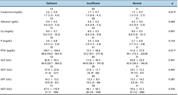
Biochemical parameters by 25(OH)D category among patients undergoing KT
Table 4 compares the biochemical parameters across 25(OH)D categories. The results revealed that only serum creatinine and PTHi significantly differed with regard to 25(OH)D levels. Specifically, we found lower serum levels of creatinine and PTHi among patients with normal 25(OH)D.
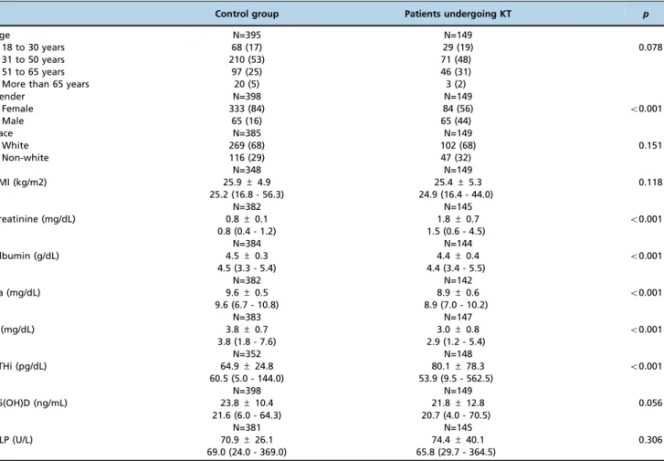
Comparison of the demographic and biochemical parameters between the control group and patients undergoing KT
Table 5 shows that the control group and patients under-going KT had different distributions based on gender. Regard-ing the biochemical data, we found significant differences in creatinine, albumin, Ca, P and PTHi between men and women. As expected, patients undergoing KT showed higher
levels of creatinine and PTHi as well as lower levels of P than the control group.
However, patients undergoing KT presented lower levels of this hormone, regardless of 25(OH)D category, when compared to the control group (p=0.001); these data are shown in Table 6.
We analyzed the correlations between the serum 25(OH)D levels and biochemical parameters but found only one nega-tive correlation with regard to serum PTHi levels (r=-0.24; po0.03).
’ DISCUSSION
Despite the growing interest in vitamin D status and its supplementation forms in both the normal population and patients with CKD, a lack of data remains regarding vitamin D levels in KT recipients. Little research has compared the 25(OH)D serum levels between patients undergoing KT and the healthy population. Our findings coincide with those of Ebbert et al., who analyzed a pediatric population,
Table 4-Biochemical parameters of patients undergoing KT according to 25(OH)D category.
Deficient Insufficient Normal p
53 60 31
Creatinine (mg/dL) 2.0±0.9 1.7±0.7 1.5±0.5 0.013*
1.7 (1.0 - 4.5) 1.5 (0.6 - 4.1) 1.3 (1.0 - 2.7)
52 60 31
Albumin (g/dL) 4.4±0.5 4.4±0.3 4.4±0.4 0.660
4.4 (3.4 - 5.3) 4.4 (3.8 - 5.2) 4.5 (3.4 - 5.5)
51 59 31
Ca (mg/dL) 8.9±0.7 8.9±0.5 8.9±0.5 0.997
9.0 (7.0 - 10.2) 8.9 (7.8 - 9.9) 8.8 (7.8 - 10.1)
55 61 31
P (mg/dL) 3.0±0.8 3.0±0.8 2.7±0.6 0.154
2.9 (1.2 - 5.4) 2.9 (1.3 - 5.3) 2.7 (1.2 - 3.8)
55 61 31
PTHi (pg/dL) 104.7±104.6 72.5±58.6 51.6±37.0 0.011*
68.6 (18.2 - 562.5) 53.2 (9.5 - 277.0) 44.1 (13.3 - 229.8)
53 60 31
ALP (U/L) 82.8±54.5 71.6±30.3 65.8±23.4 0.441
69.6 (29.7 - 364.5) 64.8 (34.2 - 191.0) 65.6 (34.5 - 166.4)
52 60 31
GOT (U/L) 27.9±22.8 27.8±11.7 23.5±12.2 0.445
21 (6 - 121) 25 (9 - 64) 19 (13 - 67)
53 60 31
GPT (U/L) 14.2±13.2 13.3±8.5 13.2±14.2 0.387
10.0 (4 - 81) 10.5 (3 - 44) 9.0 (4 - 77)
51 58 31
GGT (U/L) 67.5±116.9 44.1±50.1 35.6±37.2 0.324
31 (7 - 704) 28 (10 - 334) 22 (12 - 206)
Values are expressed as number of patients, mean±SD and median (range). KT: kidney transplant; 25(OH)D: 25 hydroxyvitamin D; Ca: calcium; P: phosphorus; PTHi: intact parathyroid hormone; ALP: alkaline phosphatase; GOT: glutamic-oxaloacetic transaminase; GPT: glutamic-pyruvic transaminase; GGT: gamma-glutamyltransferase. *Bonferroni’s post-hoc test: po0.05, Normal vs. Deficient or Insufficient.
Table 3-Sunlight exposure habits and vitamin D food source consumption frequency according to 25(OH)D category in patients undergoing KT.
25(OH)D levels Sunlight exposure* Use of sunscreen Vitamin D food source
3 or more times a week
Fewer than 3 times a week
Yes No Frequent Infrequent
Deficient 2 (5.8) 51 (44.3) 16 (37.2) 40 (38) 5 (27.7) 48 (37.2)
Insufficient 13 (38.2) 52 (45.2) 14 (32.5) 47 (44.7) 6 (33.3) 57 (44.2)
Normal 19 (55.8) 12 (10.4) 13 (30.2) 18 (17.1) 7 (38.8) 24 (18.6)
Total 34 (100) 115 (100) 43 (100) 105 (100) 18 (100) 129 (100)
reporting the presence of hypo D in patients undergoing KT as well as control groups (15). However, our findings showed that patients undergoing KT had a higher prevalence of deficiency.
The current study demonstrated that 79% of patients undergoing KT at the Hospital das Clinicas da Faculdade de Medicina da Universidade de São Paulo presented with an insufficiency or deficiency of 25(OH)D during late winter. These results are similar to those obtained in other studies conducted throughout the world and across climatic seasons, revealing a total approximate prevalence of 76-97% (7-10).
Stavroulopoulos et al. analyzed patients undergoing KT during the summer and winter and confirmed that levels of 25(OH)D are lower during winter (7). That study explains why the current evaluation was performed during late winter, i.e., to detect the highest possible number of patients
with hypo D. Researchers who plan future studies on vitamin D and clinicians who monitor and manage vitamin D deficiencies in patients undergoing KT must consider this seasonal variation.
Nutritional status did not affect serum 25(OH)D levels, and in contrast to other published data, we did not find a higher prevalence of hypo D in obese patients, even though the bioavailability of vitamin D can be influenced by its storage in adipocytes (16,17).
Hypo D is one of the major causes of secondary hyper-parathyroidism (SHPT). Unger et al. showed that healthy subjects develop SHPT in the context of vitamin D deficiency during winter but show improvements in summer (2). There-fore, this seasonal variation in 25(OH)D and PTH is likely a normal response to physiologically compensate for the drop in vitamin D levels during winter. Among patients under-going KT, studies have been conducted during different periods of the year, making it difficult to know whether they follow the trend observed in the general population. The literature commonly describes parathyroid hormone stimu-lation due to 25(OH)D deficiency during the earliest periods after KT as a consequence of the high doses of corticosteroids and persistent elevation of phosphatonins (6,18,19). Some authors have also found this correlation during later periods of KT recovery (7,20). We observed the same trends in our patients, showing a negative correlation between PTHi and 25(OH)D serum levels.
Table 5-Comparison of demographic and biochemical parameters between the control group and patients undergoing KT.
Control group Patients undergoing KT p
Age N=395 N=149
18 to 30 years 68 (17) 29 (19) 0.078
31 to 50 years 210 (53) 71 (48)
51 to 65 years 97 (25) 46 (31)
More than 65 years 20 (5) 3 (2)
Gender N=398 N=149
Female 333 (84) 84 (56) o0.001
Male 65 (16) 65 (44)
Race N=385 N=149
White 269 (68) 102 (68) 0.151
Non-white 116 (29) 47 (32)
N=348 N=149
BMI (kg/m2) 25.9±4.9 25.4±5.3 0.118
25.2 (16.8 - 56.3) 24.9 (16.4 - 44.0)
N=382 N=145
Creatinine (mg/dL) 0.8±0.1 1.8±0.7 o0.001
0.8 (0.4 - 1.2) 1.5 (0.6 - 4.5)
N=384 N=144
Albumin (g/dL) 4.5±0.3 4.4±0.4 o0.001
4.5 (3.3 - 5.4) 4.4 (3.4 - 5.5)
N=382 N=142
Ca (mg/dL) 9.6±0.5 8.9±0.6 o0.001
9.6 (6.7 - 10.8) 8.9 (7.0 - 10.2)
N=383 N=147
P (mg/dL) 3.8±0.7 3.0±0.8 o0.001
3.8 (1.8 - 7.6) 2.9 (1.2 - 5.4)
N=352 N=148
PTHi (pg/dL) 64.9±24.8 80.1±78.3 o0.001
60.5 (5.0 - 144.0) 53.9 (9.5 - 562.5)
N=398 N=149
25(OH)D (ng/mL) 23.8±10.4 21.8±12.8 0.056
21.6 (6.0 - 64.3) 20.7 (4.0 - 70.5)
N=381 N=145
ALP (U/L) 70.9±26.1 74.4±40.1 0.306
69.0 (24.0 - 369.0) 65.8 (29.7 - 364.5)
Values are expressed as number of patients (%), mean±SD and median (range). KT: kidney transplant; BMI: body mass index; Ca: calcium; P: phosphorus; PTHi: intact parathyroid hormone; 25(OH)D: 25 hydroxyvitamin D; ALP: alkaline phosphatase.
Table 6-Patients undergoing KT and the control group according to 25(OH)D category.
25(OH)D levels Control group Patients undergoing KT p
Deficient 90 (23) 56 (37) 0.001
Insufficient 209 (52) 62 (42)
Normal 99 (25) 31 (21)
In addition, regarding the biochemical associations descri-bed above in the general population, vitamin D insufficiency contributes to the development of osteoporosis. Moreover, adequate supplementation can reduce the fracture risk in approximately 20% of this population (21). However, the effect of this hormone on the prevalence of fracture during later periods of KT is unclear, which suggests that low mineral bone density and hypo D are multifactorial conditions (7). In this regard, we found no difference between patients with and without fracture.
The reasons for hypovitaminosis after KT include low sunlight exposure and the accelerated catabolism of vitamin D secondary to glucocorticoids use (22). Exposure to UV-A and UV-B radiation via sunlight can induce skin cancer through induced mutations in the DNA structure. This radia-tion is clearly the most important physical environmental carcinogen for the development of skin cancer, and non-melanoma skin cancer (NMSC) is the most common malig-nancy among patients undergoing KT (23,24). The risk of developing this complication is associated with the duration and intensity of immunosuppression and cumulative sun exposure (18). However, some guidelines suggest that sun-light exposure for short periods of time is beneficial and does not increase the risk of skin cancer (25,26).
In this sense, sunlight exposure at least 3 times a week predicted higher 25(OH)D serum levels in our patients, des-pite the use of sunscreen. However, Penny et al. found an asso-ciation between low 25(OH)D levels and a high incidence of NMSC, limiting the use of this therapeutic strategy to low-risk groups defined by skin type, age, gender and immunosup-pressive regimen (18,27).
Regarding the effect of immunosuppression on vitamin D status after KT, steroids accelerate catabolism of vitamin D. Despite not knowing the steroid doses in our patients, the development time of transplantation suggests that they recei-ved a minimal and stable glucocorticoid dose, which most likely would not explain their vitamin D deficiency. Regard-ing other immunosuppressive drugs (e.g., calcineurin inhi-bitors), we similarly did not find an association, contrary to results reported by Filipov et al. and Eyal et al., who showed that high tacrolimus doses are associated with hypo D through hepatic CYP3A4 suppression (28,29).
The potent immunomodulatory activity of 25(OH)D through the suppression of T cell receptor proliferation and its direct effect on B cells through the inhibition of immunoglobulin production are additional physiological effects to consider among patients undergoing KT (30). Sezer et al. demonstrated that patients undergoing KT showed an association between 25(OH)D deficit and renal graft failure after one year (31), whereas other authors have shown a direct relationship between 25(OH)D deficit and renal graft survival (11,32). In this respect, our patients presented with an association between low serum levels of 25(OH)D and higher creatinine serum levels. In addition to the clinical aspects described above, Bienaimé et al. found a correlation between vitamin D status and graft histological findings; specifically, 25(OH)D serum levels 3 months after transplantation independently predictor the progression of interstitial fibrosis and tubular atrophy (IF/TA) at 12 months (32).
Other studies have shown an association between hypo D and the reception of a renal graft from a cadaver donor (32). In this regard, although our study was retrospective and did not evaluate the characteristics of the renal graft, we found a higher percentage of patients with normal 25(OH)D levels
(28%) in the group receiving a renal graft from a living donor, compared to only 12% in the group receiving a renal graft from a cadaveric donor. These results suggest that the immu-nological tolerance in these patients could be due to the vitamin D status and that the concentration of this hormone depends on the type of renal graft received. Specifically, better compatibility conditions ensure a briefer cold ischemia time, and a minor ischemia-reperfusion effect predicts higher deposits of 25(OH)D.
In conclusion, vitamin D deficiency is prevalent among patients undergoing KT, even in geographic areas with high UV exposure. Additional studies are needed to clarify the role that donor type plays in vitamin D deficiency. Further-more, studies are needed to define the efficacy and safety of UVR exposure as a therapeutic strategy for these patients. However, individual sunlight exposure in moderation com-bined with vitamin D supplementation are the easiest and safest forms to guarantee adequate levels of 25(OH)D, thereby leading to the beneficial effects of this hormone.
’ ACKNOWLEDGMENTS
We acknowledge the substantial contributions of the Fundac¸ão de Amparo
à Pesquisa de Estado de São Paulo (FAPESP) (2008/50782-7) in providing support for the realization of this work.
’ AUTHOR CONTRIBUTIONS
Vilarta CF participated in study design, data collection, analysis and inter-pretation, and manuscript writing. Unger MD, dos Reis LM, Dominguez WV, David-Neto E, Moyses RM and Custodio MR participated in data interpretation and manuscript writing. Titan S and Hernandez MJ participated in statistical analysis, data interpretation and revision of the edited manuscript. Jorgetti V participated in study design, data analysis and interpretation, and manuscript writing.
’ REFERENCES
1. Holick MF. Vitamin D deficiency. N Engl J Med. 2007;357(3):266-81, http://dx.doi.org/10.1056/NEJMra070553.
2. Unger MD, Cuppari L, Titan SM, Magalhães MC, Sassaki AL, dos Reis LM, et al. Vitamin D status in a sunny country: where has the sun gone? Clin Nutr. 2010;29(6):784-8, http://dx.doi.org/10.1016/j.clnu.2010.06.009. 3. Binkley N, Novotny R, Krueger D, Kawahara T, Daida YG, Lensmeyer G, et al. Low vitamin D status despite abundant sun exposure. J Clin Endocrinol Metab. 2007;92(6):2130-5, http://dx.doi.org/10.1210/jc.2006-2250.
4. Çankaya E, Bilen Y, Keles¸M, Uyanık A, Akbas¸M¸Güngör A, et al. Comparison of serum vitamin D Levels among patients with chronic kidney disease, patients in dialysis, and renal transplant patients. Trans-plant Proc. 2015;47(5):1405-7, http://dx.doi.org/10.1016/j.transproceed. 2015.04.036.
5. Pilz S, Iodice S, Zittermann A, Grant WB, Gandini S. Vitamin D status and mortality risk in CKD: a meta-analysis of prospective studies. Am J Kidney Dis. 2011;58(3):374-82, http://dx.doi.org/10.1053/j.ajkd.2011. 03.020.
6. Sadlier DM, Magee CC. Prevalence of 25(OH) vitamin D (calcidiol) defi-ciency at time of renal transplantation: a prospective study. Clin Trans-plant. 2007;21(6):683–8, http://dx.doi.org/10.1111/j.1399-0012.2007.00696.x. 7. Stavroulopoulos A, Cassidy MJ, Porter CJ, Hosking DJ, Roe SD. Vitamin D status in renal transplant recipients. Am J Transplant. 2007;7(11):2546-52, http://dx.doi.org/10.1111/j.1600-6143.2007.01978.x.
8. Lomonte C, Antonelli M, Vernaglione L, Cazzato F, Casucci F, Chimienti D. Are low plasma levels of 25-(OH)vitamin D a major risk factor for hyperparathyroidism independent of calcitriol in renal transplant patients? J Nephrol. 2005;18(1):96-101.
9. Boudville NC, Hodsman AB. Renal function and 25-hydroxyvitamin D concentrations predict parathyroid hormone levels in renal transplant patients. Nephrol Dial Transplant. 2006;21(9):2621-4, http://dx.doi.org/ 10.1093/ndt/gfl201.
11. Eyal O, Aharon M, Safadi R, Elhalel MD. Serum vitamin D levels in kidney transplant recipients: the importance of an immunosuppression regimen and sun exposure. Isr Med Assoc J. 2013;15(10):628-33. 12. Euvrard S, Kanitakis J, Claudy A. Skin cancers after organ
transplanta-tion. N Engl J Med. 2003;348(17):1681-91, http://dx.doi.org/10.1056/ NEJMra022137.
13. Obi Y, Hamano T, Ichimaru N, Tomida K, Matsui I, Fujii N, et al. Vitamin D deficiency predicts decline in kidney allograft function: a prospective cohort study. J Clin Endocrinol Metab. 2014;99(2):527–35, http://dx.doi. org/10.1210/jc.2013-2421.
14. Levey AS, Bosch JP, Lewis JB, Greene T, Rogers N, Roth D. A more accurate method to estimate glomerular filtration rate from serum crea-tinine: a new prediction equation. Modification of Diet in Renal Disease Study Group. Ann Intern Med. 1999;130(6):461–70, http://dx.doi.org/ 10.7326/0003-4819-130-6-199903160-00002.
15. Ebbert K, Chow J, Krempien J, Matsuda-Abedini M, Dionne J. Vitamin D insufficiency and deficiency in pediatric renal transplant recipients. Pediatr Transplant. 2015;19(5):492–8, http://dx.doi.org/10.1111/petr.12527. 16. Cofán F, Vela E, Clèries M, Catalan Renal Registry. Obesity in renal transplantation: analysis of 2691 patients. Transplant Proc. 2005;37(9): 3695-7, http://dx.doi.org/10.1016/j.transproceed.2005.10.069.
17. Gore JL, Pham PT, Danovitch GM, Wilkinson AH, Rosenthal JT, Lipshutz GS, et al. Obesity and outcome following renal transplantation. Am J Transplant. 2006;6(2):357-63, http://dx.doi.org/10.1111/j.1600-6143.2005. 01198.x.
18. Penny H, Frame S, Dickinson F, Garrett G, Young AR, Sarkany R, et al. Determinants of vitamin D status in long-term renal transplant patients. Clin Transplant. 2012;26(6):E617–23, http://dx.doi.org/10.1111/ctr.12039. 19. Palmer SC, Mc Gregor DO, Strippoli GF. Interventions for preventing bone disease in kidney transplant recipients. Cochrane Database Syst Rev. 2007;18(3):CD005015, http://dx.doi.org/10.1002/14651858.CD005015.pub2. 20. Giannini S, Sella S, Silva Netto F, Cattelan C, Dalle Carbonare L, Lazzarin R, et al. Persistent secondary hyperparathyroidism and vertebral fractures in kidney transplantantion: role of calcium-sensing receptor polymorph-isms and vitamin D deficiency. J Bone Min Res. 2010;25(4):841-8, http:// dx.doi.org/10.1359/jbmr.091025.
21. Bischoff-Ferrari HA, Willett WC, Wong JB, Stuck AE, Staehelin HB, Orav EJ, et al. Prevention of nonvertebral fractures with oral vitamin D and dose dependency: a meta-analysis of randomized controlled trials. Arch Intern Med. 2009;169(6):551-61, http://dx.doi.org/10.1001/archinternmed.2008.600.
22. Hesketh CC, Knoll GA, Molnar AO, Tsampalieros A, Zimmerman DL. Vitamin D and kidney transplant outcomes: a protocol for a systematic review and meta-analysis. Syst Rev. 2014;3:64, http://dx.doi.org/10.1186/ 2046-4053-3-64.
23. Attard NR, Karran P. UVA photosensitization of thiopurines and skin cancer in organ transplant recipients. Photochem Photobiol Sci. 2012; 11(1):62–8, http://dx.doi.org/10.1039/C1PP05194F.
24. Moloney FJ, Comber H, O’Lorcain P, O’Kelly P, Conlon PJ, Murphy GM. A population-based study of skin cancer incidence and prevalence in renal transplant recipients. Br J Dermatol. 2006;154(3):498-504, http:// dx.doi.org/10.1111/j.1365-2133.2005.07021.x.
25. Reichrath J. The challenge resulting from positive and negative effects of sunlight: how much solar UV exposure is appropriate to balance between risks of vitamin D deficiency and skin cancer? Prog Biophys Mol Biol. 2006;92(1):9-16, http://dx.doi.org/10.1016/j.pbiomolbio.2006.02.010. 26. Moan J, Porojnicu AC, Dahlback A, Setlow BR. Addressing the health
benefits and risks, involving vitamin D or skin cancer, of increased sun exposure. Proc Natl Acad Sci U S A. 2008;105(2):668-73, http://dx.doi. org/10.1073/pnas.0710615105.
27. Ferreira FR, Ogawa MM, Nascimento LF, Tomimori J. Epidemiological profile of nonmelanoma skin cancer in renal transplant recipients: experience of a referral center. An Bras Dermatol. 2014;89(5):745-50, http://dx.doi.org/10.1590/abd1806-4841.20142590.
28. Filipov JJ, Zlatkov BK, Dimitrov EP, Svinarov D. Relationship between vitamin D status and immunosuppressive therapy in kidney transplant recipients. Biotechnol Biotechnol Equip. 2015;29(2):331-5, http://dx.doi. org/10.1080/13102818.2014.995415.
29. Eyal O, Aharon M, Safadi R, Elhalel MD. Serum vitamin D Levels in kidney transplant recipients: the importance of an immunosuppression regimen and Sun exposure. Isr Med Assoc J. 2013;15(10):628-33. 30. Kamen DL, Tangpricha V. Vitamin D and molecular actions on the
immune system: modulation of innate and autoimmunity. J Mol Med. 2010;88(5):441–50, http://dx.doi.org/10.1007/s00109-010-0590-9. 31. Sezer S, Yavuz D, Canoz MB, Ozdemir FN, Haberal M. Vitamin D status,
bone mineral density, and inflammation in kidney transplantation patients. Transplant Proc. 2009;41(7):2823-5, http://dx.doi.org/10.1016/ j.transproceed.2009.06.141.