Multiple sclerosis (MS) is an inflammatory de-myelinating disease of the human central nervous system (CNS) of unknown etiology but with genetic and environmental influences1. MS is postulated to
be a T cell mediated autoimmune disorder result-ant from aberrresult-ant immune responses to self-result-anti- self-anti-gens leading to myelin destruction 2,3.
Characteris-DETERMINATION OF SOLUBLE ICAM-1 AND TNF
α
R IN
THE CEREBROSPINAL FLUID AND SERUM LEVELS IN A
POPULATION OF BRAZILIAN PATIENTS WITH
RELAPSING-REMITTING MULTIPLE SCLEROSIS
Soniza Vieira Alves-Leon
1,2, Elizabeth Batista
1,3,
Regina Papais-Alvarenga
1, Thereza Quírico-Santos
4ABSTRACT - Cytokines and adhesion molecules have been implicated in the pathogenesis of multiple sclerosis (MS), a chronic inflammatory disease of the central nervous system. In this study we analyzed intrathecal (CSF) and serum levels of soluble intercellular adhesion molecule (ICAM-1) and TNFαR (60kD) from 20 patients with clinically definite MS during acute relapse or stable disease. Comparing to control groups of healthy individuals and patients with intervertebral herniated disc, MS patients showed increased levels (p< 0.001) of sICAM-1 and TNFαR in both serum and CSF samples. Regardless stage of disease there was no significant difference in the levels of sICAM-1 during acute relapse (657±124.9 ng/ml) or remission (627±36.2 ng/ml). A steady increase of TNFαR (60kD) in both serum and CSF, indicate the existence of a continuous inflammatory process within the brain tissue of MS patients despite absence of clinical signs of disease activity.
KEY WORDS: multiple sclerosis, cytokines, soluble receptor of tumor necrosis factor (TNFαR), intercellular adhesion molecule ICAM-1.
Determinação dos níveis de ICAM-1 e TNFαααααR solúvel no líquido cefalorraqueano e soro numa população de pacientes brasileiros com esclerose múltipla forma surto-remissão
RESUMO - Citocinas e moléculas de adesão estão implicadas na patogênese da esclerose múltipla (EM), uma doença inflamatória crônica do sistema nervoso central. Neste estudo, nós determinamos os níveis solúveis da molécula de adesão intercelular (sICAM-1) e TNFαR (60kD) no soro e líquido cefalorraqueano (LCR) de 20 pacientes com EM clinicamente definida durante surto ou remissão. Os pacientes com EM apresentaram, em comparação com os grupos controle formados por indivíduos sadios e com hérnia de disco intervertebral submetidos a mielografia, níveis significativamente (p< 0.001) elevados de sICAM-1 e TNFαR tanto no soro como no LCR. Independente do estágio da doença, nenhuma diferença significativa foi encontrada entre os pacientes durante o surto (657±124.9 ng/ml) ou na remissão (627±36.2 ng/ml). Um aumento consistente dos níveis de TNFαR no soro e LCR, apontam para a existência de processo inflamatório continuado no tecido cerebral dos pacientes com EM, a despeito da ausência de sinais clínicos de doença em atividade.
PALAVRAS-CHAVE: esclerose múltipla, citocinas, receptor solúvel do fator de necrose tumoral (TNFαR), molécula de adesão intercelular ICAM-1.
tic histological features of the disease include perivenular leukocyte infiltration with multifocal in-flammation accompanied by primary demyelination with relative but not absolute axonal sparing 4. The
inflammatory lesion develops in a multistep process initiated by transmigration of CNS antigen-specific autoreactive effector cells across the endothelial
Laboratory of Cellular Pathology, Department of Cellular & Molecular Biology. Institute of Biology, Federal Fluminense University. Rio de Janeiro, Brazil: 1Department of Specialized Medicine, Rio de Janeiro University, 2Department of Neurology, Hospital Universitário Clementino
Fraga Filho, Federal University of Rio de Janeiro, 3Hospital da Lagoa and 4Department of Cellular & Molecular Biology, Federal Fluminense
University, Rio de Janeiro, Brazil. This study was supported by a grant number 522273/96-3 from the Brazilian National Council Research (CNPq).
Received 25 July 2000, received in final form 6 September 2000. Accepted 15 September 2000.
blood brain barrier (BBB). Such events depend upon sequential expression of different families of ligands (adhesion molecules: ICAM, VCAM, PECAM) and corresponding receptors (e.g. integrins: LFA-1; CD11a/CD18) influenced by locally produced cytokines5-7. A circulating soluble form of ICAM
(sICAM-1) released mainly by CNS microvascular endothelial cells, has been detected in the serum of normal individuals8 although high levels have been
associated with pathological conditions9-11. The
sICAM-1 influence transendothelial migration of leu-kocyte across inflamed BBB 12 and thus may be a
useful indicator of inflammatory disease and integ-rity of the BBB12,13.
Proinflammatory cytokines such as interferon gamma (IFNγ) and tumor necrosis factor (TNFα) ex-ert its activity through specific high-affinity recep-tors present in various cell types and modulate expression of cell adhesion molecules. Indeed con-stitutive adhesion molecules expressed or induced during active immune response, can be released from cerebral endothelial cell membranes following cytokine stimulation14,15. Moreover soluble TNF
re-ceptors (sTNF-R) capable of antagonize TNF action in vivo also up regulate TNF-α levels thus inducing TNF-associated activities10,11,17,18, such as
cytotoxic-ity. Furthermore, the potential contribution of TNFα to tissue injury within the CNS2 is also dependent
upon activation of CD95, a signaling receptor mem-ber of the TNF-R superfamily expressed on the tar-get cells (e.g oligodendrocytes).
We therefore decided to determine with a high sensitive immunoassay, the levels of soluble ICAM-1 and TNFαR (60kD) in sera and CSF samples of MS patients in an attempt to correlate with disease ac-tivity.
METHOD
Subjects
It were included 20 patients (16 female and 4 male) randomized by EpiINFO program 19 with clinically definite
MS according to Poser et al. 20 criteria. The mean age was
40.05+11.28 years old (range 15 to 59), the mean age of disease onset was 27.63+10.52 years old (range 10 to 47). Relapsing-remitting (RR) course was defined 20 either
as very mild but sustained worsening or occurrence of clear episodes of disease over a short period (up to 48 h) with full or partial recovery. Each patient attending neuro-logical out-patient unit underwent full clinic-neuroneuro-logical examination and both T2-weighted and gadolinium-en-hanced magnetic resonance imaging (MRI) of the brain and spinal cord. Seventeen patients (89.5%) presented MRI imaging suggestive of myelinoclastic lesions. Determi-nation of their disability was assessed by the same
neuro-logist using the Kurtzke21Expanded Disability Status Score
(EDSS). The mean of the EDSS was 3.36+2.12, and 63.2% patients presented EDSS 0.0 to 3.5, 26.3% presented EDSS 4.0 to 6.0, and 10.6% presented EDSS 7.0 to 8.0. Selected patients had no clinical signs of concurrent infection. This study was approved by the Ethical Research Committee of the University Hospital and informed consent was ob-tained from each patient, including controls.
Sample processing
Blood and cerebrospinal fluid (CSF) samples were obtai-ned from MS patients either at remission (stable) or re-lapse (active) phase of disease. CSF samples were collec-ted from lumbar puncture before patients received immu-nosuppressive therapy. Control non-inflammatory CSF were obtained from 7 patients (3 females and 4 males) mean age of 46.1±8.93 years (range 34 to 59) with inter-vertebral herniated disc (HD) undergoing myelography at the time of study. Control sera were collected from clini-cally healthy volunteers (6 males) with mean age of 48.7±9.05 years (range 38 to 59). To prevent protein degradation, all samples were treated at the time of sample collection with a protease inhibitor 5 TIU/ml aprotinin (Sigma Chem.Co., USA), stored at -70 °C until and thawed just before testing to avoid loss of biological activity.
Immunoassay for detection of soluble ICAM-1 and TNFR (60kD)
It was used in vitro CELLFREE® enzyme-linked
immu-nosorbent assay (ELISA) for the quantitative measurement of human soluble ICAM-1 (CD54) and human soluble TNF Receptor (60kD) (sTNFR Type I) with system developed by Endogen, Inc. Wolburn, MA, USA. Duplicate samples from each patient, controls and standards were assayed at the dilution recommended by the manufacturer. Absorbance readings were carried out in a BIORAD Model 3550 micro-plate reader at 450nm or 490nm and determined the in-terpolation of unknown values relative to the standard curves.
Statistical analysis
Data are reported as mean ± standard deviation (SD). Students t-test was used to compare CSF levels of sICAM-1 or sTNF-R. Only values of p less than 0.05 were conside-red significant. Non-parametrical analysis using the Spearman Rank Correlation Coefficient (SRCC) was em-ployed to assess relationship between sICAM-1 levels and sTNF-R levels with clinical parameters.
RESULTS
Clinical data
clinical remission (stable phase) without showing subacute appearance of new symptoms or the sig-nificant worsening of existing symptoms for more than 24 hours in the absence of fever.
There was a straight correlation (r = 0.3894) of EDSS disability with length of disease and number of relapses. Those patients with onset still occur-ring duoccur-ring first decades of life (10 to 19 years) and with longer duration of disease (more than 180 months) had a poor course. Indeed, mean of re-lapses in this group was 14±2.6 (range 10-19) with evident degree of disability EDSS 7 (range 4 to 9). The patients with disease length varying from 12 up to 72 months usually had low EDSS score (range 1 to 3).
Detection of CSF and serum soluble adhesion molecule ICAM-1 (CD54)
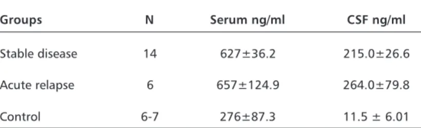
Comparing to control groups, MS patients sho-wed increased levels (p<0.001) of sICAM-1 in both CSF and blood samples (Table 1). Control group formed by individuals without clinical and/or labo-ratory signs of inflammation had mean levels of sICAM-1 in the serum of 276+87.3 ng/ml (range 187 to 387 ng/ml) considered in the normal range. In contrast, MS patients showed a marked increase of sICAM-1 in the serum: 639±194 ng/ml (range 322 to 1239 ng/ml). Likewise, CSF levels of sICAM
in MS patients were 230.5 ± 29.8 ng/ml (range 104 to 533 ng/ml) significantly higher (p< 0.001) than control group mean 11.05 (range 0 to 31.5 ng/ml) formed by IHD patients. In addition it was also ob-served a clear correlation of increased serum levels of sICAM-1 with length of disease (r = 0.46343), EDSS (r = 0.31801) and relapses (r = 0.14538).
Detection of CSF and serum soluble TNF-R1 (60kD )
Approximately a ten fold increase (p< 0.001) in the levels of sTNF-R in both serum (14.2±1.20 ng/ ml), range 8.66 to 22.2 ng/ml, and CSF (11.4±0.71 ng/ml), range 8.90 to 18.8 ng/ml, was consistently observed in MS patients. Regardless stage of dis-ease and lack of a positive correlation with degree of disability as reflected by EDSS scores (r = 0.0032), length of disease (r = 0.0029) and number of relap-ses (r = 0.0096), MS patients showed consistently (Table 2) higher concentrations of soluble TNF-R1 in comparison to respective control sera (1.59 ± 0.39 ng/ml) and CSF (1.28 ± 0.62 ng/ml),
DISCUSSION
Susceptibility to MS is genetically controlled, in-volving mainly gene related to auto-immune func-tions. These included MHC haplotypes, immuno-globulin, T cell receptor, cytokines (like TNFα), and other candidate autoantigen 22. In recent years, we
have seen growing evidence for the role of cytokines in the pathogenesis of several infectious and non-infectious inflammatory CNS disease states, inclu-ding MS. TNFα has been linked to inflammatory de-myelination of MS and animal models of immune-mediated CNS damage. Among cytokines and growth factors induced by brain damage, TNFα is well poised to modulate cellular injury as it is ex-pressed more rapidly (peak protein level within 3 to 6 hours from acute injury) than other cytokines. It has been identified in MS brain lesions, and high
Table 1. Measurement of soluble adhesion molecule ICAM-1.
Groups N Serum ng/ml CSF ng/ml
Stable disease 14 627±36.2 215.0±26.6
Acute relapse 6 657±124.9 264.0±79.8
Control 6-7 276±87.3 11.5 ± 6.01
The results expressed in ng/ml represent the mean value ± SD of serum and CSF levels of soluble sICAM-1 (CD54) assayed by ELISA. Normal values were obtained from sera pro-vided by six healthy subjects and CSF by seven patients with herniated disc.
Table 2. Measurement of soluble TNF-R1 (60 kd).
Groups N Serum CSF
Stable disease 14 14.9±1.56 11.6±0.89
Acute relapse 6 12.9±1.9 10.9±1.30
Control 6-7 1.59±0.39 1.28±0.65
concentrations have been reported in CSF and se-rum from MS patients. Moreover, CSF and systemic levels of TNFα in MS appears to reflect clinical dis-ease activity, mediating oligodendrocyte damage through the inhibition of potassium channels, in-duction of apoptosis, and upregulation of adhesion molecules, especially ICAM-1 23. It was observed
significantly increased levels of soluble ICAM-1 and soluble TNFα receptor in the CSF of 33 MS patients with acute relapsing MS during exacerbation and those with progressive disease when compared with 13 subjects with other neurological diseases 11. CSF
levels of sICAM-1 and sTNFα were positively corre-lated in patients with acute relapsing MS during an exacerbation11. This is in light of previous evidence
indicating that proinflammatory cytokines such as TNFα and adhesion molecules (sICAM-1) may rep-resent markers of inflammation3,4,11.
This study sought to evaluate with a high sensi-tive immunoassay the profile of soluble ICAM-1 and TNF-R1 in the serum and CSF samples in a popula-tion of Brazilian MS patients during active and stable phases of the disease. To the best of our knowledge this is the first report of a cytokine study and its cor-relation with clinical aspects of Brazilian MS patients. In the present study, we found a close correla-tion of increased serum and CSF levels of sICAM-1 with length of disease, EDSS and number of relapses, but not with clinical course. Indeed, MS patients either in acute relapsing or remission showed incre-ased levels of sICAM-1 in both serum and CSF. More-over, MS patients with clinically active disease showed altered MRI and very high levels of soluble ICAM-1 in both serum and CSF. The evidence of in-creased serum levels of sICAM-1 during acute re-lapses are in accordance with recently reported data11,14,24-26 showing elevated expression of
adhe-sion molecules (ICAM-1) on the luminal surface of endothelial cells in active MS lesions. In this sense, high concentrations of sICAM-1 and VCAM-1 in the peripheral blood during active disease would reflect BBB damage and may further serve as an adhesion ligand during extravasation of activated leukocytes into the brain tissue 24,27. Indeed the extent of
ICAM-1 activation may vary within different regions of the inflamed brain tissue which ultimately may ac-count for the varying degrees of neurological defi-cit2,4,28,29.
Comparing to control groups, we observed that regardless stage of disease, MS patients showed a ten fold increase of sTNF-R1 levels in the serum and CSF samples. Similarly, increased levels of soluble
TNF-R1 paralleled with circulating levels of sICAM-1 adhesion molecule. We also noticed that patients with clinical signs of active disease were those sho-wing increased levels of circulating soluble adhe-sion molecules and sTNF-R1. Soluble forms of TNFα -R1 seems to prolong the half-life of the cytokine by protecting the biological activity against proteolytic degradation 2,6,7 and ultimately influencing
neuropro-tective or neurotoxic effects2 with consequent death
or survival of oligodendrocytes and myelin damage. The presence of TNFα in inflammatory demyeli-nating lesions and its concentration in the CSF indi-cate a critical role for this cytokine in the patho-genesis of MS2,26-29 specially concerning
neurologi-cal impairment due to its myelinotoxic activity. A rise in TNFα production by blood mononuclear cells apparently precedes clinical exacerbations and is associated with active inflammatory lesions28.
In-deed, the severity of the relapse, measured by the EDSS score, highly correlate to the TNF produc-tion29,30. TNF-α up regulate expression of adhesion
molecules on endothelial cells and leukocytes thus facilitating adherence and extravasation of leuko-cytes into the CNS to initiate local tissue damage14,30.
The results reported herein indicate that regardless clinical course of disease (relapse or remission) MS patients showed a steady inflammatory process within the CNS microenvironment. In this sense, development of new therapeutic strategies should take into account that MS is a disease character-ized by a relentless inflammatory reaction within the brain tissue.
Acknowledgments - We thank Dr. Charles M. Poser for thoughtful discussions and criticism and Monica Caetano for sample processing.
REFERENCES
1. Compston DAS, Kellar Wood H, Roberstson N, Sawcer S, Wood NW. Genes and susceptibility to multiple sclerosis. Acta Neurol Scand 1995;161(Suppl.):43-51
2. Pouly S, Antel JP. Multiple sclerosis and central nervous system de-myelination. J Autoimmunity 2000;13:297-306
3. Amor S, Baker D, Layward L, McComarck K, vanNoort JM. Multiple Sclerosis: variations on a theme. Immunol Today 1997;18:368-371. 4. Hartung HP, Reiners K, Archelos JJ, et al. Circulating adhesion
mol-ecules and TNF receptor in multiple sclerosis: correlation with mag-netic resonance imaging. Ann Neurol 1995;38:186-193.
5. Ewing C, Bernard CC. Insights into the aetiology and pathogenesis of multiple sclerosis. Immunol Cell Biol 1998;76:47-54.
6. Bö L, Peterson JW, Mrk S, et al. Distribution of immunoglobulin su-perfamily members ICAM-1,-2,-3 and β2 integrin LFA-1 in multiple sclerosis lesions. J Neuropathol Exp Neurol 1996;55:1060-1072. 7. Muller WA, Randolph GJ. Migration of leukocytes across
endothe-lium and beyond: molecules involved in the transmigration and fate of monocytes. J Leuk Biol 1999; 66:698-704.
8. Cannella B, Raine C. The adhesion molecule and cytokine profile of multiple sclerosis lesion. Ann Neurol 1995; 37: 424-435.
10. Vora AJ, Kidd D, Miller DH, et al. Lymphocyte-endothelial cell inter-actions in multiple sclerosis: disease specificity and relationship to circulating tumor necrosis factor a and soluble adhesion molecules. Mult Scler 1997;3:171-179.
11. Tsukuda N, Matsuda M, Miyagi K, Yanagisawa N. Increased levels of circulating intercellular adhesion molecule (ICAM-1) and tumor ne-crosis factor receptor in the cerebrospinal fluid of patients with mul-tiple sclerosis Neurology 1993;43:2679-2682.
12. Trojano M, Abolió C, Ruggieri M, et al. Soluble intercellular adhesion molecule-I (sICAM-1) in serum and cerebrospinal fluid of demye-linating diseases of the central and peripheral nervous system. Mult Scler 1998; 4:39-44.
13. Sharief MK, Noori MA, Ciardi M, Cirelli A, Thompson EJ. Increased levels of circulating ICAM-1 in serum and cerebrospinal fluid of pa-tients with active multiple sclerosis: correlation with TNF-alpha and blood-brain barrier damage. J Neuroimmunol 1993; 43: 15-21. 14. Sacca R, Cuff CA, Ruddle NH. Mediators of Inflammation. Curr Op
Immunol 1997;9:851-857.
15. Zhang GX, Baker CM, Kolson DL, Rostami AM. Chemokines and chemokine receptors in the pathogenesis of multiple sclerosis. Mult Scler 2000;6:3-13.
16. Monteyne P, Sindic CJM. Data on cytokine mRNA expression in CSF and peripheral blood mononuclear cells from MS patients as detected by PCR. Mult Scler 1998;4:143-146.
17. Huang W-X, Huang P, Link H, Hillert J. Cytokine analysis in multiple sclerosis by competitive RT-PCR: a decreased expression of TNF in chronic progression. Mult Scler 1999;5:342-348.
18. Rohowsky-Kochan C, Molinaro D, Cook SD. Cytokine secretion pro-file of myelin basic protein T cells in multiple sclerosis. Mult Scler 2000;6:69-77.
19. Coulombier D, Hathcock L, Fajan R. EpiInfo, a word processing da-tabase and statistics program for public health from center for
dis-ease control (CDC, USA) and from World Health Organization. EpiInfo Executive Health information shell. Epi-Info 1.0. Geneva, 1994.
20. Poser CM, Paty DW, Scheinberg L, et al. New diagnostic criteria for multiple sclerosis: guidelines for research protocols. Ann Neurol 1983;13:227-231.
21. Kurtzke JF. Rating neurologic impairment in multiple sclerosis: an expanded disability status scale (EDSS). Neurology 1983;33:1444-1452.
22. Wekerle H. Immune pathogenesis of multiple sclerosis: brain au-toimmune reactivity and its control by neuronal function. Mult Scler 1998;4:136-137.
23. Sharief MK. Cytokines in multiple sclerosis: pro-inflammation or pro-remyelination? Mult Scler 1998;4:169-173.
24. Navikas V, Link H. Cytokines and the pathogenesis of MS. J Neurosci Res 1996;45:322-333.
25. Hvas J, Mclean C, Justsen J, et al. Perivascular T cells express the proinflammatory chemokine RANTES mRNA in multiple sclerosis. Scand J Immunol 1997;46:195-203.
26. Link H. Cytokine storm in multiple sclerosis. Mult Scler 1998;4:12-15. 27. Whalen MJ, Carlos TM, Dixon CE, et al. Reduced brain edema after
traumatic brain injury in mice deficient in P-selectin and intercellu-lar adhesion molecule-1. J Leuk Biol 2000;67:160-168.
28. Beck J, Rondot P, Catinot L, et al. Increased production of interferon gamma and tumor necrosis factor precedes clinical manifestation in multiple sclerosis: do cytokines trigger off exacerbations? Acta Neurol Scand 1988;78:318-323.
29. Liu J, Marino MW, Wong G,et al. TNF is a potent anti-inflammatory cytokine in autoimmune-mediated demyelination. Nature Med 1998;4:78-83.