The purposeful application of bio com-patible nanomaterials of low toxicity is a relevant problem in modern biotechnology. It is assumed that the use of nanoparticles will help to solve the problems of early disease diagnostics and targeted delivery of drugs into tissues and cells, as well as new methods of selective therapies. C60 fullerene has a prominent position among other potentially effective therapeutic agents [1]. It is known that C60 molecule normalizes cellular metabolism and neural processes, increases resistance to stress, exhibits antiviral properties, has pronounced anti-inflammatory and anti-allergic effects, enhances activity of enzymes and regenerative capacity of tissues [1]. Finally, C60 fullerene and its derivatives can be used for the treatment of cancer [2–4].
C60 fullerene can be readily dissolved in organic solvents, yet it is practically insoluble
in water [5]. Poor solubility in water severely hampers investigation of physiological and pharmacological effects of C60 fullerene. As a consequence, a number of C60 fullerene derivatives with better solubility in polar solvents have been synthesized to date [5–6]. Pristine C60 fullerene aqueous solution (С60FAS) has been received by transferring C60 molecules from toluene to water followed by sonication [7–8]. The dark brown solution was identified to be a typical colloid system containing a single C60 molecule and spherical clusters with the diameter of 2–3 nm or more (depending on the C60 fullerene concentration in water) in a hydrated state [7–9].
Due to its nanoscale dimensions, the water-soluble pristine C60 fullerene can penetrate the cell membrane [10].
Toxicity is a major concern for the use of C60 fullerene-containing drugs for biomedical
УДК 546.26.043 doi 10.15407/biotech7.03.043
C
60
FULLERENE EFFECT ON THE DYNAMICS
OF FATIGUE PROCESSES IN RAT SOLEUS MUSCLE
AFTER ISCHEMIA-REPERFUSION
Key words: C60 fullerene, skeletal muscle contraction, ischemia-reperfusion injury.
D. M. Nozdrenko1 1,2 Joint Ukrainian-German Center on Nanobiotechnology K. I. Bogutska1
Yu. I. Prylutskyy1 1 Taras Shevchenko National University of Kyiv, Ukraine U. Ritter2
P. Scharff2 2 Technical University of Ilmenau, Institute of Chemistry and Biotechnology, Germany
E-mail: [email protected]
Received 28.02.2014
applications since they can have damaged effects on human cells and animals [11]. It is important to point out that the С60FAS used does not display toxic effects towards rat erythrocyte and thymocyte cells at concentrations below 0.1 mg/ml within 24 h of incubation [12, 13].
The ability of C60 fullerene and its derivatives to inactivate reactive oxygen species has been described first by [14]. Indeed, C60 molecule is capable of attaching 34 methyl radicals. Antioxidant efficiency of C60 fullerene depends on the number of active centers in the molecule and the distance between active centers and target atoms. C60fullerene can quench superoxide anion and hydroxyl radicals in both in vivo and in vitro systems [15–17].
The ischemia-reperfusion injury of various organs or tissues has a complex pathogenesis which includes vascular dysfunction, inflammation and edema. Herewith, the key role in the pathogenesis of ischemia-reperfusion injury to tissues and organs belongs to free oxygen radicals. It is assumed that different types of antioxidants are able to mitigate ischemia-reperfusion organs or tissues injury. Thus, [18] estimated the protective effect of a water-soluble C60 fullerene derivative [С60(ONO2)7±2] in the isolated ischemic-reperfused rat lung. This drug has been shown to possess antioxidant properties and be able to furthermore release nitric oxide displaying the effects similar to those of nitroglycerine. Experimental protocol included 10 min of stabilization, 45 min of ischemia and 60 min of reperfusion. The lungs were ventilated with gas mixture containing 95% О2 and 5% СО2. Ischemia caused increase in pulmonary arterial, lung weight and filtration coefficients in controls, but C60 fullerene derivative limited this increase, and this was considered to alleviate the ischemia-reperfusion lung injury.
In canine model [19], 60 min of small intestinal ischemia were followed by 1 h of reperfusion. A water-soluble C60 fullerene derivative (fullerenol) at a dose of 1 mg/kg was administered intravenously 30 min prior to ischemia (preventively) and immediately after reperfusion (therapeutically). An increased amount of both malondialdehyde (MDA) and conjugated dienes (CD) was found in the intestinal tissue at the 30th and 60th min of reperfusion in control experiments. On the contrary, the tissue content of glutathione decreased after 60 min of reperfusion. Fullerenol administration did not alter
histological picture but caused significant decrease in tissue content of MDA and CD and furthermore increased glutathione level in both preventive and therapeutic protocols.
Yang et al. [20] investigated the influence of a water-soluble C60 fullerene derivative (hexasulfobutyl[60]fullerene) on the ischemic brain injury caused by permanent 24 h occlusion of middle cerebral artery (MCA) in gerbils. Three groups of animals were investigated: controls, and receiving low (0.5 mg/kg/day) and high (5.0 mg/kg/day intraperitoneally during 2 weeks) dose of the aforesaid compound. After 24 h of MCA occlusion the infarct size was determined with triphenylterazolium chloride staining. Prolonged hexasulfobutyl[60]fullerene therapy resulted in significant decrease in cerebral infarct size (by 42–68% in comparison with controls). The authors suggested that neuroprotective effects of this drug are secondary to its antioxidant properties.
A water-soluble C60 fullerene derivative (carboxyfullerene) was injected 30 min prior to ischemia- reperfusion either intravenously or intracerebroventricularly [21]. Carboxy-fullerene did not alter infarct size after intravenous administration, which may be due to limited permeability of blood-brain barrier for this compound. Local administration of the above-mentioned drug resulted in infarct size restriction, preservation of tissue glutathione pools and decreased amount of lipid peroxidation products in the ischemic brain cortex.
Despite the large number of studies on the interaction of С60 fullerene and its derivatives with biological objects in vitro and in vivo, a little information about the impact of pristine water-soluble C60 fullerene on the functional properties of skeletal muscles was reported [22]. Furthermore, at present time there is no information about the C60 fullerene effect on the ischemic injury of skeletal muscle. Hence, the aim of this study was to estimate the protective effect of С60FAS in ischemia-reperfusion injury of rat soleus muscle.
Materials and methods
formed were treated in ultrasonic bath. The procedure was continued until the toluene had completely evaporated and the water phase was colored yellow. Filtration of the aqueous solution allowed to separate the product from undissolved C60 fullerenes. The filter pore size during the filtration of aqueous solution was smaller than 2 µm (Typ Whatmann 602 h1/2).
The purity of prepared C60FAS samples was determined by HPLC and GC/MS techniques using standard programs. Insoluble C60FAS impurities were determined by ultra-centrifugation: their amount was identified to constitute less than 1 µg/ml. Traces of toluene after synthesis could not be detected in water by GC/MS analysis. Moreover, 1H NMR spectrum (400 MHz) of C60FAS recorded in heavy water did not reveal any residual proton signals.
The concentration of C60 fullerene in the prepared C60FAS sample was determined as the concentration of total organic carbon in aqueous solution (Analytik Jena TOC Analyser multi N/C 3100). In our experiments the C60FAS sample with 0.15 mg/ml concentration of C60 fullerene was used. The resulting atomic force microscopy images clearly indicate the presence of both individual C60 fullerenes with diameter ~0.7 nm and their aggregates with a typical diameter up to 50 nm in water [23].
Experiments were performed on 30 male Wistar rats under 3 months of 170±5 g weight. Administration of C60FAS at a dose of 1 mg/ kg was carried out in two ways: intravenously and intramuscularly 2 h prior to the start of experiment at 1, 2, 3, 4 and 5th experimental day.
The animals used in this study were treated in accordance with international principles of the European Convention for the protection of vertebrate animals used for experimental and other scientific purposes (Strasbourg, 1986).
It is important to know that the dose of C60 fullerene applied in our experiments was significantly lower than the LD50 value defined for C60 fullerene which is equivalent to 600 mg/ kg of body weight for the oral administration to mice [16].
Anesthesia of animals was performed by intraperitoneal administration of nembutal (40 mg/kg). For muscle ischemia the branch of the femoral artery of the animal, which provides blood supply of experimental muscle, was dragged by ligatures. After 2 h of ischemia, reperfusion was performed by cutting the fixing ligatures. Preparation of the experiment also included cannulation (a. carotis communis sinistra) for pharmaceuticals administration and pressure measurement,
tracheotomy and laminectomy at the lumbar spinal cord level. Rat soleus muscle was freed from the surrounding tissues. Its tendon portion was transversely cut in the distal part. The ventral roots were cut in sites of their exit from the spinal cord for modulated stimulation of efferents in L7-S1segments.
Variation of muscle contraction was measured using hypersensitive strain gauges, which measure the change in resistance of an array of single-walled carbon nanotubes (SWCNTs) by deformation [24, 25]. SWCNTs were located at the rear end of the micropipette, while its front part was attached to the investigated muscle tendon. The programmable signal generator of special form was used to form the stimulating electrical signal with duration of 6 s.
The muscle contraction strength was measured during the first 5 h and at 1, 2, 3, 4 and 5th experimental day after muscle ischemia.
The study of dynamic properties of muscle contraction was performed under conditions of muscle activation using modulated stimulation of efferents. Five filaments of ventral roots cut were fixed on stimulating electrodes and a special device was used for cyclic sequence distribution of electrical signals to stimulate the filaments. The distributed stimulation allowed monotonous and uniform muscle contraction at low stimulation frequencies of individual filaments. Stimulation of efferents in L7-S1 segments was performed by electric pulses of 2 ms, generated by pulse generator controlled through the platinum electrodes. The characteristics of stimulated signal were programmed and transmitted to generator. The external load on the muscle was controlled with the help of mechanical stimulators. The electromagnetic linear motor was used for perturbation load.
The experimental curves obtained reflect the change of the studied parameters as a percentage of intact muscle control parameters, which were considered as 100%.
The statistical analysis of the experimental data was performed using a Student t-test (the level of significance was Р ≤ 0.05).
Results and discussion
muscle necrosis and apoptosis. The primary aim in the treatment of ischemia is rapid restoration of blood flow (reperfusion) to the damaged sites. However, reperfusion therapy leads to a new pathophysiological process called reperfusion injury, which also causes significant tissue damage [27]. Ischemia-reperfusion injury of skeletal muscle caused by the acute arterial occlusion is lethal in many cases [28]. In addition, ischemia-reperfusion injury of skeletal muscle might be one of a major cause of post-traumatic pathologies after surgical procedures [29, 30]. Rapid detection of the extent of ischemic injury is crucial for the further therapy, yet there are no accurate diagnostic tests to achieve this goal at the moment [30]. Ischemia-reperfusion pathological processes were found to reduce the muscle contraction force to 40% after 1 h of ischemia and to 90% after 2 h. Its recovery was observed only at the end of the 2nd week after ischemia-reperfusion [29]. There is also a high correlation between the duration of ischemia and viability of a muscle fiber [31]. Even considering that different types of skeletal muscle fibers are characterized by significant metabolic differences, it has no significant influence on their susceptibility to ischemia-reperfusion injury [32]. Experimental data indicate that the initial pathological effects after prolonged ischemia-reperfusion may be incomplete, thereby prolonging ischemic condition for several days [33]. These data established the basis for the study and analysis of the dynamics of fatigue processes occurring in the rat soleus muscle on the background of ischemia-reperfusion pathology development within the first 5 h after reperfusion and over the 5 days following it under therapeutic administration of С60FAS.
Fig. 1 shows the curves of rat soleus muscle maximum strength change, obtained under the influence of stimulating irritation with duration of 3 min after 1, 2, 3, 4 and 5 h following the 2 h long muscle ischemia and subsequent reperfusion.
The applied relaxationless stimulation allowed to analyze the development of fatigue processes within the first hours after reperfusion. In control experiments, the level of reduction of the maximum contractile strength of muscle was from 20% to 25%. However, regardless of the method of therapeutic administration of С60FAS, its protective effect was manifested in reducing this index to 10% (Fig. 2).
The most pronounced protective effect of С60FAS was manifested at comparing
changes in the levels of maximum strength generation between the beginning and the end of stimulating irritation (Fig. 3) (in muscle without expressed pathologies this parameter is practically zero). In this case С60FAS reduced this figure to 5% vs. 20% in control regardless of the drug administration route. It should be noted that such protective effect of С60FAS has a significant therapeutic value because it’s manifested within the first hours after reperfusion, when the basic physiological
Fig. 1. The maximum strength (F) generation curves of ischemic rat soleus muscle under the influence of
stimulating irritation with 3 min duration: a — ischemic muscle without affecting the drug (control); b — intravenous administration of C60FAS; c — intramuscular administration of C60FAS.
Here and further: (n = 10; 1 mg/kg); ΔF = F(t1) – F(t2), where t1 and t2 is the time of the beginning and the end of stimulated irritation, correspondingly; 1, 2, 3, 4, and 5 are the hours after 2 h of muscle ischemia-reperfusion
a
processes take place, what leads to severe pathology as a consequence [34]. At the same time, in spite of the protective effect of С60FAS, under ischemia-reperfusion processes a progressive decrease of the strength of muscle contraction is observed for 5–6 days, after which a slow recovery process takes place [35–37].
Pathological processes in muscle after reperfusion last several days with progressive dynamics. Migration of neutrophils into the endomysium and later to perimysium already occurs within 24 h after the 2 h long ischemia-reperfusion. Structural process of regeneration starts only at the end of the first week after the 2 h long reperfusion. There is functional and morphological evidence of ischemic and reperfusion injury of muscle tissue even 1 week after reperfusion. Meanwhile, the increase of ischemia from 1 to 2 h delays regeneration processes [29]. Therefore, the next stage of
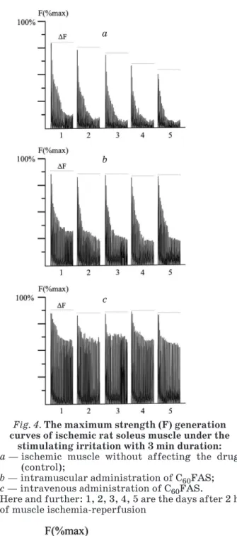
the study was to investigate the dynamics of muscle contractile process caused by the analogous stimuli at the 1, 2, 3, 4 and 5 day after 2 h long ischemia-reperfusion (Fig. 4).
Apparently, the manifestation of fatigue processes takes place throughout the experiment. Simultaneously, a protective effect of С60FAS towards the damaged muscle tissue is dramatically manifesed: namely, while the reduction of the maximum muscle contraction strength at the 5th day of the experiment reached over 30% in the control, the maximum contractile strength at the therapeutic administration of С60FAS was increased by 20% and 30% by its intramuscular and intravenous administration, respectively, which nearly eliminated the pattern of pathological disorders of muscle contraction caused by ischemia injury (Fig. 5).
The most significant protective effects of С60FAS were manifested at comparing the changes in the levels of maximum strength generation between the beginning and the end of stimulating irritation (Fig. 6). The decrease rate of this indicator was 63% in the control group (without drug administration) on day 1 of the experiment and decreased to 92% by day 5 of the study. Under intramuscular drug administration this reduction was 25% and 43%, respectively, while the intravenous administration of С60FAS reduced the difference of the strength response at the beginning and the end of stimulation to 23% and 25%, respectively. Thus, intravenous administration of the drug is more effective with regard to its impact on the development of fatigue processes in the damaged muscle.
The aforementioned effects may be observed due to the fact that the 2 h long ischemia-reperfusion of musculus soleus significantly reduces the concentration of ATP simultaneously with significantly increasing lactate. The ATP depletion at 3 h long ischemia is known to constitute approximately 95%, while glycogen is exhausted by 88% [34]. Furthermore, we can assume that a large number of macroergic phosphates is consumed by a damaged muscle cell to maintain hemostasis and, as a result of metabolic disorders, leads to increased muscle fatigue.
At the same time, the data available from literature indicate that free radicals (e. g., superoxide anion and hydroxyl groups) represent a major pathogenic factor in the process of ischemia-reperfusion tissue damage. It includes the initiation of lipid peroxidation, the direct inhibition of mitochondrial respiratory chain enzymes, inactivation of the Fig. 2. The change in the maximum strength (F) of
contraction of ischemic rat soleus muscle: ( ) — ischemic muscle without affecting the drug;
( ) — intravenous administration of C60FAS; ( ) — intramuscular administration of C60FAS.
Here and further: *P ≤ 0.05
a
c b
Fig. 3. The level of change in the maximum strength (ΔF) of contraction of ischemic rat soleus
muscle between the beginning and the end of stimulated irritation:
a — ischemic muscle without affecting the drug (control);
glyceraldehyde-3-phosphate dehydrogenase, inhibiting ATP-ase activity, the inactivation of membrane sodium channels, etc. [38]. Superoxide anion radical is produced after ischemia at the stage of reperfusion in much larger quantities and quickly reacts with NO⋅ [39]. The ascorbic acid, which is a recognized antioxidant, was shown to have a positive effect on the reduction of muscle injury caused by ischemia-reperfusion [28]. It is assumed that the modified C60 fullerene may be regarded as a potent scavenger of free radicals induced by ischemia-reperfusion small intestine injury [19]. The feasibility of C60 fullerene derivatives to reduce ischemia-reperfusion lung injury has also been shown [18, 40]. In this regard, the protective effect of С60FAS on the fatigue processes in ischemically damaged muscle can be directly linked to the strong antioxidant properties of the pristine C60 fullerene [17].
Thus, the above data obtained for the first time demonstrate a pronounced protective effect of pristine С60 fullerene by the means of injection of its colloidal aqueous solution on the fatigue processes of muscle soleus contraction during its ischemia-reperfusion injury, namely: during the study of the reduction level of maximum muscle contraction strength within the first 5 h after muscle ischemia injury, the protective effect of C60FAS constituted 10–15% of the control and did not depend on the method of drug administration; during the study of changes in the levels of maximum strength generation between the beginning and the end of the stimulus the a
c b
Fig. 4. The maximum strength (F) generation curves of ischemic rat soleus muscle under the
stimulating irritation with 3 min duration: a — ischemic muscle without affecting the drug
(control);
b — intramuscular administration of C60FAS; c — intravenous administration of C60FAS.
Here and further: 1, 2, 3, 4, 5 are the days after 2 h of muscle ischemia-reperfusion
Fig. 5. The change in the maximum strength (F) of contraction of ischemic rat soleus muscle: ( ) — ischemic muscle without affecting the drug
(control);
( ) — intramuscular administration of C60FAS; ( ) — intravenous administration of C60FAS
a
c b
Fig. 6. The level of change in the maximum strength (ΔF) of contraction of ischemic rat soleus
muscle between the beginning and the end of stimulated irritation:
a — ischemic muscle without affecting the drug (control);
protective effect of C60FAS was 15% (relative to controls) within the first 5 h after muscle ischemia injury and increased to 92% by the 5th day of the experiment. In this case, intravenous administration of therapeutic C60FAS is considered to be the most optimal route: its protective effect was 67% versus 49% under intramuscular administration.
Thus, the development of biomedical nanotechnology with the application of pristine
C60 fullerenes, based on their expressed antioxidant properties and the lack of data on induced acute and chronic intoxications, opens up new possibilities in the prevention and treatment of ischemic pathologies.
Acknowledgments. Dr. D. M. Nozdrenko is grateful to Ministry of Education and Science of Ukraine for financial support.
REFERENCES
1. Medicinal chemistry and pharmacological potential of fullerenes and carbon nanotubes. Series: Carbon materials: chemistry and physics. Cataldo F., Da Ros T. (eds.). V. 1, Springer Netherlands, 2008.
2. Zhu J., Ji Zh., Wang J., Sun R., Zhang X., Gao Y., Sun H., Liu Y., Wang Z., Li A., Ma J., Wang T., Jia G., Gu Y. Tumor-inhibitory effect and immunomodulatory activity of fullerol C60(OH)x. Small. 2008, V. 4, P. 1168–1175. 3. Prylutska S. V., Burlaka A. P., Klymenko P. P.,
Grynyuk I. I., Prylutskyy Yu. I., Schuetze Ch., Ritter U. Using water-soluble C60 fullerenes in anticancer therapy. Cancer Nanotechnol. 2011, V. 2, P. 105–110.
4. Prylutska S. V., Burlaka A. P., Prylutskyy Yu. I., Ritter U., Scharff P. Pristine C60 fullerenes inhibit the rate of tumor growth and meta sta-sis. Exp. Oncol. 2011, V. 33, P. 162–164. 5. Mchedlov-Petrossyan N. O. Fullerenes in
liquid media: an unsettling intrusion into the solution chemistry. Chem. Rev. 2013, V. 113, P. 5149–5193.
6. Hirsch A., Brettreich M. Fullerenes — chemistry and reactions. New York: John Wiley & Sons, 2005.
7. Prilutski Yu., Durov S., Bulavin L., Pogorelov V., Astashkin Yu., Yashchuk V., Ogul’chansky T., Buzaneva E., Andrievsky G. Study of structure of colloidal particles of fullerenes in water solution. Mol. Cryst. Liq. Cryst. 1998, V. 324, P. 65–70.
8. Prylutskyy Yu. I., Durov S. S., Bulavin L. A., Adamenko I. I., Moroz K. O., Geru I. I., Dihor I. N., Scharff P., Eklund P. C., Grigorian L. Structure and thermophysical properties of fullerene C60 aqueous solutions. Int. J. Thermophys. 2001, V. 22, P. 943–956.
9. Prylutskyy Yu. I., Buchelnikov A. S., Voro-nin D. P., Kostjukov V. V., Ritter U., Parkin-son J. A., Evstigneev M. P. C60 fullerene aggregation in aqueous solution. Phys. Chem. Chem. Phys. 2013, V. 15, P. 9351–9360. 10. Prylutska S., Bilyy R., Overchuk M., Bychko A.,
Andreichenko K., Stoika R., Rybalchenko V., Prylutskyy Y., Tsierkezos N. G., Ritter U.
Water-soluble pristine fullerenes C60 increase the specific conductivity and capacity of lipid model membrane and form the channels in cellular plasma membrane. J. Biomed. Nanotechnol. 2012, V. 8, P. 522–527.
11. Johnston H. J., Hutchison G. R., Christen-sen F. M., Aschberger K., Stone V. The biological mechanisms and physicochemical characteristics responsible for driving fullerene toxicity. Toxicol. Sci. 2010, V. 114, P. 162–182.
12. Prylutska S. V., Matyshevska O. P., Golub A. А., Prylutskyy Yu. I., Potebnya G. P., Ritter U., Scharff P. Study of С60 fullerenes and С60 -containing composites cytotoxicity in vitro. Mater. Sci. Engineer. C: Mater. Biolog. Appl. 2007, V. 27, P. 1121–1124.
13. Prylutska S. V., Grynyuk I. I., Grebinyk S. M., Matyshevska O. P., Prylutskyy Yu. I., Ritter U., Siegmund C., Scharff P. Comparative study of biological action of fullerenes C60 and carbon nanotubes in thymus cells. Mat.-wiss. Werkstofftech. 2009, V. 40, P. 238–241. 14. Krustic P. J., Wasserman Е., Keizer P. N.,
Morton J. R., Preston K. F. Radical reactions of С60. Science. 1991, V. 254, P. 1183–1185. 15. Scharff P., Carta-Abelmann L., Siegmund C.,
Matyshevska O. P., Prylutska S. V., Koval T. V., Golub A. A., Yashchuk V. M., Kushnir K. M., Prylutskyy Yu. I. Effect of X-ray and UV irradiation of the C60 fullerene aqueous solution on biological samples. Carbon. 2004, V. 42, P. 1199–1201.
16. Gharbi N., Pressac M., Hadchouel M., Szwarc H., Wilson S.R., Moussa F. C60 fullerene is a powerful antioxidant in vivo with no acute or subacute toxicity. Nano Lett. 2005, V. 5, P. 2578–2585.
17. Prylutska S. V., Grynyuk I. I., Matyshevska O. P., Prylutskyy Yu. I., Ritter U., Scharff P. Anti-oxidant properties of C60 fullerenes in vitro. Fullerenes, Nanotubes, Carbon Nanostruct. 2008, V. 16, P. 698–705.
19. Lai H. S., Chen W. J., Chiang L. Y. Free radical scavenging activity of fullerenol on the ischemia-reperfusion intestine in dogs. World J. Surg. 2000, V. 24, P. 450–454. 20. Yang D. Y., Wang M. F., Chen I. L., Chan Y. C.,
Lee M. S., Cheng F. C. Systemic administration of water-soluble hexasulfonated C60 (FC(4) S) reduces cerebral ischemia induced infarct volume in gerbils. Neurosci. Lett. 2001, V. 311, P. 121–124.
21. Lin A. M. Y., Fang S. F., Lin S. Z., Chou C. K., Luh T. Y., Ho L. T. Local carboxyfullerene protects cortical infarction in rat brain. Neurosci. Res. 2002, V. 43, P. 317–321. 22. Andreichenko K. S., Prylutska S. V.,
Medyns-ka K. O., BogutsMedyns-ka K. I., Nurishchenko N. E., Prylutskyy Yu. I., Ritter U., Scharff P. Ef-fect of fullerene C60 on ATPase activity and superprecipitation of skeletal muscle acto-myosin. Ukr. Biokhim. Zh. 2013, V. 85, P. 20–26.
23. Prylutskyy Yu. I., Petrenko V. I., Ivankov O. I., Kyzyma O. A., Bulavin L. A., Litsis O. O., Evstigneev M. P., Cherepanov V. V., Naumo-vets A. G., Ritter U. On the origin of C60fullerene solubility in aqueous solution. Langmuir. 2014, V. 30, P. 3967–3970. 24. Prylutskyy Yu. I., Ogloblya O. V., Eklund P.,
Scharff P. Electronic properties of carbon nanotubes with defects. Synth. Met. 2001, V. 121, P. 1209–1210.
25. Ogloblya O. V., Prylutskyy Yu. I., Strzhemech-ny Yu. M. Peculiarities of conductance of carbon nanotube-based quantum dots. Int. J. Quantum Chem. 2010, V. 110, P. 195–201.
26. Murdock M., Murdoch M. M. Compartment syndrome: a review of the literature. Clin. Podiatr. Med. Surg. 2012, V. 29, P. 301–310. 27. Wang W. Z., Baynosa R. C., Zamboni W. A. Therapeutic interventions against reper-fusion injury in skeletal muscle. J. Surg. Res. 2011, V. 171, P. 175–182.
28. Erkut B., Özyazıcıoğlu A., Karapolat B. S., Koçoğulları C. U., Keles S., Ateş A., Gundogdu C., Kocak H. Effects of ascorbic Acid, alpha-tocopherol and allopurinol on ischemia-reperfusion injury in rabbit skeletal muscle: an experimental study. Drug Target Insights. 2007, V. 2, P. 249–258.
29. Rácz I. B, Illyés G., Sarkadi L., Hamar J. The functional and morphological damage of ischemic reperfused skeletal muscle. Eur. Surg. Res. 1997, V. 29, P. 254–263.
30. Bortolotto S. K., Morrison W. A., Messina A. The role of mast cells and fibre type in ischemia reperfusion injury of murine skeletal muscles. J. Inflamm. (Lond). 2004, V. 1, P. 1–7.
31. Turóczi Z., Arányi P., Lukáts Á., Garbaisz D., Lotz G., Harsányi L., Szijártó A. Muscle fiber viability, a novel method for the fast detection of ischemic muscle injury in rats. PLoS One. 2014, V. 9, P. e84783.
32. Sternbergh W. C., Adelman B. Skeletal muscle fiber type does not predict sensitivity to postischemic damage. J. Surg. Res. 1992, V. 53, P. 535–541.
33. Loerakker S., Oomens C. W., Manders E., Scha-kel T., Bader D. L., Baaijens F. P., Nicolay K., Strijkers G. J. Ischemia-reperfusion injury in rat skeletal muscle assessed with T2-weighted and dynamic contrast-enhanced MRI. Magn. Reson. Med. 2011, V. 66, P. 528–537.
34. Carvalho A. J., McKee N. H., Green H. J. Metabolic and contractile responses of fast and slow twitch rat skeletal muscles to ischemia and reperfusion. Plast. Reconstr. Surg. 1997, V. 99, P. 163–171.
35. Grace P. A. Ischemia-reperfusion injury. Br. J. Surg. 1994, V. 81, P. 637–647.
36. Carvalho A. J., Hollett P., McKee N. H. Recovery of synergistic skeletal muscle function following ischemia. J. Surg. Res. 1995, V. 59, P. 527–533.
37. Tidball J. G. Mechanisms of muscle injury, repair, and regeneration. Compr. Physiol. 2011, V. 1, P. 2029–2062.
38. Cuzzocrea S., Riley D. P., Caputi A. P., Sal ve-mini D. Antioxidant therapy: a new pharma-co logical approach in shock, inflammation, and ischemia-reperfusion injury. Pharmacol. Rev. 2001, V. 53, P. 135–159.
39. Matheis G., Sherman M. P., Buckberg G. D., Habron D. M., Young H. H., Ignarro L. J. Role of L-arginine-nitric oxide pathway in myocardial reoxygenation injury. Am. J. Physiol. 1992, V. 262, P. H616–620.
ВПЛИВ C60-ФУЛЕРЕНУ НА ДИНАМІКУ ВТОМЛЮВАЛЬНИХ ПРОЦЕСІВ У КАМБАЛОПОДІБНОМУ М’ЯЗІ ЩУРА
ПІСЛЯ ІШЕМІЇ-РЕПЕРФУЗІЇ
Д. М. Ноздренко1 К. І. Богуцька1 Ю. І. Прилуцький1
У. Ріттер2 П. Шарфф2
1, 2 Спільний Українсько-Німецький центр
з нанобіотехнології
1 Київський національний університет
імені Тараса Шевченка, ННЦ «Інститут біології», Україна
2 Teхнічний університет Ілменау,
Інститут хімії і біотехнології, ФРН
E-mail: [email protected]
З використанням тензометричного методу досліджено вплив водного колоїдного розчи-ну немодифікованого С60-фулерену (1 мг/кг) на динаміку втомлювальних процесів у кам-балоподібному м’язі щура після ішемічно-ре-перфузійного ушкодження. Експерименти проводили упродовж перших 5 год і 5 діб після ішемії. Аналізували зміни максимальної сили скорочення м’яза та рівня його генерації між початком і кінцем стимулювального подраз-нення тривалістю 3 хв за внутрішньовенного і внутрішньом’язового введення водного колоїд-ного розчину немодифіковаколоїд-ного С60-фулерену. Вперше виявлено виражений захисний ефект препарату на динаміку м’язового скорочення. Захисна дія водного колоїдного розчину немо-дифікованого С60-фулерену за порівняння змін у рівнях генерації максимальної сили скоро-чення скелетного м’яза між початком і кінцем стимулювального подразнення становила 15% у перші 5 год після ішемії і зростала до 92% на 5-ту добу експерименту. При цьому внутріш-ньовенне введення водного розчину фулерену було найбільш оптимальним: захисний ефект досяг 67% проти 49% за внутрішньом’язового введення. Отже, розвиток біомедичних нано-технологій із застосуванням немодифіковано-го С60-фулерену як сильного антиоксиданта відкриває нові можливості у профілактиці та лікуванні ішемічних ушкоджень скелетних м’язів.
Ключові слова: С60-фулерен, ішемічно-репер-фу зійне ушкодження.
ВЛИЯНИЕ C60-ФУЛЛЕРЕНА НА ДИНАМИКУ УСТАЛОСТНЫХ ПРОЦЕССОВ В КАМБАЛОВИДНОЙ МЫШЦЕ КРЫСЫ ПОСЛЕ
ИШЕМИИ-РЕПЕРФУЗИИ
Д. Н. Ноздренко1 К. И. Богуцкая1 Ю. И. Прилуцкий1
У. Риттер2 П. Шарфф2
1, 2 Совместный Украинско-Немецкий центр
нанобиотехнологии
1 Киевский национальный университет
имени Тараса Шевченко,
ННЦ «Институт биологии», Киев, Украина 2 Teхнический университет Илменау,
Институт химии и биотехнологии, ФРГ
E-mail: [email protected]
С использованием тензометрического мето-да исследовано влияние водного коллоидного раствора немодифицированного С60-фуллерена (1 мг/кг) на динамику усталостных процессов в камбаловидной мышце крысы после ишеми-чески-реперфузионного повреждения. Опыты проводили в течение первых 5 ч и 5 сут после ишемии. Анализировали изменения макси-мальной силы сокращения мышцы и уровня ее генерации между началом и концом стиму-лирующего раздражения длительностью 3 мин при внутривенном и внутримышечном введе-нии водного коллоидного раствора немодифи-цированного С60-фуллерена. Впервые обнару-жен выраобнару-женный защитный эффект препарата на динамику мышечного сокращения. Защит-ное действие водного раствора фуллерена при сравнении изменений в уровнях генерации максимальной силы сокращения скелетной мышцы между началом и концом стимулирую-щих раздражений составило 15% в первые 5 ч после ишемии и увеличилось до 92% к 5-м сут эксперимента. При этом внутривенное введе-ние водного коллоидного раствора немодифи-цированного С60-фуллерена оказалось наиболее оптимальным: защитный эффект составил 67% против 49% при внутримышечном введении. Таким образом, развитие биомедицинских нанотехнологий с применением немодифици-рованного С60-фуллерена в качестве сильного антиоксиданта открывает новые возможности в профилактике и лечении ишемических по-вреждений скелетных мышц.