Antagonist G-mediated targeting and
cytotoxicity of liposomal doxorubicin in
NCI-H82 variant small cell lung cancer
Laboratory of Pharmaceutical Technology, Faculty of Pharmacy and Center for Neuroscience and Cell Biology, University of Coimbra, Coimbra, Portugal
J.N. Moreira and R. Gaspar
Abstract
The aim of the present study was to characterize the interactions of antagonist G (H-Arg-D-Trp-NmePhe-D-Trp-Leu-Met-NH
2)-targeted sterically stabilized liposomes with the human variant small cell lung cancer (SCLC) H82 cell line and to evaluate the antiprolifera-tive activity of encapsulated doxorubicin against this cell line. Variant SCLC tumors are known to be more resistant to chemo-therapy than classic SCLC tumors. The cellular association of antagonist G-targeted (radiolabeled) liposomes was 20-30-fold higher than that of non-targeted liposomes. Our data suggest that a maximum of 12,000 antagonist G-targeted liposomes were inter-nalized/cell during 1-h incubation at 37ºC. Confocal microscopy experiments using pyranine-containing liposomes further confirmed that receptor-mediated endocytosis occurred, specifically in the case of targeted liposomes. In any of the previously mentioned experiments, the binding and endocytosis of non-targeted lipo-somes have revealed to be negligible. The improved cellular asso-ciation of antagonist G-targeted liposomes, relative to non-tar-geted liposomes, resulted in an enhanced nuclear delivery (evalu-ated by fluorimetry) and cytotoxicity of encapsul(evalu-ated doxorubicin for incubation periods as short as 2 h. For an incubation of 2 h, we report IC50 values for targeted and non-targeted liposomes con-taining doxorubicin of 5.7 ± 3.7 and higher than 200 µM doxorubi-cin, respectively. Based on the present data, we may infer that receptors for antagonist G were present in H82 tumor cells and could mediate the internalization of antagonist G-targeted lipo-somes and the intracellular delivery of their content. Antagonist G covalently coupled to liposomal drugs may be promising for the treatment of this aggressive and highly heterogeneous disease. Correspondence
J.N. Moreira
Laboratory of Pharmaceutical Technology, Faculty of Pharmacy University of Coimbra Rua do Norte, 3000-295 Coimbra
Portugal
Fax: +351-239827126 E-mail: jmoreira@ci.uc.pt
J.N. Moreira was the recipient of a Portuguese grant from Praxis XXI (Ref. BD/5600/95).
Received July 15, 2003 Accepted April 26, 2004
Key words
•Pegylated liposomes •Doxorubicin •Targeting •Antagonist G •Lung cancer
Introduction
Small cell lung cancer (SCLC) is an ag-gressive form of lung cancer that is highly metastatic in humans (1). SCLC accounts for 25% of all pulmonary cancers and,
impor-tant role in the aggressiveness of this disease (3,4). Substances that interrupt the mitogen-ic signals triggered by these neuropeptides provide a new way of treating SCLC. The hexapeptide, H-Arg-D-Trp-Nme
Phe-D-Trp-Leu-Met-NH2, known as antagonist G, is one
such substance. It works by blocking the action of multiple neuropeptides at the recep-tor level (5,6) and has been shown to inhibit the growth of SCLC cells both in vitro and in vivo (4,6).
Entrapment of anticancer drugs in Stealth®
liposomes sterically stabilized with poly (eth-ylene glycol) (Mr 2000) distearoylphosphati-dylethanolamine conjugates, results in in-creased tumor accumulation and improved therapeutic efficacy. Coupling cancer cell-specific ligands like monoclonal antibodies to the surface of Stealth liposomal doxorubicin (DXR) has proved to be an efficient means of improving the cytotoxicity and therapeutic efficacy of the encapsulated drug (7,8). Small ligands like antagonist G may be more advantageous than antibodies for targeting purposes because they are chemically de-fined and can be manufactured in large quan-tities in pure form without biological con-taminants. We have shown that the use of antagonist G as a targeting ligand for Stealth liposomes improved the intracellular delivery and cytotoxicity of encapsulated DXR against the classic SCLC H69 cell line compared to non-targeted liposomes (9). This interaction was shown to be peptide- and cell-specific (9). However, SCLC is believed to undergo a progression from a classical to a variant form. This transformation is associated with possible changes in surface proteins (recep-tors), increased cell proliferation, amplifica-tion of the c-myc proto-oncogene, and resis-tance to chemotherapy (10,11). Patients pre-senting variant SCLC are known to respond less well to chemotherapy and have shorter survival times (10).
The interaction between antagonist G and variant SCLC cell lines has not been charac-terized. Among the several variant SCLC cell
lines available, the H82 cell line is the one that presents growing features (namely, growing as floating cellular aggregates) that are iden-tical to the ones presented by the classic SCLC H69 cell line previously studied (9). Therefore, here we studied the interaction of antagonist G-targeted liposomes (SLG) with the human variant SCLC H82 cell line and evaluated the antiproliferative activity of en-capsulated DXR.
Material and Methods
Material
Antagonist G (H-Arg-D-Trp-Nme
Phe-D-Trp-Leu-Met-NH2) and substance P [1-9]
(H-Arg-Pro-Lys-Pro-Gln-Gln-Phe-Phe-Gly-NH2) were synthesized by the Alberta
Pep-tide Institute (Edmonton, AB, Canada). Fully hydrogenated soy phosphatidylcholine (HSPC), methoxy poly(ethylene glycol) (Mr 2000) distearoyl-phosphatidylethanolamine (mPEG-DSPE) and N-(3'-(pyridyldithio) propionoyl)amino-poly(ethylene glycol) (Mr 2000) distearoylphosphatidylethanolamine (PDP-PEG-DSPE) were generous gifts of Alza Corp. (Mountain View, CA, USA). All other chemicals were of analytical grade purity.
Cell line
The human variant SCLC cell line NCI-H82 (ATCC HTB-175) was purchased from the American Type Culture Collection and cultured in RPMI 1640 supplemented with 10% (v/v) heat-inactivated fetal bovine se-rum, 100 U/mlpenicillin, 100 µg/ml strepto-mycin (Gibco-BRL, Grand Island, NY, USA) and maintained at 37ºC in a humidified incu-bator containing 5% CO2.
Preparation of liposomes
2:1:0.08:0.02 molar ratio, were prepared by lipid film hydration at 65ºC. For 8-hydroxy-pyrene-1,3,6-trisulfonic acid, trisodium salt (HPTS)-containing liposomes, the aqueous-space label was added during the hydration step (12). The resulting multivesicular prepa-rations were then extruded at 65ºC sequen-tially through 0.2 down to 0.08 µm polycar-bonate membranes (Nucleopore, Pleasanton, CA, USA) using a Lipex extruder (Lipex Biomembranes, Vancouver, BC, Canada), to provide vesicles averaging 100 nm in diam-eter (13), as ddiam-etermined by dynamic light scattering. Liposomes containing DXR (Faulding Inc., Vaudreuil, PQ, Canada) were prepared by the ammonium sulfate gradient method (14). The loading efficiency of DXR was greater than 95% and the liposomes (with a mean diameter of 100 nm) routinely contained approximately 200 µg DXR/µmol phospholipid.
Antagonist G-targeted liposomes were prepared by chemical coupling of the peptide to the end of the PEG chain of PDP-PEG-DSPE, according to a previously described method (9). The amount of coupled peptide was approximately 1 µg antagonist G/µmol phospholipid. The same procedure was used to couple a non-specific peptide, substance P [1-9].
Phospholipid concentration was deter-mined from either the specific activity counts of the [1α,2α(n)-3H] cholesteryl hexadecyl
ether ([3H]-CHE) tracer or by the
colorimet-ric assay of Bartlett (15).
Association of liposomes with H82 cells
[3H]-CHE-liposomes were incubated with
1 x 10⁶ cells/well on Falcon 48-well plates for
1 h at 37ºC, as described (7). In competition experiments, the cells were incubated with either free antagonist G (0-29 µg antagonist G/well, for 30 min at 4º or 37ºC) or antago-nist G-coupled non-radiolabeled liposomes (0-0.6 µg antagonist G/well), or just lipo-somes for 30 min at 37ºC before the addition
of [3H]-CHE-SLG (0.1 mM phospholipid/
well). After incubation, the cells were washed three times with cold phosphate-buffered saline (PBS), pH 7.4. The amount of lipo-somes associated with cells was calculated from the initial specific activity of [3
H]-CHE-liposomes by scintillation counting and is reported as nmol phospholipid/106 cells.
In some experiments, cells were plated onto 24-well plates at 2 x 10⁶ cells/well.
HPTS-containing liposomes, with or with-out coupled antagonist G, or coupled to substance P [1-9], HPTS-SLP [1-9], were added to each well (0.8 mM phospholipid/ well, a total volume of 0.4 ml) and maintained at 37ºC in an atmosphere with 95% humidity and 5% CO2 for 1 h. After washing three
times with PBS, the cells were visualized and optically sectioned with an LSM-510 laser-scanning confocal microscope (Carl Zeiss, Jena, Germany) using an ultraviolet laser with emission at 364 nm for scanning.
Doxorubicin uptake
The kinetics of DXR uptake was exam-ined for the H82 cells as a function of time both in whole cell extracts and in nuclei isolated from 50 x 106 cells exposed to free
DXR or DXR-containing liposomes, with (DXR-SLG) or without coupled antagonist G (DXR-SL), or coupled with the non-specific peptide substance P (DXR-SLP [1-9]). The procedure for isolating nuclei and determining their DXR content has been previously described (9).
Cytotoxicity
In vitro cytotoxicity of free DXR and various DXR-containing liposome formula-tions was determined for H82 cells using the 3-(4,5-dimethylthiazol-2-yl)2,5-diphenyltet-razolium bromide in vitro proliferation assay
(16). Briefly, H82 cells were seeded onto 96-well plates at 3 x 10⁴ cells/well and incubated
DXR-SL, DXR-SLG, free antagonist G, empty SLG or free DXR mixed with empty SLG (at 200 µg DXR/µmol phospholipid), for 2, 24 or 48 h at 37ºC in an atmosphere with 95% humidity and 5% CO2. At the end
of incubation the cells were gently washed twice with PBS to remove the drug. The cells were then maintained in fresh medium at 37ºC in an atmosphere of 95% humidity and 5% CO2 for up to 3 days from the beginning
of the study. Cell viability was then assessed as described (17). Results are reported as IC50 (µM DXR, unless otherwise stated),
determined from the dose-response curves.
Statistical analysis
The Student t-test was used to identify statistically significant differences between pairs of samples. Multiple comparisons of IC50 (Table 1) were performed by analysis of
variance (ANOVA). The level of significance was set at P < 0.05 for all analyses.
Results and Discussion
Small cell lung cancer proliferation is driven by multiple autocrine and paracrine growth loops involving, among others, sev-eral neuropeptides including vasopressin, bradykinin and gastrin-releasing peptide. Binding of these neuropeptides to their re-ceptors triggers a cascade of intracellular signals (including an increase in intracellular calcium) that culminates with DNA
synthe-sis and cell proliferation (4). SCLC is known to undergo a transformation from a classical form that responds to chemotherapy to a variant form that grows rapidly, overex-presses the proto-oncogene c-myc and is refractory to treatment (10). The proto-oncogene c-myc is thought to play an impor-tant role in the regulation of cell division (18). Chemotherapy itself can be one of the causes for such differentiation (19). It has been previously reported that classic SCLC cell lines exhibit an increase of intracellular cal-cium upon the binding of several autocrine growth factors (namely, vasopressin) at the receptor level, while most variant cell lines (including the H82 cell line) show no increase in intracellular free calcium (11,20). It was hypothesized that one of the reasons for this refractorinesscould be the absence in variant cells of receptors for many small mitogenic neuropeptides (20). The presence or ab-sence of these receptors could be critical for the therapeutic success of target-based strat-egies against SCLC.
In the present study we have shown that the covalent attachment of antagonist G at the end of PEG-grafted (Stealth) liposomes gave results that were consistent with recep-tor-mediated internalization of liposomes by the human variant SCLC H82 cell line. This resulted in improved intracellular delivery and cytotoxic activity of encapsulated DXR relative to non-targeted liposomes.
The differences in cellular association in the experiments carried out at 4º and 37ºC
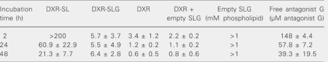
Table 1. Cytotoxicity of various doxorubicin formulations against H82 cells.
Incubation DXR-SL DXR-SLG DXR DXR + Empty SLG Free antagonist G
time (h) empty SLG (mM phospholipid) (µM antagonist G)
2 >200 5.7 ± 3.7 3.4 ± 1.2 2.2 ± 0.2 >1 148 ± 4.4
24 60.9 ± 22.9 5.5 ± 4.9 1.2 ± 0.2 1.1 ± 0.2 >1 57.8 ± 7.2
48 21.3 ± 7.7 6.4 ± 2.8 0.6 ± 0.5 0.8 ± 0.6 >1 39.3 ± 19.5
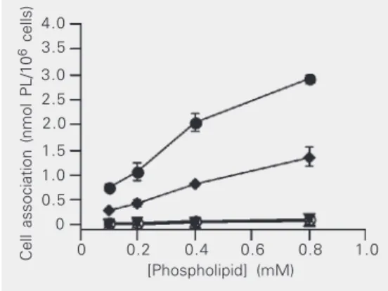
with [3H]-CHE-SLG (Figure 1) suggests that
antagonist G-targeted liposomes were being internalized by H82 cells. At 37ºC, where both binding and internalization take place, SLG associated with the cells approximately 20-30-fold more than SL, SLP [1-9] or SL in the presence of free antagonist G. At 4ºC, a 2.2- to 2.7-fold decrease in the cellular asso-ciation of SLG was observed, which is to be expected if the component of cell-liposome association due to liposome internalization by the tumor cells was inhibited at this low temperature. Based on the assumption that there are 7.7 x 10¹² liposomes/µmol
phos-pholipid (7), we estimate that, after a 1-h incubation, within the phospholipid concen-tration range tested, there were 3,600-12,000 SLG liposomes internalized/cell.
Images of optically sectioned cells ob-tained by confocal microscopy showed that after 1 h at 37ºC SLG liposomes containing the fluorescent dye HPTS (HPTS-SLG) were distributed both on the cell surface and intra-cellularly (Figure 2A). Under the same con-ditions, no staining was detectable when the cells were treated with either HPTS-SL (Fig-ure 2B) or HPTS-SLP [1-9] (Fig(Fig-ure 2C).
Association of [3H]-CHE-SLG with H82
cells was competitively inhibited only when cells were pre-incubated with non-radiola-beled SLG (Figure 3B) at a concentration that was 48-fold lower than the highest amount of free antagonist G that showed no inhibi-tion of binding either at 4º or at 37ºC (Figure 3A). This result is also consistent with the explanation that the association of SLG with H82 cells was taking place mainly through a receptor-mediated process, possibly involv-ing multivalent bindinvolv-ing sites. The amount of coupled antagonist G needed to reach 50% of cell-liposome association inhibition was 0.03 µg (Figure 3B). In a control experiment, liposomes pre-incubated with the tumor cells did not interfere with the cell-liposome asso-ciation of [3H]-CHE-SLG (data not shown).
The covalent linkage of antagonist G to the PEG-grafted liposomes appears to have
in-Cell association (nmol PL/10
6 cells)
4.0 3.5
3.0 2.5 2.0
1.5 1.0 0.5 0
0 0.2 0.4 0.6 0.8 1.0
[Phospholipid] (mM)
Figure 1. Association of [3 H]-CHE-labeled liposome formula-tions with SCLC H82 cells. H82 cells (1 x 10⁶ cells) were incu-bated with different liposomal formulations (0.1-0.8 mM phos-pholipid (PL)/well) containing SL (open circles) or SLP [1-9] (inverted triangles) at 37ºC or SLG at 4ºC (lozenges) or 37ºC for 1 h (filled circles) or SL in the presence of free antagonist G at an antagonist G/phospholipid molar ratio of 1:200 (open tri-angles). Each point is the mean ± SD for 3-4 samples. SCLC = small cell lung cancer; SL = non-targeted liposomes; SLG = antagonist G-targeted liposomes; SLP = substance P-coupled liposomes.
Figure 2. Association of HPTS-containing liposomes with SCLC H82 cells. H82 cells (2 x 10⁶ cells) were incubated with liposomes (0.8 mM phospho-lipid/well) at 37ºC for 1 h. H82 cells were incubated with SLG (A), SL (B) or SLP [1-9] (C). After washing with cold PBS, the cells were visualized with an LSM-510 laser-scanning confocal microscope. HPTS = 8-hydroxypyrene-1,3,6-trisul-fonic acid, trisodium salt; SCLC = small cell lung cancer; SLG = antagonist G-targeted somes; SL = non-targeted lipo-somes; SLP = substance P-coupled liposomes. Scale bar for all panels = 10 µm. A
B
C
creased the avidity of the peptide for its receptors due to the multivalent presentation of the peptide, that is, the presence of several attached-peptide molecules allows one liposome to bind more than one receptor at once. DeFrees et al. (21) have shown that the use of sialyl Lewis glycolipid coupled to liposomes was a stronger inhibitor of E-selectin-dependent cell adhesion than free glycolipid. Overall, these studies reinforce
the idea that liposomes might be a powerful tool for the efficient presentation of ligands to cell surface receptors that require multiva-lent contact.
The increased levels of cell-liposome as-sociation of SLG relative to SL or SLP [1-9] resulted in a more efficient intracellular deliv-ery of DXR when it was encapsulated in antagonist G-targeted formulations, both to the whole cells and to the nuclei (Figure 4A,B). When delivered by SLG, accumula-tion of DXR, both into whole cells and nuclei, was faster and higher (10- to 20-fold) than when the drug was delivered either by SL or SLP [1-9]. Interestingly, in whole cell ex-tracts DXR from SLG accumulated more rapidly and plateaued at 6 h compared to drug accumulation in the nucleus, which pla-teaued at around 12 h. This may be related to either uptake of some free DXR released from the liposomes prior to internalization or to the release of the drug from endosomes (22). The accumulation of free DXR both in the whole cell extracts and in the nuclear fractions, was more rapid and occurred to a greater extent than that of the liposomal samples. This does not necessarily lead to improved tumor accumulation in vivo due to the higher volume of distribution of free DXR relativeto that of liposomal DXR (23). We then determined whether the im-proved intracellular drug delivery by the targeted formulation would translate into an improved antiproliferative activity against the H82 cell line (Table 1). Interestingly, after an incubation of only 2 h, DXR-SLG was as toxic as free DXR (P > 0.05) and approxi-mately 35 times more toxic than DXR-SL (P < 0.001). After 24- and 48-h incubations, the differences in cytotoxicity between DXR-SLG and free DXR were not significant (P > 0.05), and while the differences between DXR-SLG and DXR-SL decreased, they were still statistically significant (P < 0.001). The absence of any differences between free DXR and free DXR in the presence of empty SLG at 200 µg DXR/µmol phospholipid (P >
Whole cell uptake (DXR fluorescence
units x 10
2/50 x 10 6 cells)
1200 900 600 300 200 150 50 0 100
Nuclear uptake (DXR fluorescence
units x 10
2/50 x 10 6 cells)
85 60 35 10 6 4 0 2
0 5 10 15 20 25
Time (h)
0 5 10 15 20 25
Time (h) A
B Figure 4. Kinetics of DXR or
DXR-containing liposome up-take by SCLC H82 cells. Twenty µM of free DXR (lozenges) or DXR-containing liposomes (SL, open circles; DXR-SLG, filled circles, and DXR-SLP [1-9], triangles), were incubated with 50 x 106 H82 cells at 37ºC for 24 h. DXR was measured in whole cell extracts (A) or in iso-lated nuclei (B). The back-ground was subtracted from the values at each time point. Each point is the mean ± SD of 3 samples. DXR = doxorubicin; SL = liposomes; SLG = antago-nist G-targeted liposomes; SLP = substance P-coupled lipo-somes.
Cell association (nmol PL/10
6 cells) 1.2
1.0 0.8 0.6 0.4 0.2 0 0.6 0.5 0.4 0.3 0.2 0.1 0
Cell association (nmol PL/10
6 cells)
0 5 10 15 20 25 30
Free antagonist G (µg)
0 0.1 0.2 0.3 0.4 0.5 0.6
A
B Figure 3. Competitive inhibition
of the association of [3 H]-CHE-SLG with SCLC H82 cells. H82 cells (1 x 10⁶ cells) were pre-incubated for 30 min with: A, 0-29 µg of free antagonist G, ei-ther at 4ºC (lozenges) or 37ºC (circles); B, 0-0.6 µg of antago-nist G covalently linked to non-radiolabeled SLG at 37ºC (circles). Competitive inhibition was determined by adding [3 H]-CHE-SLG (0.1 mM phospholipid (PL)/well), either at 4º or 37ºC and incubating for 1 h. Each point is the mean ± SD for 3 samples. [3H]-CHE-SLG = [1α, 2α(n)-3H] cholesteryl hexadecyl ether-antagonist G-targeted li-posomes; SCLC = small cell lung cancer; SLG = antagonist G-targeted liposomes.
0.05), independent of incubation time, sug-gested that there was no contribution of antagonist G to the observed cytotoxicities. The IC50 values for empty SLG and free
antagonist G were much higher than the concentrations of phospholipid and antago-nist G at the IC50 for DXR-SLG at all
incu-bation times, confirming that the cytotoxic activity of this sample was exclusively due to the more efficient delivery of the encapsu-lated drug by SLG.
The cytotoxicity results demonstrate that binding and internalization of DXR-SLG con-tribute to an increased level of cytotoxicity against the H82 cell line compared to DXR-SL. The results for DXR uptake into whole cells plateaued before that in isolated nuclei, suggesting that the release of DXR from the endosomes may be delayed. How this delay in DXR release from endosomes, which will affect the rate at which the drug reaches intracellular sites of action, impinges on the overall cytotoxicity of targeted formulations remains to be assessed. In spite of a possible delay in the release of DXR from the endo-somes, however, we observed that the tar-geted formulations had a cytotoxicity that was similar to that of the free drug in vitro. Overall, based on the data presented here, it may be inferred that receptors for antago-nist G in H82 cells were present and func-tionally active, and were not turned off by the
differentiation process that took place. These results, along with the high affinity of an-tagonist G for the vasopressin receptor (6), which is expressed on the surface of variant SCLC cells (11,24), suggest that this could be the main receptor involved in the internal-ization of SLG. Moreover, the small differ-ences between the amounts of coupled an-tagonist G necessary to inhibit 50% of cellu-lar association of SLG, both in the H82 variant and the classical H69 SCLC cell lines (9), suggested a similar density of receptors for antagonist G between these two subtypes of SCLC cells. This was confirmed by the similar cellular association patterns of either fluorescent radiolabeled liposomes or DXR-containing liposomes (9).
The similar cytotoxic activity of DXR-SLG against the H82 variant and the H69 SCLC cell lines (9), where c-myc proto-oncogene is either not detectable or present at only trace levels (18), suggests that the delivery of liposomal DXR through receptor-mediated endocytosis could be an efficient way to overcome the problems of c-myc overexpression by variant cells. Moreover, the long circulation half-lives of SLG (9), a crucial feature for in vivo applications, give antagonist G-targeted liposomes containing DXR, or antisense oligonucleotides against c-myc, potential for the treatment of variant small cell lung cancer.
References
1. Moore SM, Rintoul RC, Walker TR, Chilvers ER, Haslett C & Sethi T (1998). The presence of a constitutively active phosphoinositide 3-kinase in small cell lung cancer cells mediates anchorage-inde-pendent proliferation via a protein kinase B and p70s6k-dependent pathway. Cancer Research, 58: 5239-5247.
2. Worden FP & Kalemkerian GP (2000). Therapeutic advances in small cell lung cancer. Expert Opinion on Investigational Drugs, 9: 565-579.
3. Moody TW & Cuttitta F (1993). Growth factor and peptide receptors in small cell lung cancer. Life Sciences, 52: 1161-1173.
4. Sethi T, Langdon S, Smyth J & Rozengurt E (1992). Growth of small cell lung cancer cells: stimulation by multiple neuropeptides and inhibition by broad spectrum antagonists in vitro and in vivo. Cancer
Research, 52s: 2737s-2742s.
5. Seckl MJ, Newman RH, Freemont PS & Rozengurt E (1995). Substance P-related antagonists inhibit vasopressin and bombesin but not 5'-3-O-(thio)triphosphate-stimulated inositol phosphate production in Swiss 3T3 cells. Journal of Cellular Physiology, 163: 87-95.
6. Woll PJ & Rozengurt E (1990). A neuropeptide antagonist that inhibits the growth of small cell lung cancer in vitro. Cancer Re-search, 50: 3968-3973.
7. Lopes de Menezes DE, Pilarski LM & Allen TM (1998). In vitro and
in vivo targeting of immunoliposomal doxorubicin to human B-cell lymphoma. Cancer Research, 58: 3320-3330.
immunolipo-somes: enhanced efficacy attributable to targeted delivery. Clinical Cancer Research, 8: 1172-1181.
9. Moreira JN, Hansen CB, Gaspar R & Allen TM (2001). A growth factor antagonist as a targeting agent for sterically stabilized lipo-somes in human small cell lung cancer. Biochimica et Biophysica Acta, 1514: 303-317.
10. Gazdar AF, Carney DN, Nau MM & Minna JD (1985). Characteriza-tion of variant subclasses of cell lines derived from small cell lung cancer having distinctive biochemical, morphological, and growth properties. Cancer Research, 45: 2924-2930.
11. Fay MJ, Friedmann AS, Yu XM & North WG (1994). Vasopressin and vasopressin-receptor immunoreactivity in small-cell lung carci-noma (SCCL) cell lines: disruption in the activation cascade of V1a -receptors in variant SCCL. Cancer Letters, 82: 167-174. 12. Daleke DL, Hong K & Papahadjopoulos D (1990). Endocytosis of
liposomes by macrophages: binding, acidification and leakage of liposomes monitored by a new fluorescence assay. Biochimica et Biophysica Acta, 1024: 352-366.
13. Olson F, Hunt CA, Szoka FC, Vail WJ & Papahadjopoulos D (1979). Preparation of liposomes of defined size distribution by extrusion through polycarbonate membranes. Biochimica et Biophysica Acta, 557: 9-23.
14. Bolotin EM, Cohen R, Bar LK, Emanuel N, Ninio S, Lasic DD & Barenholz Y (1994). Ammonium sulphate gradients for efficient and stable remote loading of amphipathic weak bases into lipo-somes and ligandolipolipo-somes. Journal of Liposome Research, 4: 455-479.
15. Bartlett GR (1959). Phosphorus assay in column chromatography.
Journal of Biological Chemistry, 234: 466-468.
16. Mosmann T (1983). Rapid colorimetric assay for cellular growth and survival: application to proliferation and cytotoxicity assays. Journal of Immunological Methods, 65: 55-63.
17. Lopes de Menezes DE, Kirchmeier MJ, Gagne JF, Pilarski LM & Allen TM (1999). Cellular trafficking and cytotoxicity of anti-CD19-targeted liposomal doxorubicin in B lymphoma cells. Journal of Liposome Research, 9: 199-228.
18. Rygaard K, Vindeløv LL & Spang-Thomsen M (1993). Expression of myc family oncoproteins in small-cell lung-cancer cell lines and xenografts. International Journal of Cancer, 54: 144-152. 19. Brambilla E, Moro D, Gazzeri S, Brichon PY, Nagy-Mignotte H, Morel
F, Jacrot M & Brambilla C (1991). Cytotoxic chemotherapy induces cell differentiation in small-cell lung carcinoma. Journal of Clinical Oncology, 9: 50-61.
20. Bunn Jr PA, Chan D, Dienhart DG, Tolley R, Tagawa M & Jewett PB (1992). Neuropeptide signal transduction in lung cancer: clinical implications of bradykinin sensitivity and overall heterogeneity.
Cancer Research, 52: 24-31.
21. DeFrees SA, Phillips L, Guo L & Zalipsky S (1996). Sialyl Lewis x liposomes as a multivalent ligand and inhibitor of E-selectin medi-ated cellular adhesion. Journal of the American Chemical Society, 118: 6101-6104.
22. Kirchmeier MJ, Ishida T, Chevrette J & Allen TM (2001). Correla-tions between the rate of intracellular release of endocytosed liposomal doxorubicin and cytotoxicity as determined by a new assay. Journal of Liposome Research, 11: 15-29.
23. Gabizon A, Catane R, Uziely B, Kaufman B, Safra T, Cohen R, Martin F, Huang A & Barenholz Y (1994). Prolonged circulation time and enhanced accumulation in malignant exudates of doxorubicin en-capsulated in polyethylene-glycol coated liposomes. Cancer Re-search, 54: 987-992.
![Figure 3. Competitive inhibition of the association of [ 3 H]-CHE-SLG with SCLC H82 cells](https://thumb-eu.123doks.com/thumbv2/123dok_br/15808463.650573/6.918.269.537.163.564/figure-competitive-inhibition-association-che-slg-sclc-cells.webp)
