Immunizatio n by subcutane o us
implants o f po lye ste r-po lyure thane
spo nge s co uple d with antige n
Departamentos de 1Bioquímica e Imunologia, 2Patologia and 3Fisiologia, Instituto de Ciências Biológicas,
Universidade Federal de Minas Gerais, Belo Horizonte, MG, Brasil 4Fundação Ezequiel Dias, Belo Horizonte, MG, Brasil
S.L. Lima1,4, C.B. Machado1, M.A.C. Pereira1, D.C. Cara2, D.T. Velarde4, S.P. Andrade3 and C.M. Gontijo1
Abstract
A new protocol is described for immunization of outbred Swiss mice. The procedure is based on subcutaneous implantation of antigen-coupled polyester-polyurethane sponges cut into disks of 10 mm in diameter vs 2 mm in thickness. Antigen coupling was performed by overnight incubation of the sponge with a solution of ovalbumin (Ova) (2 mg/ml) diluted in sodium carbonate buffer, pH 9.6. The amount of ovalbumin that was taken up by the sponge was between 71.4 to 82.5 µg. This was estimated by comparing the Ova absorbance at 280 nm in coating buffer solutions before and after incubation. To compare the efficiency of the proposed method, experimental groups immunized with the antigen in the presence of adjuvants (10 µg in Al(OH)3 or 100 µg in complete Freunds adjuvant (CFA)) were run in parallel. The data obtained after the 3rd week of immunization indicate that both cellular and humoral immune responses were achieved. These were assayed by antigen-induced footpad swelling and ELISA (specific antibodies), respectively. The levels of both immune responses elic-ited were similar to the responses observed in mice immunized with ovalbumin in the presence of Al(OH)3. The method might represent an advantage when immunizing with pathogenic antigens. Preliminary experiments have suggested that the antigen remains immobilized or bound to the sponge for a long period of time, since there is an increment on the cell population inside the sponges after boosting the animals. If so, the undesirable effects of immunization would be reduced.
Co rre spo nde nce
C.M. Gontijo
Departamento de Bioquímica e Imunologia, ICB, UFMG Caixa Postal 486
30161-970 Belo Horizonte, MG Brasil
Fax: + 55-31-441-5963
E-mail: cgontijo@ mono.icb.ufmg.br Presented at the XIII Annual Meeting of the Federação de Sociedades de Biologia Experimental, Caxambu, MG, Brasil, August 26-29, 1998. Research supported by FAPEMIG (No. 2295/96) and PRPq-Fundep-UFMG (No. 2913/33). C.B. Machado is supported by PRO BIC scholarship program (FAPEMIG).
Received April 9, 1998 Accepted January 13, 1999
Ke y words ·Sponge ·Implant ·Immunization ·Vaccine
There is a great interest in the develop-ment of new methods for immunizing exper-imental animals with lethal antigens such as toxins and/or venoms in order to obtain spe-cific immune sera for medical purposes. The use of adjuvants such aluminum hydroxide (Al(OH)3) and complete Freunds adjuvant
(CFA) in schemes of immunization has been
for a long period of time often show skin lesions followed by a reduction in their abil-ity to produce antibodies (1,2). Al(OH)3,
which does not induce these effects, is not as potent an adjuvant as CFA (3). Protein iodi-nation (4) and encapsulation of the antigen into liposomes (5,6) are examples of meth-ods that have been proposed to avoid these problems. These methods reduce toxicity but not immunogenicity. However, losses may occur and accidents have been reported (4-7).
Looking for an alternative way of immu-nizing, we became interested in an experi-mental model developed for studying angio-genesis in rats (8) and mice (9,10). In this model, subcutaneous implants of polyester-polyurethane sponges are performed in the dorsal region of the animals where they act as a matrix for the development of blood vessels and connective tissue. These im-plants stimulate an inflammatory infiltrate rich in mononuclear cells by the end of the first week. Antigens, on the other hand, can be associated with inorganic materials such as polystyrene plates (Nunc, Copenhagen, Denmark) that are used for ELISA reactions. Using a similar protocol, polyester-polyure-thane sponges were treated with chicken egg albumin (ovalbumin - Ova) grade III (Sigma Chemical Co., St. Louis, MO, USA) in so-dium carbonate buffer, pH 9.6 (coating buf-fer), and then implanted into experimental mice. Ova is not a pathogenic antigen and its immunogenicity is not considered to be high. Polyester-polyurethane sponges were cut into disks (10 mm in diameter vs 2 mm in thick-ness), treated with 70% ethanol for 1 h and then boiled in distilled water for 30 min. The sponges were then dried under sterile condi-tions and immersed in coating buffer (1 ml/ sponge) containing Ova at different concen-trations (0.2, 2 and 20 mg/ml), centrifuged to assure that all the internal surface was in contact with the solution and then incubated overnight at 4o
C. The sponges were washed 3 times in sterile PBS before being
subcuta-neously implanted into 6-8-week-old out-bred female Swiss mice. We estimated the incorporation of 71.4-82.5 µg of Ova/sponge by comparing the Ova absorbance at 280 nm in coating buffer solutions before and after incubation.
For positive control of immunization, mice were injected either intraperitoneally (ip) with 200 µl of saline containing 10 µg Ova mixed with 1 mg Al(OH)3 or
subcutane-ously (sc) with 40 µl of saline containing 100 µg Ova mixed with CFA (Sigma). Negative control mice were implanted with a sponge that was not pretreated with Ova, or were injected as described above, but without the antigen. In some experiments, mice were boosted 3 weeks later with 10 µg Ova in 200 µl saline, ip. Blood samples were collected weekly from the tail of mice and diluted in PBS (1:3). After clotting and centrifugation, the supernatants were collected and stored at -20o
C.
Anti-Ova antibodies were assayed by ELISA. Briefly, polystyrene plates (Nunc) were coated overnight at 4o
C with 2 µg Ova diluted in 100 µl coating buffer per well, washed with saline containing 0.05% (w/v) Tween-20, saturated with 0.25% (w/v) casein in PBS, washed again and then coated with serial dilution of mouse antiserum starting at 1:100. After 1 h at 37o
C, plates were washed, incubated for 1 h at 37o
C with peroxidase-conjugated goat anti-mouse globulin antise-rum (Southern Biotechnology, Birmingham, AL), washed and developed by the addition of H2O2 and ortho-phenylene-diamine (OPD;
Sigma). The reaction was interrupted at 10 min by the addition of H2SO4 at 1/20 dilution
and the values obtained at 1/25600 dilution always reached the blank level. This way of reporting the results is equivalent to report-ing titration curves or selectreport-ing the absorb-ance at one particular serum dilution as rep-resentative. The significance of the differ-ence between experimental and control groups was assessed by the Kruskal-Wallis test for nonparametric data. Positive and negative control samples were run on every plate. A mouse was considered to be immu-nized if its ELISA* score was higher than the mean of normal control mice plus 3 times the SD.
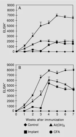
The kinetics of anti-Ova antibody pro-duction for a period of 8 weeks is shown in Figure 1A for mice immunized with im-plants of sponges containing Ova. Positive control groups were prepared by immuniz-ing mice with the antigen in the presence of adjuvants (CFA and Al(OH)3). Another group
received 10 µg of Ova in saline, ip, without adjuvant. No antibody was produced by the animals of this last group, which actually exhibited the same profile as negative non-immunized control groups (data not shown). Although requiring a longer time, sponge-implanted mice produced specific antibod-ies at the same level as those immunized with Al(OH)3. When the activity of CFA and
Al(OH)3 as adjuvants is compared, we
ob-serve that a higher level of antibody produc-tion was obtained with CFA. This may be explained by the difference in the amount of Ova injected. Figure 1B shows the profiles of the groups when animals received 10 µg of Ova in saline, ip, as a booster on day 21. One week after the booster, sponge-implanted mice reached the antibody level of the group immunized with Al(OH)3. The levels reached
by these 2 groups in Figure 1B were higher than those observed in Figure 1A. On the other hand, with the exception of the first week after the booster (week 4), no differ-ence was observed between the profiles of mice immunized with Ova in CFA as adju-vant, when primary and secondary responses
were compared.
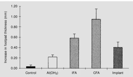
We also tested whether the implant of Ova-coated sponges also elicited a cellular immune response. As shown in Figure 2, we have compared this procedure between groups that were immunized with Ova di-luted in saline with or without the presence of CFA, incomplete Freunds adjuvant (IFA) or Al(OH)3 as adjuvants. During the third
week after implant or immunization, the groups were injected sc with 30 µl of a 2% aggregated Ova solution into the left foot-pad. The same volume of PBS was injected
sc into the right footpad as control (11). The data in Figure 2 are reported as the differ-ence in thickness between footpads observed 48 h after injection. With the exception of the group immunized with Ova in the ab-sence of adjuvants, in all other groups a significant increase of footpad thickness was observed. Differences (P<0.05) were
ob-E
L
IS
A
*
9000 8000 7000
6000
5000 4000 3000 2000 1000 0
A
0 1 2 3 4 5 6 7
Weeks after immunization
E
L
IS
A
*
9000 8000 7000
6000 5000 4000
3000 2000 1000 0
B
Control
Implant
AI(OH)3
CFA
Figure 1 - Humoral immune re-sponses elicited by Ova-conju-gated implanted sponges. A, For the primary immune response, Sw iss mice w ere immunized w ith Ovain Al(OH)3 (10 µg, ip) or
served between the three groups immunized with adjuvants (CFA, IFA and Al(OH)3). No
difference was observed between mice im-planted with an Ova-treated sponge and those immunized with Ova in the presence of IFA or Al(OH)3 as adjuvants.
The subcutaneous implantation of an an-tigen-coated sponge provided an alternative method for immunization. Both humoral and cellular immune responses were achieved at levels similar to those obtained when Al(OH)3
was used as adjuvant. It is known that poly-ester-polyurethane sponge implants cause a granulomatous reaction characterized by an inflammatory infiltrate rich in polymorpho-nuclear cells, macrophages and giant cells around the trabeculae of the sponge matrix (9). A similar reaction is observed in granu-lomas formed when antigen is subcutane-ously injected in the presence of Al(OH)3
(12). The similarity of our proposed method and the use of Al(OH)3 as adjuvant should be
further analyzed. For instance, we are cur-rently characterizing the isotypes that are formed when mice are implanted with anti-gen-coated sponges (13). It is known that, differently from CFA that usually elicits im-mune responses with a predominance of the TH1 subset of T-lymphocytes, Al(OH)3 is
thought to elicit TH2-type immune responses
(14).
Antigen delivery from the sponge is an-other fact that deserves attention. Prelimi-nary data from histological studies indicate that antigen delivery from the sponge should be slow. Histological changes occur in sponges when mice are boosted, indicating that antigens are probably still present at the site after at least 3 weeks. This possibility is very attractive since the method could repre-sent an alternative for fixing pathogenic an-tigens at a site, with the consequent reduc-tion of toxic effects during immunizareduc-tion procedures.
In
c
re
a
s
e
i
n
f
o
o
tp
a
d
t
h
ic
k
n
e
s
s
(
m
m
)
1.20
1.00
0.80
0.60
0.40
0.20
0.00
Control Al(OH3) IFA CFA Implant
Figure 2 - Cellular immune response elicited by Ova-conjugated implanted sponges. Sw iss mice w ere immunized w ith Ovain Al(OH)3 (10 µg, ip), complete Freund’s adjuvant (CFA)
(100 µg, sc) or incomplete Freund’s adjuvant (IFA) (100 µg, sc), or w ere implanted subcuta-neously w ith Ova-coupled sponges. Controls w ere either injected w ith 10 µg of Ova in saline, ip, or implanted w ith sponges treated w ithout Ova. During w eek 3, mice w ere injected subcutaneously w ith 30 µl of either 2% aggregated Ova (left footpad) or PBS (right footpad). Data are reported as the difference in thickness (mean ± SEM , N = 5-7) betw een both footpads observed 48 h after injection.
Re fe re nce s
1. Oropeza RM , Perez GC & M anes SC (1972). Esquem a m odif icado para la obtención de sueros antiofídicos. Revista de Investigación en Salud Pública, 32: 179-186.
2. Bolanos R & Cerdas L (1978). The produc-tion and control of anti-venous sera. De-velopments in Biological Standardization, 41: 907-911.
3. Raw I, Guidolin R, Higashi HG & Kelem EM A (1991). Antivenins in Brazil: prepara-tion. In: Tu AT (Editor), Reptile Venoms and Toxins, Handbook of Natural Toxins.
M arcel Dekker, New York.
4. Daniel JP, Heneine LGD, Tavares CAP, Nascimento M CS & Heneine IF (1987). Generation of protective immune sera by
Crotalus durissus terrificus venom detoxi-fied by controlled iodination. Brazilian Journal of M edical and Biological Re-search, 20: 713-720.
5. Freitas TV & Frézard F (1997). Encapsula-tion of native crotoxin in liposomes: a safe approach for the production of antivenom and vaccination against Crotalus durissus terrificus venom. Toxicon,35: 91-100. 6. Chavez-Olortegui C, Ait-Amara D, Rochat
H, Diniz CR & Granier C (1991). In vivo
protection against scorpions by liposomal immunization. Vaccine, 9: 713-720. 7. Freitas TV, Fortes-Dias CL, Diniz CR,
Velarde DT & Freitas CF (1991). Immuni-zation of horses w ith Crotalus durissus terrificus (South American Rattlesnake) venom. A comparison of four different procedures. Brazilian Journal of M edical and Biological Research, 24: 281-290. 8. Andrade SP, Fan TPD & Lew is GP (1987).
experi-mental para estudo da angiogênese em camundongos. M aster’s thesis, Departa-mento de Fisiologia, Universidade Fed-eral de M inas Gerais, Belo Horizonte. 10. Andrade SP, M achado RDP, Teixeira AS,
Belo AV, Tarso AM & Beraldo WT (1997). Sponge-induced angiogenesis in mice and the pharmacological reactivity of the neo-vasculature quantified by a fluorimetric method. M icrovascular Research, 54: 253-261.
11. Titus RG & Chiller JM (1981). A simple
and effective method to assess murine delayed-type hypersensitivity to proteins.
Journal of Immunological M ethod, 45: 65-78.
12. Gupta RK, Rost BE, Relyveld E & Siber GR (1995). Adjuvant properties of aluminum and calcium compounds. In:Pow ell M F & New man M J (Editors), Vaccine Design: The Subunit and Adjuvant Approach. Ple-num Publishing Corporation, New York. 13. Gontijo CM , Lima SL, Cara DC, Velarde
DT & Andrade SP (1998). Subcutaneous
im plant s of polyest er-polyuret hane sponges coupled w ith antigen as a model for immunizing experimental animals. In: Talw ar GP, Nath I, Ganguly NK & Rao KVS (Editors), Proceedings of the 10th Interna-tional Congress of Immunology, New Delhi, India, November 1-6, 1998. M on-duzzi Editore, Bologna, 1415-1419. 14. Vogel FR (1995). The role of adjuvants in
Brasil
Carlos Eduardo Rocha-Miranda
Vice President, Academia Brasileira de Ciências Rua Anfilófio de Carvalho, 29, 3º andar 20030-060 Rio de Janeiro, RJ, Brasil Tel.: 220-4794/Fax: 240-4695 E-mail: cerm@ abc.org.br
Argentina
Israel Algranati
Instituto de Investigaciones Bioquímicas Fundación Campomar Av. Patricias Argentinas, 435 1405 Buenos Aires Tel: 863-4018/Fax: 865-2246 e-mail: algra@ iris.iib.uba.ar
Mexico
Hugo Aréchiga
Facultad de Medicina UNAM Ciudad Universitaria 04510 Mexico, D.F.
Tel: 622-0725/Fax: 550-8859 e-mail: arechiga@ servidor.unam.mx
Chile
Manuel A. Kukuljan, Ph.D.
Universidad de Chile
Departamento de Fisiologia y Biofisica Casilla 70005 Santiago 7, Chile Tel: 2-678-6310/Fax: 2-777-6916 e-mail: kukuljan@ bitmed.med.uchile.cl
All other countries
Silvia Montano de Jiménez
The Pew Latin American Fellows Program 3333 California Street, Suite 410 San Francisco, CA 94118
Deadline for applications: O ctober 1, 1999
The Pew Latin Am erican Fellows Program in the Biom edical Sciences is providing support for young scientists from Latin Am erica for post-doctoral training in the United States.
Ten Fellows will be selected in
1999. An award of $50,000 will be provided as a salary stipend for the fellow during the period of training (2 years) and will be administered by the sponsoring U.S. institution. The sponsoring institution is expected to supplement the stipend with at least $5,000 a year and provide medical benefits for the fellow. Following the two year fellowship, the Program will issue an additional $35,000 award to the sponsoring institution to purchase equipment and supplies for the fellow to establish a laboratory in his or her home country.
Applicants must have held a Ph.D. and/or M.D. degrees, or equivalent, for no more than five years as of July 1, 1999. Strong preference will be given to those applicants with no previous postdoctoral training outside of their home country. Applicants are not required to have a commitment of a position and laboratory space after the fellowship. However,
applicants must submit a written statement of intent to return to Latin America. Fellows must have a confirmed position and laboratory space in their home country by the end of the fellowship period in order to obtain the $35,000 portion of the award.
Fellows will be selected on the basis of their promise as outstanding investigators, as well as the scientific merit of their research proposal, their record of training and how well their interests coincide with the laboratory of their sponsor in the United States. If potential applicants need assistance with the identification of an appropriate sponsoring laboratory in the United States, they may contact the Program O ffice before August 1, 1999. The Program will accept applicants from Mexico, Central and South America. Applications may be obtained from the Regional Committee contact listed here for your country or from our website at
http://futurehealth.ucsf.edu/pewlatin.htm l
The application deadline is O ctober 1, 1999. Winners will be notified in April 2000 and the fellowship should begin no later than August 2000.
FELLOWS
PROGRAM
in the
BIOMEDICAL
S . C . I . E . N. C . E . S
PEW
Latin American