ISSN 0100-879X
BIOMEDICAL SCIENCES
AND
CLINICAL INVESTIGATION
www.bjournal.com.br
www.bjournal.com.br
Volume 45 (1) 1-92 January 2012
Braz J Med Biol Res, January 2012, Volume 45(1) 13-19
doi: 10.1590/S0100-879X2011007500161
Hypoxic-ischemic injury decreases anxiety-like behavior in rats
when associated with loss of tyrosine-hydroxylase immunoreactive
neurons of the substantia nigra
Ming-Yan Hei, Ya-Li Luo, Xiao-Chun Zhang, Hong Liu, Ru Gao and Jing-Jiang Wu
Institutional Sponsors
The Brazilian Journal of Medical and Biological Research is partially financed by
Faculdade de Medicina de Ribeirão Preto Campus
Ribeirão Preto
Ex plor e H igh - Pe r for m a n ce M S Or bit r a p Te ch n ology I n Pr ot e om ics & M e t a bolom ics
Hypoxic-ischemic injury decreases anxiety-like
behavior in rats when associated with loss
of tyrosine-hydroxylase immunoreactive
neurons of the substantia nigra
Ming-Yan Hei, Ya-Li Luo, Xiao-Chun Zhang, Hong Liu, Ru Gao and Jing-Jiang Wu
Department of Pediatrics, the Third Xiangya Hospital, Central South University, Changsha, Hunan, China
Abstract
Neonatal Sprague-Dawley rats were randomly divided into normal control, mild hypoxia-ischemia (HI), and severe HI groups (N = 10 in each group at each time) on postnatal day 7 (P7) to study the effect of mild and severe HI on anxiety-like behavior and the expression of tyrosine hydroxylase (TH) in the substantia nigra (SN). The mild and severe HI groups were exposed to hypoxia (8% O2/92% N2) for 90 and 150 min, respectively. The elevated plus-maze (EPM) test was performed to assess anxiety-like behavior by measuring time spent in the open arms (OAT) and OAT%, and immunohistochemistry was used to determine the expression of TH in the SN at P14, P21, and P28. OAT and OAT% in the EPM were significantly increased in both the mild (1.88-, 1.99-, and 2.04-fold, and 1.94-, 1.51-, and 1.46-fold) and severe HI groups (1.69-, 1.68-, and 1.87-fold, and 1.83-, 1.43-, and 1.39-fold, respectively; P < 0.05). The percent of TH-positive cells occupying the SN area was significantly and similarly decreased in both the mild (17.7, 40.2, and 47.2%) and severe HI groups (16.3, 32.2, and 43.8%, respectively; P < 0.05). The decrease in the number of TH-positive cells in the SN and the level of protein expression were closely associated (Pearson correlation analysis: r = 0.991, P= 0.000 in the mild HI group and r = 0.974, P= 0.000 in the severe HI group)with the impaired anxiety-like behaviors. We conclude that neonatal HI results in decreased anxiety-like behavior during the juvenile period of Sprague-Dawley rats, which is associated with the decreased activity of TH in the SN. The impairment of anxiety and the expression of TH are not likely to be dependent on the severity of HI.
Key words: Anxiety; Tyrosine hydroxylase; Substantia nigra; Hypoxia-ischemia
Introduction
Correspondence: Ming-Yan Hei, Department of Pediatrics, the Third Xiangya Hospital, Central South University, Hunan, Changsha, 410013 China. E-mail: [email protected]
Received May 22, 2011. Accepted November 28, 2011. Available online December 9, 2011. Published January 16, 2012.
Perinatal asphyxia is one of the major causes of neonatal morbidity and mortality. The incidence of perinatal asphyxia ranges from 1 to 8 per 1000 live births in the US (1); however,
no national data are currently available in China. During
the early stage of life, the brain is particularly vulnerable to hypoxic-ischemic (HI) insults and the outcome varies from mild behavioral dysfunctions to severe mental retardation and motor disturbances. Mild exposure to hypoxia in the early stage of brain development results in subtle behavioral
abnormalities, including attention deficit combined with anxi -ety, in spite of the lack of detectable brain damage in adult
life (2,3). Early brain damage can influence development
by changing the expression of genetic traits that interact with other characteristics of children. On this basis, an early hypoxic event resulting in neurological dysfunction
can influence development directly and indirectly, leading
to different developmental pathways in children.
Perinatal HI-induced behavioral disturbances have been grouped into two categories, hyperactivity and learning/
memory deficits (4). Evidence has shown that locomotor
activity disorders are associated with an abnormal level of dopamine content in the rat brain with simultaneous substan-tia nigra (SN) injury (5). Anxiety-like behavior is associated with the levels of dopamine and its metabolites in the mouse brain following ischemia (6). Dopamine is regulated by the activity of tyrosine hydroxylase (TH) and the expression of
14 Ming-Yan Hei et al.
Neonatal HI encephalopathy is brain damage caused by the combination of hypoxia and ischemia, and it is mostly attributed to perinatal asphyxia (9). It has been shown that less anxiety-like behavior is observed in animals subjected to HI (10). Current animal studies regarding ischemia or
hypoxia are complex, and even conflicting findings of
anxiety-like behaviors are obtained. It was reported that increased anxiety (11) or no changes in anxiety (12) was found in animals subjected to global ischemia, and increased anxiety (2) or no changes in anxiety (13) has been found after cerebral hypoxia (14,15).
Clinical studies have reported an elevated rate of anxiety in children with mild and moderate neonatal encephalopathy (NE) following perinatal asphyxia (16). Furthermore, more problems were found to be related to anxiety at school age in a mixed group of children with NE compared to a control group (17), indicating that the perinatal asphyxia insult indeed has a developmental impact that lasts at least until middle school age. Clinical data have shown that behavior impairment was more often present in children with moder-ate to severe NE, but not in children with mild NE following asphyxia (18). Therefore, it is necessary to clarify the effect of the severity of HI on anxiety and related mechanisms.
In the present study, we used a Rice-Vannucci HIBD rat model (19) with different hypoxic exposure to determine whether a mild or severe HI insult caused different anxiety-like behaviors, and whether the change of behavior was related to TH activity in the SN.
Material and Methods
Animal preparation
All animal procedures were approved by the Animal Ethics Committee of Central South University. Sprague-Dawley (SD) rats on postnatal day 7 (P7; both genders, body weight 11-16 g) were randomly divided into normal control, severe HI, and mild HI groups. In the HI groups,
pups were lightly anesthetized with isoflurane (4% induc -tion, 1.5% maintenance). The Rice-Vannucci method was used to induce HI brain damage (19). Ligation of the right common carotid artery was performed under a surgical microscope. After the wound was sutured, animals were placed on a warm heating pad (34°C) for recovery from anesthesia for 30 min. In the mild and severe HI groups,
animals were exposed to hypoxia (8% O2/92% N2) for 90
and 150 min (20), respectively.
Elevated plus-maze (EPM) test
The EPM test was performed on P14, P21, and P28 (N = 10 in each group at each time). The EPM test is widely used to observe the behaviors, which are relevant for understanding anxiety in animal studies (21,22). The EPM apparatus consists of two open arms (50 x 10 x 1 cm) and two enclosed arms (50 x 10 x 40 cm) originating from a common central platform (10 x 10 cm) to form a
plus shape. The entire apparatus was elevated to a height
of 50 cm above the floor. A video camera and illumination
lamps were mounted on the ceiling. The anxiety-related behaviors of each animal were recorded for a period of 5 min by a VCR-recording system on P19. At the beginning of the test, the rat was placed on the central platform with its
head facing an open arm. The arm entry was defined as all
four paws into an open or closed arm. The total time each animal spent in various sections of the maze (open arms, center, or enclosed arms) was recorded. The results are reported as open arm time (OAT), percent of OAT (OAT%, time spent in open arms divided by the sum of time spent in both closed and open arms).
TH immunohistochemistry (IHC)
At P14, P21 and P28 (N = 10 in each group at each
time), rat pups were anesthetized with 4% isoflurane and sacrificed by transcardial perfusion with physiological saline
followed by 4% paraformaldehyde. After being cryoprotected with 30% sucrose in phosphate-buffered solution (PBS), pH 7.4, the brain was cryosectioned into 10-µm thick coronal sections and collected onto Chrome-Album-coated slides and air-dried. The coronal sections of the midbrain were cut
at a level 1/3 rostrallyfrom the lambda to the bregma. The
sections were incubated with monoclonal mouse anti-rat antibody to TH (1:200, Chemicon, USA) at 4°C overnight, washed in PBS, and then incubated with biotinylated goat anti-mouse secondary antibody (1:400, Vector, USA) at room temperature for 2 h, followed by incubation by the avidin-biotin complex method (ABC elite kit, Vector, USA). Finally, the sections were visualized with 3,3V-diaminoben-zidine tetrachloride (DAB, Sigma, USA). A Nikon Eclipse 80i microscope was used for observation, and a Nikon DS digital camera equipped with the NIS-Elements AR Ver-sion 3.0 software was used to photograph and study the images. The data are reported as percent of TH-positive cells occupying the area in the SN, which was calculated as positive-staining area divided by the total area of the section. The result was the average of the three sections in
each brain. We took the first section in every 3 consecutive
sections with an interval of 30 µm.
Statistical analysis
The SPSS 16.0 software was used to analyze the data (supplied by Department of Medical Statistics, Institute of Public Health, Central South University, China). To avoid bias, all histological and behavioral analyses were carried out by an operator who did not know from which hypoxia treatment group the tissue or video was obtained. Data are reported as means ± SD and were analyzed by one-way ANOVA followed by the Student-Newman-Keuls test.
Results with a P< 0.05 were considered to be statistically
significant. Pearson correlation analysis was used to study
Results
Mild and severe HI significantly increased OAT and OAT% detected by the EPM test
The EPM test was used to assess the level of anxiety. It is known that a longer OAT and a higher OAT% indicate a lower level of anxiety (23). In the present study, longer OAT and higher OAT% (Table 1) were observed in both the mild and severe HI groups compared to the normal control group at all times examined. We found that time spent in the open arms by
rats in the mild HI group was significantly increased by 1.88-,
1.99-, and 2.04-fold (P < 0.05), OAT% was also increased by 1.94-, 1.51-, and 1.46-fold (P < 0.05), when compared to control at P14, P21, and P28. Similarly, in the severe HI group,
OAT was significantly increased by 1.69-, 1.68-, and 1.87-fold (P < 0.05), and OAT% was significantly increased by 1.83-,
1.43-, and 1.39-fold (P< 0.05). Although both OAT and OAT%
were slightly increased in the mild HI group compared to the severe HI group at all times examined, statistical analysis
demonstrated no significant difference between these two
groups (P> 0.05).
HI induced down-regulation of TH expression
TH immunohistochemistry has been used to detect the damage to dopaminergic neurons (8), which is found to be associated with anxiety-like behavior (6). To examine whether HI induced a change in TH expression, we determined its expression in the SN after HI at different postnatal times. An abundant number of TH-positive cells in the SN were found in
the normal control group, while the number was significantly
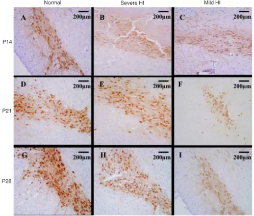
decreased in both the mild and severe HI groups (Figure 1).
This change can also be observed under a higher magnifi
-Figure 1. Immunohistochemical stain-ing of tyrosine hydroxylase (TH) in the substantia nigra (SN) of rats subjected to hypoxia-ischemia (HI) on postnatal day (P) 7 at P14, P21, P28 (N = 10). There were abundant TH-positive cells (brown color) in the SN of the normal control group at all times (A, D, G). The number and intensity of TH-positive cells significantly decreased in both severe (B, E, H) and mild (C, F, I) HI groups. Magnification: 100X.
Table 1. Effect of hypoxia-ischemia (HI) on anxiety-like behavior measured by the elevated plus-maze test.
OAT (s) OAT%
P14 P21 P28 P14 P21 P28
Control 28.33 ± 9.21 48.33 ± 33.53 45.27 ± 22.22 21.63 ± 10.61 31.50 ± 9.20 32.06 ± 16.74 Severe HI 48.00 ± 10.00* 81.33 ± 10.51* 84.97 ± 13.99* 39.66 ± 12.69* 45.17 ± 7.61* 44.75 ± 17.04* Mild HI 53.33 ± 11.04* 96.33 ± 25.31* 92.33 ± 12.44* 42.07 ± 13.69* 47.67 ± 8.96* 46.81 ± 11.42*
16 Ming-Yan Hei et al.
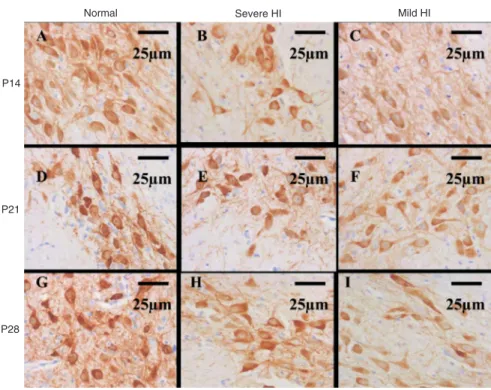
cation microscope (Figure 2), in which the positive staining was mainly distributed in the cytoplasm. TH-positive cells were morphologically normal with an intact cell membrane and normal nuclei. Based on their morphology, these cells were neurons. The percent of TH-positive cells occupying the area in the SN in the mild HI group was markedly
de-creased by 17.7, 40.2, and 47.2% (P< 0.05), respectively,
when compared to control at P14, P21, and P28. Similarly, in the severe HI group, the percent was also decreased by
16.3, 32.2, and 43.8%, respectively (P< 0.05). There was
no significant difference (P> 0.05) between the mild and severe HI groups (Table 2).
Correlation between the change of OAT and percent
TH expression
To examine whether a correlation existed between the change of OAT and TH expression in IHC, Pearson cor-relation analysis was performed. The results showed that
the coefficient index of the change of OAT in the EPM test
and the percent of the area occupied by TH-positive cells
in IHC was r = 0.991 (P= 0.000) in the mild HI group and r
= 0.974 (P= 0.000) in the severe HI group, indicating that
the down-regulation of TH expression was associated with the decreased anxiety-like behaviors.
Discussion
The major findings of the present study were that both
mild and severe HI insults during the neonatal period result in a decreased anxiety-like behavior during the juvenile
pe-riod in SD rats.The decrease of the number of TH-positive
cells in the SN and of the level of protein expression were associated with the decreased anxiety-like behaviors. How-ever, neither the impairment of anxiety nor the expression
of TH were dependent on the severity of HI. These findings
provide further information on the mechanism underlying hypoxic-ischemic brain injury and the anxiety-like behavior abnormality caused by perinatal HI.
The majority of studies concerning perinatal asphyxia focus on the detection of major developmental abnormalities at a very young age (24) or biopathophysiological changes within several days after birth (25,26), while behavioral changes such as anxiety during the juvenile period are one of the important focuses in long-term neurological outcomes after perinatal HI insult (27). The present study
Figure 2. Higher magnification of the
immunohistochemical TH staining in Figure 1. The rats were subjected to hypoxia-ischemia (HI) on postna-tal day (P) 7 and data are reported for P14, P21, and P28 (N = 10). The number of TH-positive cells in the normal control group at all times (A, D, G) was higher than that in both the severe (B, E, H) and mild (C, F, I) HI groups. TH-positive cells were mor-phologically normal neurons with pos-itive cytoplasm staining (brown color). Magnification: 400X. TH = tyrosine hydroxylase; SN = substantia nigra.
Table 2. Percent of tyrosine hydroxylase-positive cells occupying the area of the substantia nigra after hypoxia-ischemia (HI) was applied on day P7.
P14 P21 P28
Normal 4.58 ± 1.03 5.69 ± 1.40 5.78 ± 1.68 Severe HI 3.83 ± 0.82* 3.86 ± 0.62* 3.25 ± 1.02* Mild HI 3.77 ± 0.91* 3.40 ± 0.95* 3.05 ± 0.84*
was novel because it targeted the effect of a neonatal HI insult on anxiety during the period from 1 week after the HI insult to 3 weeks after HI, a time that corresponds to the human juvenile period. For SD rats, P28 is the weaning time when male and female rats mate with each other. P21 has been used as a time point for studying the neurobehav-ioral performance of juvenile SD rats (14,28). In addition,
findings for a series of times in this study (P14, P21, and
P28) were considered to be the dynamic changes during the juvenile period.
Knyazev et al.(29) wrote that “anxiety is seen as
be-ing most often generated by concurrent and equivalent activation of fear (or frustration) and reward systems. Therefore, anxiety should be most evident in a situation of uncertainty when chances of winning and losing are about equal. If uncertainty disappears, anxiety should give way to satisfaction or joy (if winning) or some negative emo-tions ranging from sadness to fear”. Exposure to novelty has been shown to induce anxiety responses in a variety of behavioral paradigms. Most current models of anxiety consist of exposure of animals to novelty in an anxiogenic environment (30). EPM is a widely used animal test of anxi-ety based on the curiosity of rats and their familiarization with the environment induced by previous experiences in the EPM, which could lead to a reduction in the approach/
avoidance of conflict (31). Results from the present EPM
test showed that the HI insult increased the number of OAT and OAT%. The behaviors measured are relevant for un-derstanding anxiety. A reduction in HI-induced anxiety-like behavior has also been reported in other animal models of perinatal asphyxia (14,15). In the present study, there
was no significant difference in OAT or OAT% between the
mild and severe HI groups, indicating the possibility that the duration of hypoxic exposure or the severity of the HI insult might not contribute the most to the impairment of
anxiety-like performance. On the other hand, Fan et al.(28)
reported that neonatal HI resulted in not only persistent white matter injury, but also in a higher proportion of open arm entry in the EPM test. In addition, they found that both brain damage and behavioral impairment were dependent on the duration of hypoxic exposure. The possible explanations for the differences are: 1) P4 rat pups were used in Fan’s study, whereas P7 rat pups were used in the present study, and 2) the HI insult was induced by bilateral carotid artery occlusion followed by exposure to hypoxia in Fan’s study, whereas the HI insult was induced by unilateral ligation of the common carotid artery followed by exposure to hypoxia in the present study.
Basal ganglia injury is common in hypoxic-ischemic brain damage (HIBD). Clinical autopsy data have proven that up to 43% of HI exposed neonates have basal ganglia injury (32). It has been reported that behavioral problems are
associated with an abnormal level of dopamine content in the rat brain concomitant with SN injury (5). It is well known that dopamine is one of the cholinergic neurotransmitters regulated by TH. In the present study, in both the mild and severe HI groups, the expression of TH in the SN was re-duced from P14 to P28, indicating a persistent damage of
dopaminergic neurons after HI. This finding was consistent with previous studies showing that HI significantly reduced
the number of TH-positive cells in SD rats on P21 (28) and in Wistar rats on P9 (33) after a neonatal HI insult, and this reduction during the neonatal period was reported to result in a decreased adult number of SN neurons (34). Moreover, in the present study, Pearson correlation analysis indicated that the down-regulation of TH expression was associated with the decreased anxiety-like behaviors. The expression of TH represents the function of dopaminergic neurons, and it has been commonly used to study dopamine-related neuro-degenerative diseases such as Parkinson disease (7). The altered TH activity in the SN of juvenile rats in the current study may be part of the neural mechanisms contributing to the reduced anxiety-like behavior induced by neonatal HI. However, the present study showed neither any time-dependent change of TH activity during the juvenile period of SD rat, nor any difference in the change of TH activity between the mild and severe HI groups. This is inconsistent
with the findings reported by Burkeet al. (34), who showed that both the reduction in the number of TH-positive neurons in the SN and the extent of hyperactivity in the juvenile rat (P21) following HI depended on hypoxia duration. One of the possible explanations is that the duration of hypoxia differed between the present study and the Burke et al. study (34). The HI model in the Burke et al. study consisted of applying unilateral carotid ligation followed by 3- to 4-h exposure to
8% O2 to P7 rats, while the HI model in the present study
consisted of applying unilateral carotid ligation followed by
90- (1.5 h) or 150-min (2.5 h) exposure to 8% O2 in P7 rats.
Further studies are needed to clarify whether more precise control of the duration of hypoxia would affect the changes of TH-positive neurons in the SN and the hyperactivity in the juvenile rat after HI.
Acknowledgments
18 Ming-Yan Hei et al.
References
1. American College of Obstetrics and Gynecology: Task force on neonatal encephalopathy. Neonatal encephalopathy and cerebral palsy: Defining the pathogenesis and pathophysi-ology. Washington: American College of Obstetrics and Gynecology; 2003.
2. Grojean S, Schroeder H, Pourie G, Charriaut-Marlangue C, Koziel V, Desor D, et al. Histopathological alterations and functional brain deficits after transient hypoxia in the new-born rat pup: a long term follow-up. Neurobiol Dis 2003; 14: 265-278.
3. Casolini P, Zuena AR, Cinque C, Matteucci P, Alema GS, Adriani W, et al. Sub-neurotoxic neonatal anoxia induces subtle behavioural changes and specific abnormalities in brain group-I metabotropic glutamate receptors in rats. J Neurochem 2005; 95: 137-145.
4. Nyakas C, Buwalda B, Luiten PG. Hypoxia and brain devel-opment. Prog Neurobiol 1996; 49: 1-51.
5. Bakos J, Duncko R, Makatsori A, Pirnik Z, Kiss A, Jezova D. Prenatal immune challenge affects growth, behavior, and brain dopamine in offspring. Ann N Y Acad Sci 2004; 1018: 281-287.
6. Winter B, Juckel G, Viktorov I, Katchanov J, Gietz A, Sohr R, et al. Anxious and hyperactive phenotype following brief ischemic episodes in mice. Biol Psychiatry 2005; 57: 1166-1175.
7. Shi J, Yu WJ, Sun GY, Zhu SW. Organotypie brain slice triple culture of neocortex-striatum-substa-nigra of neonatal SD rats. Progr Anat Sci 2006; 12: 18-20.
8. Testa CM, Sherer TB, Greenamyre JT. Rotenone induces oxidative stress and dopaminergic neuron damage in orga-notypic substantia nigra cultures. Brain Res Mol Brain Res 2005; 134: 109-118.
9. Armstrong-Wells J, Bernard TJ, Boada R, Manco-Johnson M. Neurocognitive outcomes following neonatal encephal-opathy. NeuroRehabilitation 2010; 26: 27-33.
10. Yan XB, Wang SS, Hou HL, Ji R, Zhou JN. Lithium improves the behavioral disorder in rats subjected to transient global cerebral ischemia. Behav Brain Res 2007; 177: 282-289. 11. Dhooper A, Young C, Reid KH. Ischemia-induced anxiety
following cardiac arrest in the rat. Behav Brain Res 1997; 84: 57-62.
12. Roberge MC, Hotte-Bernard J, Messier C, Plamondon H. Food restriction attenuates ischemia-induced spatial learn-ing and memory deficits despite extensive CA1 ischemic injury. Behav Brain Res 2008; 187: 123-132.
13. Buwalda B, Nyakas C, Vosselman HJ, Luiten PG. Effects of early postnatal anoxia on adult learning and emotion in rats. Behav Brain Res 1995; 67: 85-90.
14. Fan LW, Lin S, Pang Y, Rhodes PG, Cai Z. Minocycline attenuates hypoxia-ischemia-induced neurological dysfunc-tion and brain injury in the juvenile rat. Eur J Neurosci 2006; 24: 341-350.
15. Caputa M, Rogalska J, Wentowska K, Nowakowska A. Peri-natal asphyxia, hyperthermia and hyperferremia as factors inducing behavioural disturbances in adulthood: a rat model. Behav Brain Res 2005; 163: 246-256.
16. van Handel M, Swaab H, de Vries LS, Jongmans MJ. Be-havioral outcome in children with a history of neonatal en-cephalopathy following perinatal asphyxia. J Pediatr Psychol
2010; 35: 286-295.
17. Moster D, Lie RT, Markestad T. Joint association of Apgar scores and early neonatal symptoms with minor disabilities at school age. Arch Dis Child Fetal Neonatal Ed 2002; 86: F16-F21.
18. Marlow N, Rose AS, Rands CE, Draper ES. Neuropsycho-logical and educational problems at school age associated with neonatal encephalopathy. Arch Dis Child Fetal Neonatal Ed 2005; 90: F380-F387.
19. Rice JE III, Vannucci RC, Brierley JB. The influence of im-maturity on hypoxic-ischemic brain damage in the rat. Ann Neurol 1981; 9: 131-141.
20. Matchett GA, Fathali N, Hasegawa Y, Jadhav V, Ostrowski RP, Martin RD, et al. Hydrogen gas is ineffective in moder-ate and severe neonatal hypoxia-ischemia rat models. Brain Res 2009; 1259: 90-97.
21. Vargas KM, Da Cunha C, Andreatini R. Amphetamine and pentylenetetrazole given post-trial 1 enhance one-trial toler-ance to the anxiolytic effect of diazepam in the elevated plus-maze in mice. Prog Neuropsychopharmacol Biol Psychiatry 2006; 30: 1394-1402.
22. Stock H, Foradori C, Ford K, Wilson MA. A lack of tolerance to the anxiolytic effects of diazepam on the plus-maze: comparison of male and female rats. Psychopharmacology 2000; 147: 362-370.
23. Pellow S, Chopin P, File SE, Briley M. Validation of open:closed arm entries in an elevated plus-maze as a measure of anxiety in the rat. J Neurosci Methods 1985; 14: 149-167.
24. van Handel M, Swaab H, de Vries LS, Jongmans MJ. Long-term cognitive and behavioral consequences of neonatal encephalopathy following perinatal asphyxia: a review. Eur J Pediatr 2007; 166: 645-654.
25. Calvert JW, Zhou C, Nanda A, Zhang JH. Effect of hyper-baric oxygen on apoptosis in neonatal hypoxia-ischemia rat model. J Appl Physiol 2003; 95: 2072-2080.
26. Sung DK, Chang YS, Kang S, Song HY, Park WS, Lee BH. Comparative evaluation of hypoxic-ischemic brain injury by flow cytometric analysis of mitochondrial membrane poten-tial with JC-1 in neonatal rats. J Neurosci Methods 2010; 193: 232-238.
27. Katz LY, Fotti SA, Postl L. Cognitive-behavioral therapy and dialectical behavior therapy; adaptations required to treat adolescents. Psychiatr Clin North Am 2009; 32: 95-109. 28. Fan LW, Lin S, Pang Y, Lei M, Zhang F, Rhodes PG, et al.
Hypoxia-ischemia induced neurological dysfunction and brain injury in the neonatal rat. Behav Brain Res 2005; 165: 80-90.
29. Knyazev GG, Savostyanov AN, Levin EA. Uncertainty, anxiety, and brain oscillations. Neurosci Lett 2005; 387: 121-125.
30. Ennaceur A, Michalikova S, Chazot PL. Models of anxiety: responses of rats to novelty in an open space and an en-closed space. Behav Brain Res 2006; 171: 26-49. 31. Carobrez AP, Bertoglio LJ. Ethological and temporal
analy-ses of anxiety-like behavior: the elevated plus-maze model 20 years on. Neurosci Biobehav Rev 2005; 29: 1193-1205. 32. Bai XL, Meng SZ, Gao DN, Han YK. Pathological features,
D2 receptor in neonatal hypoxic-ischemic basal ganglia injury. Chin J Neonatol 2001; 16: 255-258.
33. Oo TF, Henchcliffe C, Burke RE. Apoptosis in substantia nigra following developmental hypoxic-ischemic injury. Neu-roscience 1995; 69: 893-901.