Ercan YilmazI, Rauf MelekogluII, Osman CiftciIII, Sevil EraslanIV, Asli CetinV, Nese BasakVI
The therapeutic effects of curcumin and capsaicin
against cyclophosphamide side effects on the uterus in
rats
1Acta Cir Bras. 2018;33(6):499-507 Abstract
Purpose: To evaluate the impact of systemic cyclophosphamide treatment on the rat uterus
and investigate the potential therapeutic effects of natural antioxidant preparations curcumin and capsaicin against cyclophosphamide side effects.
Methods: A 40 healthy adult female Wistar albino rats were used in this study. Rats were
randomly divided into four groups to determine the effects of curcumin and capsaicin against Cyclophosphamide side effects on the uterus (n=10 in each group); Group 1 was the control group (sham-operated), Group 2 was the cyclophosphamide group, Group 3 was the cyclophosphamide + curcumin (100mg/kg) group, and Group 4 was the cyclophosphamide + capsaicin (0.5 mg/kg) group.
Results: Increased tissue oxidative stress and histological damage in the rat uterus were
demonstrated due to the treatment of systemic cyclophosphamide chemotherapy alone. The level of tissue oxidant and antioxidant markers and histopathological changes were improved by the treatment of curcumin and capsaicin.
Conclusion: Cytotoxic effects of natural alkylating chemotherapeutic agents like
cyclophosphamide on the uterus can be prevented by curcumin and capsaicin.
Key words: Cyclophosphamide. Curcumin. Capsaicin. Antioxidant. Rats.
DOI: http://dx.doi.org/10.1590/s0102-865020180060000004
IAssociate Professor, Medicine Faculty, Inonu University, Department of Obstetrics and Gynecology, Malatya, Turkey.
Manuscript writing.
IIAssistant Professor, Medicine Faculty, Inonu University, Department of Obstetrics and Gynecology, Malatya, Turkey.
Acquisition of data.
IIIFull Professor, Medicine Faculty, Pamukkale University, Department of Medical Pharmacology, Denizli, Turkey. Analysis
of data.
IVMD, Elbistan State Hospital, Department of Obstetrics and Gynecology, Kahramanmaras, Turkey. Statistical analysis. VAssistant Professor, Medicine Faculty, Inonu University, Department of Histology, Malatya, Turkey. Histopathological
analysis.
VIMD, Pharmacy Faculty, Inonu University, Department of Pharmeceutical Toxicology, Malatya, Turkey. Acquisition of
topical medicines all over the world and has
inflammatory, antioxidant and anti-tumoral properties5.
Although the antitumoral properties of both compounds are known, how they prevent complex tumorigenic pathways characterized by uncontrolled cell proliferation, angiogenesis, invasion, and metastasis is still unclear. It is believed that this effect is achieved by inhibiting the transcription and growth factors, blocking the receptor, inactivating the kinase enzyme system, and inhibiting cytokine release which causes tumorigenesis. Similarly, the catastrophic effect of chemotherapeutic agents on the body is thought to be inhibited
by the mechanisms outlined above6.
In this study, we aimed to show the beneficial effect of CRC and CPS treatment against the tissue damage due to systemic cyclophosphamide chemotherapy in rat uterus.
■
Methods
The experiments were performed
according to the animal ethics guidelines
of the Inonu University Institutional Animal Ethics Committee (Ethical approval no: 2016/A-87).
A total of 40 healthy adult female Wistar albino rats (aged 3–4 months) obtained from The Experimental Animal Institute (Malatya, Turkey) were used in the experiment. Animals were housed in sterilized polypropylene rat cages under a 12/12-h light/dark cycle at an ambient temperature of 21°C. Food and water
were provided ad libitum. Rats were randomly divided into four groups to determine the effects of curcumin and capsaicin against cyclophosphamide side effects on the uterus (n=10 in each group); Group 1 was the control group (sham-operated), Group 2 was the cyclophosphamide group, Group 3 was the cyclophosphamide + curcumin (100mg/
■
Introduction
It is now accepted that the incidence of cancers of the organs and tissues has increased significantly in recent years throughout
the world1. As is known, adjuvant and/or
neoadjuvant chemotherapeutic agents have an essential role in addition to appropriate surgical treatment in cancer therapy. Besides the ability of chemotherapeutic agents to neutralize tumor cells, they also have significant adverse effects on many cells in the organism, especially those with anti-mitotic potency2.
Cyclophosphamide is an alkylating chemotherapeutic agent, and it has an extensive use for adjuvant or neoadjuvant purposes in the treatment of hematological malignancies, neuroblastoma, retinoblastoma,
rhabdomyosarcoma, breast, lung,
endometrium and ovarian cancers. Despite
its frequent use, catastrophic side effects on the genitourinary, reproductive and gastrointestinal system organs are still not
preventable today3. While the effect of CYC on
ovary in the female genital system has been well described, there is limited data about the effect of CYC on the uterus.
The use of natural antioxidants in order to minimize the side effects of chemotherapeutic agents such as CYC has increased in recent years. Curcumin (CRC) and capsaicin (CPS) are the most prominent of these. CRC is derived from “Curcuma longa”. Its homeland is East India and is defined as “the queen of spices”. It includes natural polyphenol derivatives, gives a yellow color to natural turmeric plant, has anti-oxidant, anti-inflammatory, and apoptosis-inducing features and naturally inhibits cancer cell proliferation4. CPS is a
kg) (CYC+CRC) group, and Group 4 was the cyclophosphamide + capsaicin (0.5 mg/kg) (CYC+CPS) group. Chemotherapy induction was carried out by intraperitoneal injection of 200 mg/kg CYC (Endoxan, EIP ECZACIBASI, Istanbul, Turkey) on day 1 and then 8 mg/ kg/day for the following 14 days to the rats in group 2, 3, and 4. After 14 days curcumin (100mg/kg) and capsaicin (0.5 mg/kg) were orally administered for 14 days by gavages. The doses of CRC and CPS were chosen based on
previous studies7,8. No treatment was given to
the control group. The animals were sacrificed
without pain or distress using increasing
anaesthesia doses (intraperitoneal lethal doses of pentobarbital; Bioveta Inc., Ankara, Turkey) after the treatment course, and the uterine horns were removed for histopathological analyses. Blood samples were collected under anaesthesia from the left ventricle using a syringe. Tissue samples were stored at −45°C until analysed.
Biochemical assays
Tissues were homogenized using a Teflon glass homogenizer in 150 mM KCl (pH 7.4) at a 1:10 (w/v) dilution of the
whole homogenate. The homogenates were
centrifuged at 18.000 × g and 4°C for 30 min
to determine malondialdehyde (MDA) and reduced glutathione (GSH) concentrations and the superoxide dismutase (SOD) and catalase
(CAT) activities or at 25.000 × g for 50 min to
determine the glutathione peroxidase (GPx) activity.
Histopathological examinations
For light microscopic evaluation, ovarian tissues were fixed in 10% formalin and embedded in paraffin wax. Paraffin wax-embedded specimens were cut into five μm
thick sections, mounted on slides, and stained with hematoxylin and eosin (H-E). The tissue samples were examined using a Leica DFC280 light microscope and a Leica Q-Win Image Analysis system (Leica Micros Imaging Solutions Ltd., Cambridge, UK). Histopathological examination of the tissue damage was performed regarding each parameter, such as inflammatory cells in the lumen, vascular congestion, epithelial degeneration
and vacuoles in epithelial cells and dilated
endometrial gland. At least five microscopic regions were examined to score the specimens semi-quantitatively. Each sample was scored for each criterion using a scale ranging from 0 to 3 (0, none; 1, mild; 2, moderate; 3, severe).
Total scores were calculated according to these parameters.
Statistical analysis
All values were presented as the
mean ± standard deviation. Differences were considered significant at p < 0.01. The SPSS software (ver. 18.0; SPSS Inc., Chicago, IL, USA) was used for statistical analyses. Biochemical values were analyzed using
one-way ANOVA and post hoc Tukey’s honestly
significant difference test. Histological results were compared using Kruskal-Wallis variance analysis. When differences among the groups were detected, group means were compared using the Mann-Whitney U-test.
■
Results
Biochemical results
GPx, CAT, and SOD activities were significantly increased in uterine tissues in the CYC+CRC and
Table 1 - MDA, GSH, SOD, GPx and CAT levels in tissues of the control group and the study group.
Groups MDA GSH SOD GPx CAT
Control 5.97±0.57a 44.0±2.02a 114.9±4.84a 495.5±45.8a 0.031±0.002a
CYC 8.08±0.79b 39.2±2.50b 92.7±8.25b 480.4±140.6b 0.017±0.004b
CYC+CPS 4.46±1.11c 53.5±5.60c 146.6±27.3c 486.3±122.9c 0.029±0.005c
CYC+CRC 4.58±0.68c 45.2±3.15ac 123.3±28.9ac 487.5±103.1c 0.032±0.003ac
*MDA: malonaldehyde, GSH: glutathione, SOD: superoxide dismutase, GPx: glutathione peroxidase, CAT: catalase, CYC: cyclophospha-mide, CPS: Capsaicin, CRC: Curcumin.
**** Mean values bearing different superscript letters within the same column are significantly different (p< 0.01).
the CYC+CPS groups compared with the control
group.
Histological results
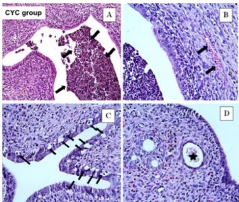
We observed some histopathological changes in uterine tissue in the CYC group.
These histopathological changes were
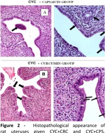
inflammatory cells in the lumen (Figure 1A), vascular congestion (Figure 1B), increased epithelial degeneration, vacuoles in epithelial cells (Figure 1C) and dilated endometrial glands (Figure 1D). Figure 2 shows little inflammatory cells in the lumen and normal endometrial gland. Improved epithelial degeneration and normal endometrial gland were shown in A in CYC+CPS group. And B shows few epithelial degeneration and vacuolization in epithelial cells in the CYC+CRC group. Histopathologic scores for all four groups are demonstrated in Table 2. A significant increment in vascular congestion, inflammatory cells in the lumen, epithelial degeneration, vacuoles in epithelial cells and
dilated endometrial gland were observed
in the CYC group compared with the other
groups. All histological parameters were
significantly improved with the administration of CRC and CPS.
Figure 1 - Histopathological evaluation of rat uteruses
given CYC. A: inflammatory cells were observed
in lumen (black arrows), B: vascular congestion
(black arrows), C: significantly increased epithelial
degeneration and vocuolization in epithelial cells (black arrows), D: dilated endometrial gland
(black asterix). A; H-E x20, B,C,D: H-E x40. CYC;
Figure 2 - Histopathological appearance of
rat uteruses given CYC+CRC and CYC+CPS.
A: Significantly decreased inflammatory cell
deposition, decreased epithelial degeneration and normal endometrial glandular tissue (black arrows)
compared to the group given CYC, B: minimal
epithelial degeneration, decreased epithelial vocuolization (black arrows), H-E x40. CYC;
cyclophosphamide, CRC; curcumin, CPS; capsaicin, H-E; Hematoxylene-Eosine.
Table 2 - Histopathological score of groups. Groups Histopathologic Damage
(Mean± SD)
Control 0.45 ± 0.07a
CYC 2.99 ± 0.21b
CYC+ CPS 1.35 ± 0.25c
CYC + CRC 1.28 ± 0.10c
*CYC: cyclophosphamide; CRC: curcumin; CPS: capsaicin ** Mean values bearing different superscript letters within the same column are significantly different (p< 0.01).
■
Discussion
Physicians are sometimes overwhelmed in the treatment of cancer due to adverse
environmental factors despite technological
advances. Adjuvant and/or neoadjuvant chemotherapeutic agents considered crucial in cancer treatment to enhance patient survival. Alkylating agents such as CYC inhibit DNA synthesis in tumor cells as well as have cytotoxic effects in the gastrointestinal system, liver and cardiac tissue cells, and
especially in the genital system9. Although the
effect of CYC on ovary in the female genital system has been well defined, the effect of CYC on the uterus has not been evaluated. To our knowledge, this is the first report to investigate the effects of CYC on the uterus, and this study demonstrated the detrimental effect of CYC on rat uterus by biochemical and histopathological analyses.
CYC, a compound of N, N-bis
(2-chloroethyl)
tetrahydro-2H-1,3,2-oxazaphosphorin 2-amine 2-oxide, is widely used in the treatment of autoimmune diseases such as systemic lupus erythematosus and rheumatoid arthritis in addition to its important place in cancer treatment. It is primarily metabolized by cytochrome P450 enzyme system in the liver after systemic administration and is converted into two
basic compounds as phosphoramide mustard
and acrolein. Phosphoramide mustard is responsible for basic antineoplastic activity and acts as a DNA alkylating agent independent from the cell cycle, while acrolein is responsible from cytotoxic effects by causing the formation of reactive oxygen species (ROS) in the
organism10. In normal cell cycle, endogenous
ROS is produced at the stages of mitochondrial respiration, NADPH oxidase activity, and peroxisome β- oxidation. Besides, ROS may be produced exogenously (ionizing or ultraviolet radiation, chemotherapeutic agents, and environmental stimuli) at a cellular level11.
whereas high ROS activity negatively affects intracellular homeostasis by causing cell cycle arrest, cell dysfunction and consequent DNA
damage12. This is especially shown as the
cause of chemotherapy-induced cytotoxicity. This situation has directed physicians to the use of natural antioxidants to prevent cytotoxicity, an important health problem associated with chemotherapy. The results of this study suggest that both curcumin and capsaicin have a protective effect against CYC-induced uterine damage. An animal study showed that selenium, which has natural antioxidant property, decreased total oxidant status (TOS) and increased total antioxidant status (TAS) in the kidney tissue of rats given CYC10. Another in vitro
study with Carvacrol, a monoterpenic phenol compound with antioxidant activity found in aromatic plants such as thyme, showed that TOS and oxidative stress index decreased and antioxidant markers such as GSH, SOD, GPx, and CAT significantly increased in kidney
tissues of CYC administered rats13.
Two important natural substrates
used as antioxidants and also used in our study are CRC and CPS. CPS provides basic activity by pro-apoptotic action in the cell cycle. It inhibits the Nuclear factor-kappa
beta pathway14, which plays an essential role
in the aetiology of tumorigenesis and at the same time affects the cell cycle homeostasis negatively. It produces this effect indirectly by inhibiting the synthesis of transient receptor potential vanilloid receptors15. CRC produces
antioxidant and anti-inflammatory activity by inhibiting the synthesis of the inducible nitric oxide synthase enzyme and inflammatory cytokines (such as interleukin-1β and 6). Also, CRC contributes to the inhibition of ROS by increasing activities of enzymes and genes such as NAD (P) H, quinone oxidoreductase 1 and glutathione S-transferase that are responsible
for detoxification16. In vitro studies examining
the antioxidant effect of CRC showed promising results. An in vitro rat study demonstrated that renal toxicity due to cyclosporine decreased significantly by CRC administration and TOS and MDA levels in this group were significantly
low17. Another animal study detected that
in renal tissues of cisplatin and methotrexate administered rats MDA level was increased and GSH level was decreased; however in CRC administered rats level of oxidant MDA significantly decreased and level of antioxidant
GSH significantly increased18. Another in vitro
study which evaluated cisplatin toxicity and CRC effectiveness in female rats in which experimental breast cancer was induced by 12-dimethyl benz[a]anthracene; TNF-α, interleukin 6 and interleukin-8 activities significantly decreased in the group which was given cisplatin and CRC compared with the group which was given only CRC. The same study also evaluated the effect of CRC on cell homeostasis. Histopathological evaluation of breast tissue showed that cellular degeneration, necrosis, and hemorrhage were severe-very severe in the cisplatin administered group. However, none of these pathological findings occurred in the CRC+ cisplatin group19. Our
study also showed that oxidant activity (MDA) was high and antioxidant activity (GSH, SOD, GPx, and CAT) was low in the uterus of rats treated with CYC; however in the uterus of the rats in the CRC+CYP group oxidant activity was low and antioxidant activity was high. In the histopathological examination, inflammation, congestion, epithelial degeneration and vacuolization were detected in the rat uterus exposed to CYC chemotherapy. However, in the CRC and CYP group, we found that these pathological findings were improved.
Although several studies showed cell
limited studies to show the preventative
effect of CPS against increased cytotoxicity
due to chemotherapy. A study by Huang et
al.20 showed the neuroprotective effect of
CPS in cortical neurons of rats after ischemia/ reperfusion injury. In another study on rats, it has been demonstrated that the oxidative damage caused by alcohol in liver tissues of CPS-treated animals is minimized and that MDA and hydrogen peroxide-dependent oxidative processes are inhibited by CPS-related antioxidant activity21. As mentioned,
studies investigating CPS activity in order to prevent the increased cytotoxic effect due to chemotherapy are limited, but in an animal
study performed Manjunathaet al.22 detected
histopathological examination of the tissues showed a marked decrease in inflammatory cell accumulation and epithelial degeneration in the hepatic tissues of the group given CRC and CPC. In our study also showed decreased inflammation and prevention of tissue degeneration in histopathological evaluation of uterus tissues of rats given CPS.
Chemotherapeutic agents, which are indispensable for treatment in increasing cancer cases around the world, have positive effects on patient survival. Especially alkylating agents like CYC, prevents DNA synthesis in tumor cell not specific to mitosis stages and therefore blocks uncontrolled divisions of cancer cells. In addition to these effects, chemotherapeutic agents cause serious cytotoxic effects due to increased ROS, especially in liver, kidney and gastrointestinal system. These effects on the genital system have become a critical problem especially for chemotherapeutic agents applied in cancer cases diagnosed in younger ages in recent years. This caused focusing of physicians on specific antioxidant preparations especially natural ones. These antioxidant preparations also include CRC and CPS which are entirely
natural. In our study, CPS and CRC were shown to have protective effects on the uterus in rats treated with CYC, taking both oxidant and antioxidant parameters into consideration. Besides, histopathological evaluations also confirmed these tissue protective effects of CRC and CPS. To the best of our knowledge, this study is the first one to demonstrate the cytotoxic effects of cyclophosphamide on the uterus in the female genital system, and also the results suggest that these cytotoxic
effects can be treated by CRC and CPS. We
believe that this study would have a guiding role for further studies in that it demonstrated that damage of chemotherapy on the genital system is reversible and preventable.
■
Conclusion
Cytotoxic effects of natural
alkylating chemotherapeutic agents like cyclophosphamide on the uterus can be prevented by curcumin and capsaicin
■
References
1. Yilmaz E, Coskun EI, Sahin N, Ciplak B,
Ekici K. MPV, NLR, and platelet count:
new hematologic markers in diagnosis of
malignant ovarian tumour. Eur J Gynaecol
Oncol. 2017;38:346-9. PMID: 29693870. 2. Pyun CW, Kim JH, Han KH, Hong GE, Lee CH.
In vivo protective effects of dietary curcumin and capsaicin against alcohol-induced oxidative stress. Biofactors. 2014;40:494-500. doi: 10.1002/biof.1172.
3. Pascuali N, Scotti L, Di Pietro M, Oubiña G, Bas D, May M, Gómez Muñoz A, Cuasnicú PS,
Cohen DJ, Tesone M, Abramovich D, Parborell
F. Ceramide-1-phosphate has protective
properties against
cyclophosphamide-induced ovarian damage in a mice model
of premature ovarian failure. Hum Reprod. 2018;33:844-59. doi: 10.1093/humrep/ dey045.
its antitumor effects in a context dependent fashion. J Proteomics. 2018;182:65-72. doi: 10.1016/j.jprot.2018.05.007.
5. El-Kott AF, Bin-Meferij MM.
Suppressive effects of capsaicin
against N-nitrosomethylurea-induced
mammary tumorigenesis in rats. Biomed
Pharmacother. 2018;98:673-9. doi:
10.1016/j.biopha.2017.12.036.
6. Wojcik M, Krawczyk M, Wojcik P, Cypryk K, Wozniak LA. Molecular mechanisms underlying curcumin-mediated therapeutic effects in type 2 diabetes and cancer. Oxid Med Cell Longev. 2018;20:1-14. doi: 10.1155/2018/9698258.
7. Ciftci O, Ozdemir I, Tanyildizi S, Yildiz S, Oguzturk H. Antioxidative effects of curcumin, β-myrcene and 1,8-cineole
against
2,3,7,8-tetrachlorodibenzo-p-dioxin-induced oxidative stress in rats liver. Toxicol Ind Health. 2011;27:447-53. doi: 10.1177/0748233710388452.
8. Huang M, Cheng G, Tan H, Qin R, Zou Y, Wang Y, Zhang Y. Capsaicin protects cortical neurons against ischemia/reperfusion injury via down-regulating NMDA receptors. Exp Neurol. 2017;295:66-76. doi: 10.1016/j. expneurol.2017.05.001.
9. Kurauchi K, Nishikawa T, Miyahara E, Okamoto Y, Kawano Y. Role of metabolites of cyclophosphamide in cardiotoxicity. BMC Res Notes. 2017;10:1-10. doi: 10.1186/ s13104-017-2726-2.
10. Gunes S, Sahinturk V, Uslu S, Ayhanci A, Kacar S, Uyar R. Protective effects of selenium on cyclophosphamide-induced oxidative stress and kidney injury. Biol Trace Elem Res. 2017;30:1-8. doi: 10.1007/ s12011-017-1231-8.
11. Wang GF, Dong Q, Bai Y, Yuan J, Xu Q, Cao
C, Liu X. Oxidative stress induces mitotic arrest by inhibiting Aurora A-involved mitotic spindle formation. Free Radic Biol Med. 2017;103:177-87. doi: 10.1016/j. freeradbiomed.2016.12.031.
12. Turhan U, Yilmaz E, Gul M, Melekoglu R, Turkoz Y, Ozyalin F, Parlakpinar H, Simsek Y. Investigation of the effect of gestational diabetes on fetal cardiac tissue in streptozotocin ınduced in rats. Acta Cir Bras. 2018;33:306-13. doi: 10.1590/s0102-865020180040000002.
13. Gunes S, Ayhanci A, Sahinturk V, Altay DU, Uyar R. Carvacrol attenuates cyclophosphamide-induced oxidative stress in rat kidney. Can J Physiol Pharmacol.
2017;95:844-9. doi: 10.1139/cjpp-2016-0450.
14. Yilmaz E, Coskun EI, Gul M, Sahin N, Tuncay
G, Simsek Y. Nuclear factor- k a p p a
beta pathway and endometrial cancer: a pilot study. Eur J Gynecol Oncol. 2017;38:536-40. doi: 10.12892/ejgo3471.2017.
15. Melekoglu R, Ciftci O, Eraslan S, Cetin A, Basak N. Beneficial effects of curcumin and capsaicin on cyclophosphamide-induced
premature ovarian failure in a rat model. J
Ovarian Res. 2018;11:33:1-8. doi: 10.1186/ s13048-018-0409-9.
16. Jaja-Chimedza A, Graf BL, Simmler C, Kim Y, Kuhn P, Pauli GF, Raskin I. Biochemical characterization and anti-inflammatory properties of an isothiocyanate-enriched moringa (Moringa oleifera) seed extract. PLoS One. 2017;12:1-21. doi: 10.1371/ journal.pone.0182658.
17. Huang J, Yao X, Weng G, Qi H, Ye X. Protective effect of curcumin against cyclosporine A-induced rat nephrotoxicity. Mol Med Rep. 2018;17:6038-44. doi: 10.3892/ mmr.2018.8591.
18. Said Salem NI, Noshy MM, Said AA.
Modulatory effect of curcumin against genotoxicity and oxidative stress induced by cisplatin and methotrexate in male mice. Food Chem Toxicol. 2017;105:370-6. doi: 10.1016/j.fct.2017.04.007.
19. Kumar P, Barua CC, Sulakhiya K, Sharma
RK. Curcumin ameliorates cisplatin-induced nephrotoxicity and potentiates its anticancer activity in sd rats: potential role of curcumin in breast cancer chemotherapy. Front Pharmacol. 2017;8:1-12. doi: 10.3389/ fphar.2017.00132.
20. Huang M, Cheng G, Tan H, Qin R, Zou Y, Wang Y, Zhang Y. Capsaicin protects cortical neurons against ischemia/reperfusion injury via down-regulating NMDA receptors. Exp Neurol. 2017;295:66-76. doi: 10.1016/j. expneurol.2017.05.001.
2014;40:494-500. doi: 10.1002/biof.1172.
22. Manjunatha H, Srinivasan K. Protective effect of dietary curcumin and capsaicin on induced oxidation of low-density
lipoprotein, iron-induced hepatotoxicity and carrageenan-induced inflammation in experimental rats. FEBS J. 2006;273:4528-37. PMID: 16956363.
Correspondence:
Ercan Yilmaz
Inonu University, Medicine Faculty, Obstetrics and Gynecology Department Malatya Turkey
Phone: +0905369556180 ercanyilmazgyn@yahoo.com
Received: Feb 22, 2018 Review: Apr 25, 2018 Accepted: May 23, 2018
Conflict of interest: none Financial source: none
1Research performed at Medicine Faculty,