Detrusor overactivity in diabetic and non-diabetic
patients: Is there a difference?
Tomasz Golabek, Eamonn Kiely, Barry O’Reilly
Cork University Hospital (TG,EK) and Maternity Hospital (BOR), Cork, Republic of Ireland
ABSTRACT ARTICLE INFO
_________________________________________________________ ___________________
Key words:
Diabetes mellitus; Urodynamics; Urothelium
Int Braz J Urol. 2012; 38: 652-60
__________________
Submitted for publication: March 13, 2012
__________________
Accepted after revision: July 27, 2012
Purpose: To compare urodynamic characteristics in patients with idiopathic detrusor overactivity (IDO) with those of an age matched cohort with diabetes mellitus (DM) and detrusor overactivity (DO). Secondly, to determine whether urodynamic features could help distinguish these two groups of patients.
Materials and Methods: Urodynamic data was collected on 58 female patients; 29 with IDO and 29 with DM and detrusor overactivity. Eight urodynamic parameters were selected for analysis: amplitude of the first overactive contraction (AOFC), the volume at the first contraction, cystometric capacity, maximal detrusor pressure, maximal flow rate, voiding pressure at maximal flow, voided volume and postvoid residual (PVR) urine volume. Finally, sensitivity analysis for distinguishing urodynamic parameters between studied groups was performed.
Results: AOFC, volume at AOFC and maximal detrusor pressure were statistically gre-ater in diabetic patients, compared with the non-diabetic group of women (16.00 cm H2O versus 9.00 cm H2O, 309.00 mL versus 167.00 mL and 76.48 cm H2O versus 55.41 cm H2O respectively). A specificity of 72.41% and positive predictive value of 71.43% were achieved for AOFC with cutoff value of 12 cm H2O. These parameters were fur-ther improved with cutoff value of 258 mL for volume at AOFC and were 75.86% and 73.08% respectively.
Conclusions: Certain urodynamic parameters in diabetic female patients with DO are shown to be significantly different than those in women with IDO. Further prospective study should provide additional information about the pathogenesis and progression of DO in diabetic patients as well as the validity of diabetic screening in patients with IDO.
INTRODUCTION
Diabetes mellitus (DM) has been shown to alter vesicourethral function in a number of ways, from decreased detrusor contractility to bladder overactivity present in up to 61% of dia-betic patients (1).
The etiology of DO in diabetic patients is not fully understood and is most likely multifac-torial. Both central and peripheral mechanisms have been implicated; namely, diabetic cerebral
vasculopathy and peripheral nerve stimulation as well as changes in the detrusor muscle and uro-thelium (2-4).
all individuals with idiopathic overactive bladder require treatment, the condition has been shown to significantly impact on patient quality of life; often leading to isolation and depression (8,9).
Given that lower urinary tract symptoms are not disease specific and it is still unclear what can initiate IDO, the aim of the present study was to determine if there are differences in urodynam-ic characteristurodynam-ics between patients with overactive bladder secondary to diabetes mellitus and pa-tients with overactive bladder without diabetes or any known neurologic abnormalities. Sensitivity and specificity analyses were performed to assess their ability to predict diabetic overactive bladder according to various urodynamic parameters.
MATERIALS AND METHODS
Urodynamic data of all female patients who underwent urodynamic studies in Depart-ments of Urology and Urogynaecology of the Cork University Hospital over the period 2004 and 2008 were reviewed retrospectively. Patients with objective signs of overactive bladder during the study, defined as an involuntary rise in detru-sor pressure of greater than 5 cm H2O during fill-ing associated with urgency or bladder fullness, were selected. Then, medical charts of all select-ed patients were reviewselect-ed and a database with patients’ blood glucose levels and HBA1c were searched. Only patients with a known diagnosis of diabetes mellitus were included in the data-base of patients with DM while non-neurogenic patients with no history of diabetes and normal glucose levels were selected for the study in the idiopathic bladder overactivity group.
Exclusion criteria included patients with urodynamic evidence of bladder outlet obstruc-tion, defined as maximum flow rate less than 12 mL/min. and detrusor pressure at maximum flow of more than 45 cm H2O. Those with presence of concurrent neurologic disorders such as stroke, Parkinson disease, spinal cord injury, and multiple sclerosis were also excluded. Lastly, patients with medical conditions that could interfere with void-ing function such as prior pelvic surgery, ante-rior pelvic prolapse of stage 2 or greater (Baden-Walker) or those on medication that could affect
bladder function such as diuretics, calcium chan-nel blockers and narcotics were excluded from the study. A total of 58 patients fulfilled the inclusion criteria and of these, 29 were diabetic with DO and 29 patients had IDO.
Urodynamic studies were performed by two experienced urodynamic nurses using the So-lar Silver (MMS, Enschede, The Netherlands) and Dantec Menuet (Medtronic Functional Diagnostics A/S, Slovlunde, Denmark) urodynamic measure-ment systems. A standard protocol was employed in accordance with the guidelines established by the International Continence Society (ICS) (10). All anticholinergic medications were stopped at least 72 hours before study and all patients who under-went urodynamic evaluation had confirmed nega-tive urinalysis findings prior the procedure. The studies were performed with patients in the seated position. Urinary bladders were filled at 50 mL/ min. rate using room temperature sterile saline. A dual lumen 8F vesical catheter and 4.5 F rectal catheter were used. Eight urodynamic parameters were selected for analysis: amplitude of the first contraction (AOFC), the volume at the first con-traction, cystometric capacity, maximal detrusor pressure (Pdetmax), maximal flow rate (Qmax), voiding pressure at maximal flow (PdetQmax), voided volume and postvoid residual (PVR) urine volume. Also Bladder Voiding Efficiency (BVE), an index that defines bladder voiding function, was calculated as described previously by Abrams, and then statistically analyzed (11). BVE was obtained by the formula: BVE=100% x voided volume/ (voided volume+PVR).
All measurements were repeated three times by the same investigator to avoid bias.
Statistical analyses were performed using SPSS package version 11.5. The Shapiro-Wilk test was used to examine for normal distribution. Re-sults were presented as mean values ± standard deviation when data were normally distributed, otherwise as median, 25th and 75th percentile (AOFC, volume at AOFC, PVR and BVE). Non-parametric t test and Mann-Whitney U tests were applied as appropriate.
a set of variables distinguishing diabetic patients with DO from IDO.
The associations between age and urody-namic parameters were examined using Pearson’s correlation analyses for normally distributed data, otherwise Spearman’s rank correlation coefficient was used. Sensitivity and specificity analyses for the ability to predict diabetic DO on the basis of AOFC, volume at AOFC and Pdetmax were also performed. For all statistical tests p < 0.05 was considered significant.
RESULTS
A total of 97 urodynamic studies were car-ried out on female diabetic patients referred from urology or urogynaecology departments in our university hospital over the period 2004 -2008. Of these, 41 women had DO. Strict inclusion criteria were fulfilled only in 29 patients. Three patients had type 1 DM and 26 type 2 of at least 3 years duration. Their average age was 53.84 ± 16.0 years. Average HBA1c level measured over one year preceding uro-dynamics was determined in 26 patients and was 6.05 ± 2.38% (5.1-12.1%). Five patients had HBA1c level checked after or more than a year before the study. In 15 diabetic women (51% of total), the ma-jor reported complaints were frequency and urge incontinence; 8 cases reported urgency without in-continence (28%); mixed urinary inin-continence was the main problem in a further 4 (14%) and recurrent bladder infections in 2 more cases (7%).
Urodynamic data were also collected from 29 female patients complaining of symptoms sug-gestive OAB and who had no previous history of diabetes mellitus or neurological disorder and who were referred for evaluation of their lower urinary tract. The average age of this group was 50.42 ± 20.23years. The most common symptoms of women with IDO were urge incontinence in 11 cases (38%), mixed urinary incontinence in 7 (24%), urgency without incontinence in 5 (17%), voiding symp-toms (hesitancy, dribbling, incomplete emptying) in 4 (14%) and stress incontinence in 2 cases (7%).
Table-1 shows characteristics of the groups studied and comparison of the analyzed urody-namic parameters between the two investigated groups. Greater amplitude of the first overactive
contraction was observed in patients with DM than in females with IDO (18.31 cm H2O versus 11.03 cm H2O). Also, these patients had a stronger maximal detrusor contraction compared to those with IDO (76.48 cm H2O versus 55.41 cm H2O). The initial contraction occurred later during the filling phase in diabetic women than in those patients with IDO (333.83 mL versus 208.72 mL).
The remaining analyzed parameters were not statistically different in both groups under investigation. Also, BVE was within the normal range and showed no statistical difference in both diabetic DO and IDO group.
Multiple logistic regression analysis us-ing the forward stepwise regression with Wald test method showed that a set of three urodynamic pa-rameters (AOFC, volume at AOFC and Pdetmax) distinguished diabetic overactive bladder from IDO. AOFC showed to be the most independently aspect with ability to differ diabetic overactive bladder and IDO with classification accuracy of 70.7%. The combination of AOFC, volume at AOFC and Pdet-max improved accuracy to 79.3%.
To examine associations between age and parameters distinguishing diabetic DO and IDO, Pearson’s correlation analyses or Spearman’s rank correlation coefficient were performed. No rela-tionship between age and parameters under inves-tigation was found.
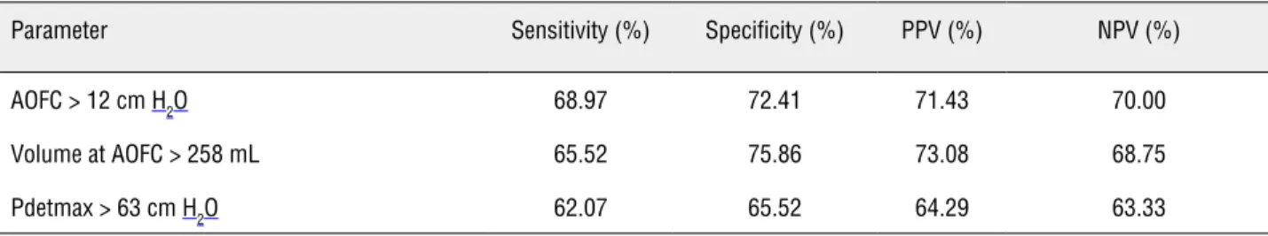
Since AOFC, volume at AOFC and maximal detrusor pressure between diabetic patients and women with IDO differed significantly, cutoff val-ues were established to evaluate sensitivity, speci-ficity, positive (PPV) and negative predictive val-ues (NPV). A cutoff value of 12 cm H2O or greater for AOFC produced sensitivity, specificity and positive predictive value of 68.97%, 72.41% and 71.43% respectively. Whereas analyses of using a cutoff value of 258 or greater for volume at AOFC resulted in further improvement of specificity and PPV (75.86% and 73.08% respectively). Evaluation of maximal detrusor pressure did not led to reason-able results (Treason-able-2).
DISCUSSION
dia-betic cystopathy and 29 female patients with DO without DM or known neurological abnormalities. All of them were referred from urology or urogy-naecology departments at our university hospital for an evaluation of lower urinary tract function. In our patient population, we demonstrated that the amplitude of the first detrusor contraction was greater in diabetic patients than in women without known glycaemic and neurologic abnor-malities. Similarly, the maximum detrusor
pres-sure generated was higher in the group of patients with DM. These findings may suggest less con-trollable symptoms of DO in diabetic individuals. Although comparable data on the lower urinary tract symptoms between patients with IDO and diabetic DO are not available, a recent large ob-servational study reported on an increase in urge incontinence frequency in women with DM (12). This symptom has been shown to have a profound effect on patients’ quality of life (13,14).
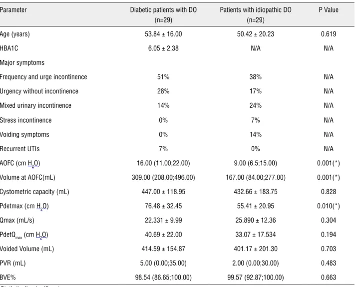
Table 1 - Characteristics of the groups and comparison of urodynamic parameters and index of bladder voiding function in diabetic patients with DO and IDO.
Parameter Diabetic patients with DO (n=29)
Patients with idiopathic DO (n=29)
P Value
Age (years) 53.84 ± 16.00 50.42 ± 20.23 0.619
HBA1C 6.05 ± 2.38 N/A N/A
Major symptoms
Frequency and urge incontinence 51% 38% N/A
Urgency without incontinence 28% 17% N/A
Mixed urinary incontinence 14% 24% N/A
Stress incontinence 0% 7% N/A
Voiding symptoms 0% 14% N/A
Recurrent UTIs 7% 0% N/A
AOFC (cm H2O) 16.00 (11.00;22.00) 9.00 (6.5;15.00) 0.001(*)
Volume at AOFC(mL) 309.00 (208.00;496.00) 167.00 (84.00;277.00) 0.001(*)
Cystometric capacity (mL) 447.00 ± 118.95 432.66 ± 183.75 0.828
Pdetmax (cm H2O) 76.48 ± 32.45 55.41 ± 20.95 0.010(*)
Qmax (mL/s) 22.331 ± 9.99 25.890 ± 12.36 0.304
PdetQmax (cm H2O) 40.69 ± 22.00 33.07 ± 17.534 0.194
Voided Volume (mL) 414.59 ± 154.87 401.17 ± 201.30 0.703
PVR (mL) 5.00 (0.00;35.00) 2.00 (0.00;30.00) 0.483
BVE% 98.54 (86.65;100.00) 99.57 (92.87;100.00) 0.663
* Statistically significant.
Key: HBA1C = glycosylated haemoglobin level, N/A= not applicable; UTIs = urinary tract infections, DO = detrusor overactivity; IDO = idiopathic detrusor overactivity; AOFC = amplitude of first overactive contraction; Pdetmax = maximal detrusor pressure; Qmax = maximal flow rate; PdetQmax = detrusor pressure at maximal flow; PVR = postvoid residual (urine volume); BVE = Bladder Voiding Efficiency.
Interestingly in our study, detrusor con-tractions occurred later during filling in the DM group than in patients with IDO. However, PVR in both patients with diabetes and DO and IDO was not increased thus functional capacity in diabetic patients was not reduced and bladder contractions were occurring at different intervals in both investigated groups.
These findings are different from other reports that noticed increased PVR and decreased functional capacity in diabetic DO individuals (2,15); but similar to those suggesting that condi-tions affecting nervous system may induce stron-ger overactive contractions at higher volume (16).
The greater amplitude of the first detru-sor contraction, volume at first contraction, and maximum detrusor pressure observed in patients with diabetes can be explained by diuresis-induced bladder wall tissue remodeling and neuropathy. Several studies in the past have demonstrated that the high rate of bladder filling during cystometry may result in an increase in intravesical pressure and threshold volume (17-19). In addition, such non-physiological filling rates may mechani-cally damage the afferent limb of the micturition reflex resulting in the later generation of action potentials and, consequently, an urge to void at volumes which are larger then normal. Although the mechanisms involved in triggering bladder tis-sue hypertrophy and hyperplasia in patients with diabetes are not very clear, it seems that a high filling rate is a primary factor in the stimulation of hyperplasia of bladder smooth muscle, urothelium and connective tissue (20,21). An increase in blad-der weight is also related to alterations in bladblad-der
volume as well as the rate of stretch of the blad-der wall, both caused by polyuria, a consequence in itself of diabetes (22). Increases in fluid output likely contribute to faster and greater increases in bladder weight in diabetic patients with DO than in patients with idiopathic detrusor overactivity.
Both peripheral autonomic neuropathy and central nervous system dysfunction due to ce-rebral vasculopathy are implied in the aetiology of DO in diabetic patients (2,23). However, this kind of diabetic bladder dysfunction can also be pres-ent in the absence of CNS lesions (2). In our study, patients with history of stroke were excluded, thus peripheral pathology as the cause of development of DO need to be considered. In addition, altera-tions in bladder innervation, the bladder smooth muscle cells and urothelium have been proposed to be involved in early stages of diabetic bladder dysfunction (4). M2 receptors up-regulation with partial autonomic denervation leading to the de-creased cholinergic transmission are involved in the aetiology of DO with altered contractility in early stages of diabetic bladder dysfunction (24).
Although the chronology of bladder dys-function in DM and its correlation with diabetic control is not fully known, one group led by Run-dles investigated the initial manifestation of the diabetic neurogenic bladder (25). In their series, 83% of diabetic patients with neuropathy had an abnormal cystometrogram and enlarged bladder indicating neurogenic bladder. However, most of them had no residual urine. This differs from ad-vanced diabetic neurogenic bladder with paralysis. These findings were consistent with those in recent reports garnered from laboratory animals (21).
Table 2 - Sensitivity, specificity, positive predictive value and negative predictive value for AOFC, Volume at AOFC and Pdet-max at developed cutoff values.
Parameter Sensitivity (%) Specificity (%) PPV (%) NPV (%)
AOFC > 12 cm H2O 68.97 72.41 71.43 70.00
Volume at AOFC > 258 mL 65.52 75.86 73.08 68.75
Pdetmax > 63 cm H2O 62.07 65.52 64.29 63.33
Up to 61% of patients with diabetic blad-der dysfunction have DO (1). Currently diagnostic evaluation of patients with IDO does not include determination of diabetes (26). Although routine assessment prior to urodynamics includes ques-tions about DM there is no additional check for diabetes except for urine testing for glycosuria. However, urine testing for glycosuria as screening for DM is not recommended particularly in patients with type 2 diabetes, because of the low sensitivity of the test (27-29). Glucose tolerance test and fast-ing glucose measures have been the standard tests for screening and diagnosing of diabetes mellitus. Recently hemoglobin A1c testing for the diagnosis of type 2 diabetes has been recommended (30).
The utility of fasting glucose and HBA1c measurements have not yet been evaluated in pa-tients with IDO.
In our study we used a cutoff value of 12cm H2O for the amplitude of the first overactive contraction in diabetic patients with DO. Specific-ity was 72.41%, however positive predictive value was 71.43%. These parameters were further im-proved when analyzed for volume at AOFC. Using a cutoff value of 258 mL specificity and PPV were 75.86% and 73.08% respectively. Therefore, we suggest diabetic screening in the IDO patient with greater amplitude and bladder volume at the first overactive contraction.
Our study was a retrospective, two-unit analysis of patients and as such is subjected to biases and limitations that surround these study types. Data obtained from medical charts may not have revealed undiagnosed neurologic condition which could affect all women, however to our knowledge none had been diagnosed at this point. In addition, study participants may not be a repre-sentative cohort from the greater community; thus limiting the ability to generalize findings. Finally, amplitude of the first overactive contraction is not an ideal parameter quantifying detrusor overactiv-ity as it may be affected by various factors during bladder filling. Although all urodynamic studies were performed under the same conditions and in accordance with the strict guidelines established by the ICS to maintain their objectivity, accuracy and reliability it is possible that some discrete fac-tors could affect AOFC. However, combining AOFC,
volume at AOFC and Pdetmax improved accuracy for identifying diabetic female with DO and mini-mized potential bias. Further prospective study in a larger cohort of patients would be useful to stratify certain subgroups based on type and duration of diabetes, symptom levels and glycaemic control.
CONCLUSIONS
Certain urodynamic parameters are im-portant for the detection of diabetes-related DO. It seems that stronger overactive contractions in the presence of larger threshold volume at which they occur characterize the DO in diabetic female pa-tients and suggest different pathogenesis then that involved in IDO. Also diabetic screening of women with IDO and greater amplitude of the first over-active contraction may have a role in identifying patients who do not have a true IDO.
Further prospective studies will provide additional information about pathogenesis and progression of DO in diabetic patients as well as validity of diabetic screening in patients with IDO who have high amplitude and volume at first over-active contractions. Comparison of urodynamic parameters in diabetic patients with and without urodynamically demonstrable DO as well as in pa-tients with neurogenic detrusor overactivity may provide important information on chronology of bladder dysfunction in DM and mechanisms in-volved in the development of diabetic cystopathy.
ABBREVIATIONS
AOFC = amplitude of first overactive contraction
BE = bladder voiding efficiency
CNS = the central nervous system
DM = diabetes mellitus
DO = detrusor overactivity
HBA1c = glycosylated haemoglobin level
IDO = idiopathic detrusor overactivity
ICS = The International Continence Society
NPV = negative predictive value
Pdetmax = maximal detrusor pressure
PdetQmax = detrusor pressure at maximal flow
PPV = positive predictive value
PVR = postvoid residual (urine volume)
ACKNOWLEDGEMENTS
We thank Dolores O’Donnell, clinical nurse specialist at CUH and Elaine Dilloughery, conti-nence nurse specialist at CUMH for performing the urodynamic studies.
CONFLICT OF INTEREST
None declared.
REFERENCES
1. Starer P, Libow L: Cystometric evaluation of bladder dysfunction in elderly diabetic patients. Arch Intern Med. 1990; 150: 810-3. 2. Yamaguchi C, Sakakibara R, Uchiyama T, Yamamoto T, Ito
T, Liu Z, et al.: Overactive bladder in diabetes: a peripheral or central mechanism? Neurourol Urodyn. 2007; 26: 807-13. 3. Lee WC, Wu HP, Tai TY, Yu HJ, Chiang PH: Investigation of
urodynamic characteristics and bladder sensory function in the early stages of diabetic bladder dysfunction in women with type 2 diabetes. J Urol. 2009; 181: 198-203.
4. Yoshimura N, Chancellor MB, Andersson KE, Christ GJ: Recent advances in understanding the biology of diabetes-associated bladder complications and novel therapy. BJU Int. 2005; 95: 733-8.
5. Stewart WF, Van Rooyen JB, Cundiff GW, Abrams P, Herzog AR, Corey R, et al.: Prevalence and burden of overactive blad-der in the United States. World J Urol. 2003; 20: 327-36. 6. Milsom I, Abrams P, Cardozo L, Roberts RG, Thüroff J, Wein
AJ: How widespread are the symptoms of an overactive blad-der and how are they managed? A population-based preva-lence study. BJU Int. 2001; 87: 760-6. Erratum in: BJU Int 2001; 88: 807.
7. Ahlberg J, Edlund C, Wikkelsö C, Rosengren L, Fall M: Neu-rological signs are common in patients with urodynamically verified “idiopathic” bladder overactivity. Neurourol Urodyn. 2002; 21: 65-70.
8. Brown JS, Posner SF, Stewart AL: Urge incontinence: new health-related quality of life measures. J Am Geriatr Soc. 1999; 47: 980-8.
9. Dugan E, Cohen SJ, Bland DR, Preisser JS, Davis CC, Sug-gs PK, et al.: The association of depressive symptoms and urinary incontinence among older adults. J Am Geriatr Soc. 2000; 48: 413-6.
10. Abrams P, Cardozo L, Fall M, Griffiths D, Rosier P, Ulmsten U, et al.: The standardisation of terminology of lower urinary tract function: report from the Standardisation Sub-commit-tee of the International Continence Society. Neurourol Uro-dyn. 2002; 21: 167-78.
11. Abrams P: Bladder outlet obstruction index, bladder con-tractility index and bladder voiding efficiency: three simple indices to define bladder voiding function. BJU Int. 1999; 84: 14-5.
12. Jackson RA, Vittinghoff E, Kanaya AM, Miles TP, Resnick HE, Kritchevsky SB; et al.: Urinary incontinence in elderly wom-en: findings from the Health, Aging, and Body Composition Study. Obstet Gynecol. 2004; 104: 301-7.
13. Kobelt G, Kirchberger I, Malone-Lee J: Review. Quality-of-life aspects of the overactive bladder and the effect of treatment with tolterodine. BJU Int. 1999; 83: 583-90.
14. Patel AS, O’Leary ML, Stein RJ, Leng WW, Chancellor MB, Patel SG, et al.: The relationship between overactive bladder and sexual activity in women. Int Braz J Urol. 2006; 32: 77-87.
15. Lee WC, Wu HP, Tai TY, Liu SP, Chen J, Yu HJ: Effects of dia-betes on female voiding behavior. J Urol. 2004; 172: 989-92. 16. Lemack GE, Frohman EM, Zimmern PE, Hawker K, Ramnara-yan P: Urodynamic distinctions between idiopathic detrusor overactivity and detrusor overactivity secondary to multiple sclerosis. Urology. 2006; 67: 960-4.
17. Hamaide AJ, Verstegen JP, Snaps FR, Onclin K, Balligand MH: Validation and comparison of the use of diuresis cystometry and retrograde filling cystometry at various infusion rates in female Beagle dogs. Am J Vet Res. 2003; 64: 574-9. 18. Klevmark B: Volume threshold for micturition. Influence of
filling rate on sensory and motor bladder function. Scand J Urol Nephrol Suppl. 2002; 210: 6-10.
19. Robertson AS, Griffiths CJ, Ramsden PD, Neal DE: Bladder function in healthy volunteers: ambulatory monitoring and conventional urodynamic studies. Br J Urol. 1994; 73: 242-9. 20. Saito M, Longhurst PA, Murphy M, Monson FC, Wein AJ, Levin
RM: Effect of slow and rapid cystometry on in vitro rat urinary bladder DNA synthesis. Gen Pharmacol. 1994; 25: 1021-5. 21. Daneshgari F, Liu G, Imrey PB: Time dependent changes in
diabetic cystopathy in rats include compensated and decom-pensated bladder function. J Urol. 2006; 176: 380-6. 22. Liu G, Daneshgari F: Temporal diabetes- and diuresis-induced
remodeling of the urinary bladder in the rat. Am J Physiol Regul Integr Comp Physiol. 2006; 291: R837-43.
23. Rapidi CA, Karandreas N, Katsifotis C, Benroubi M, Petro-poulou K, Theodorou C: A combined urodynamic and elec-trophysiological study of diabetic cystopathy. Neurourol Uro-dyn. 2006; 25: 32-8.
24. Wein AJ: Lower urinary tract dysfunction in neurological im-aging and disease. In: Cambell-Walsh Urology, 9th ed. Edited by AJ Wein, LR Kavoussi, AC Novik, AW Partin and CA Peters. Philadephia: WB Saunders Co 2007; pp. 2011-45.
25. Rundles RW: Diabetic neuropathy. General review with report of 125 cases. Medicine. 1945; 24:111-60.
27. Friderichsen B, Maunsbach M: Glycosuric tests should not be employed in population screenings for NIDDM. J Public Health Med. 1997; 19: 55-60.
28. No authors listed: Diabetes mellitus. Report of a WHO Study Group. World Health Organ Tech Rep Ser. 1985; 727: 1-113.
29. Lawton J, Peel E, Douglas M, Parry O: ‘Urine testing is a waste of time’: newly diagnosed Type 2 diabetes patients’ percep-tions of self-monitoring. Diabet Med. 2004; 21: 1045-8. 30. International Expert Committee: International Expert
Commit-tee report on the role of the A1C assay in the diagnosis of diabetes. Diabetes Care. 2009; 32: 1327-34.
EDITORIAL COMMENT
This is a nice, small study that deserves some considerations. The authors described urody-namic over activity particularities related to diabe-tes mellitus. In my opinion, more important than isolated numbers themselves, are the implications of such increasing data relating diabetes mellitus and urinary tract disorders.
We cannot forget the potential impact that systemic illness and its repercussions can lead to the urinary tract, and how its treatment could change things, over time. Although small, the numbers showed by the authors bring some ques-tions in mind.
1. Diabetic patients in the study have had this diagnosis for at least, 3 years, but we do not know for how long, ex-actly, they have this condition. Prob-ably, the fact that all others variables analyzed did not show statistical dif-ferences when compared to the control group, can be related to the fact that these patients don´t have very lasting disease timing.
Clinical and experimental data confirm that detrusor over activity, both neuro-genic and myoneuro-genic can be present in diabetic neuropathic bladders. More-over: these findings are normally seem in earlier stages of the disease, whereas detrusor under activity appears to be linked to later stages of DM. (Does dia-betes mellitus-induced bladder remodel-ing affect lower urinary tract function? Kirschner-Hermanns R, Daneshgari F, Vahabi B, Birder L, Oelke M, Chacko S.Neurourol Urodyn. 2012; 31(3): 359-64. ICI-RS 2011). So, if the sample was
bigger, and the lasting time of the dis-ease analyzed, could the results associ-ated to the other variables be different? This is a nice question to be answered, in the future. The same doubt can be ex-tended to the glycemic control. Do the patients that have betters long lasting glycemic controls show less symptoms and urodynamic changes over time? If so, could we act as prophylactic agents of chronic urinary tract disturbances in such group?
2. The authors also raise the question about diabetic screening of patients with IDO and some urodynamic find-ings. Thinking on the clinical nature of the diagnosis of hyperactive bladder, I agree with such screening but I rather do it on all patients with this clinical picture, instead of doing it only in pa-tients submitted to such study with critical findings. It looks like the practi-cal application of an increasing body of knowledge on the complex etiology of urinary tract disorders.
I expect that more studies like this can help us to understand the real relationship of chronic illness like diabetes and urinary tract disorders in order to open new perspectives of, perhaps, prophylactic treatments in the future.
Dr. Caio Cintra