Letters to the Editor
Radiol Bras. 2015 Set/Out;48(5):333–340
339
Marília Henrique Destefani1, Alessandro Spanó Mello2, Ricardo Santos de Oliveira3, Gustavo Novelino Simão2
1. Cedirp – Radiologia e Diagnóstico por Imagem, Ribeirão Preto, SP, Brazil. 2. Hospital das Clínicas – Faculdade de Medicina de Ribeirão Preto da Universidade de São Paulo (HCFMRP-USP), and Cedirp – Radiologia e Diagnóstico por Imagem, Ribeirão Preto, SP, Brazil. 3. Hospital das Clínicas – Faculdade de Medicina de Ribei-rão Preto da Universidade de São Paulo (HCFMRP-USP), RibeiRibei-rão Preto, SP, Brazil. Mailing Address: Dra. Marília Henrique Destefani. Avenida Professor João Fiusa, 2055, Jardim Irajá. Ribeirão Preto, SP, Brazil, 14024-260. E-mail: [email protected].
http://dx.doi.org/10.1590/0100-3984.2014.0125 REFERENCES
1. Ortega-Martínez M, Cabezudo JM, Bernal-García LM, et al. Glioma cordoide del III ventrículo. Nuevo caso y revisión de la literatura. Neuro-cirugía. 2007;18:115–22.
2. Desouza RM, Bodi I, Thomas N, et al. Chordoid glioma: ten years of a low-grade tumor with high morbidity. Skull Base. 2010;20:125–38. 3. Ni HC, Piao YS, Lu DH, et al. Chordoid glioma of the third ventricle:
four cases including one case with papillary features. Neuropathology. 2013;33:134–9.
4. Pomper MG, Passe TJ, Burger PC, et al. Chordoid glioma: a neoplasm unique to the hypothalamus and anterior third ventricle. AJNR Am J Neuroradiol. 2001;22:464–9.
5. Smith AB, Smirniotopoulos JG, Horkanyne-Szakaly I. From the radio-logic pathology archives: intraventricular neoplasms: radioradio-logic-patho- radiologic-patho-logic correlation. Radiographics. 2013;33:21–43.
6. Zarghouni M, Vandergriff C, Layton KF, et al. Chordoid glioma of the third ventricle. Proc (Bayl Univ Med Cent). 2012;25:285–6.
7. Glastonbury CM, Osborn AG, Salzman KL. Masses and malformations of the third ventricle: normal anatomic relationships and differential diagnoses. Radiographics. 2011;31:1889–905.
Enteroenteric intussusception in an adult caused by an ileal angiomyolipoma
Intussuscepção entero-entérica em um adulto causada por um angiomiolipoma ileal
Dear Editor,
A white, 32-year-old man was admitted to the emergency de-partment with severe pain principally in the right inferior quad-rant of the abdomen, abdominal distension and vomiting for one day.
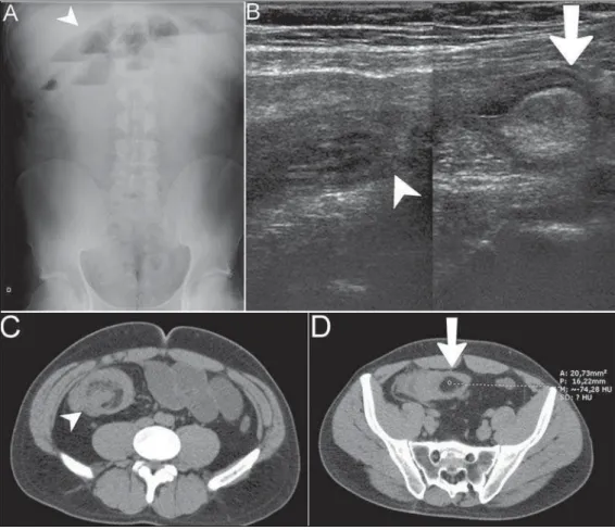
Abdominal radiography, ultrasonography and computed to-mography demonstrated small bowel loops distension (Figure 1A) and signs of ileo-ileal invagination associated with intraluminal
nodule with fat content compatible with “intussusception head” (Figures 1B, 1C and 1D). Option was made for surgical treatment. Anatomopathological study in association with immunohis-tochemical analysis diagnosed angiomyolipoma (AML) as follows:
Macroscopy: Bowel loop containing a non-encapsulated de-limited, submucosal, polypoid yellowish lesion measuring 3.0 × 2.5 × 2.3 cm, with no sign of malignancy.
Microscopy: Masson’s trichrome staining diagnosed AML compromising the entire intestinal wall, from the serosa to the mucosa.
Immunohistochemical analysis: Desmin, HHF 35, CD31, CD34, protein S100, smooth muscle actin 1 to 4 = positive.
Intussusception is the invagination of a proximal intestinal segment with its mesenteric fold with the corresponding
Letters to the Editor
Radiol Bras. 2015 Set/Out;48(5):333–340
340
http://dx.doi.org/10.1590/0100-3984.2014.0143
larization into the lumen of the distal intestinal portion, which may lead to obstruction, inflammatory process and segmental is-chemia(1,2).
In adult individuals, this condition corresponds to about 5% of the general cases, out of which only 1% of cases cause ob-struction(1). In this age group, it is estimated that in 90% of cases
one finds organic intraluminal causes called “intussusception heads” (for example, benign neoplasms such as lipoma, or ma-lignant; adenomatous polyps or other polyp types; hamartomas) or extraluminal causes (for example, adhesions, Meckel’s diver-ticulum)(1,3).
Intussusception heads in the small bowel are most frequently associated with benign lesions, while in the colon they are most associated with either primary or secondary malignant neopla-sias(2). The treatment is generally surgical for organic causes,
complications such as obstruction and intestinal ischemia(2,3).
The clinical signs of intussusception are related to the oc-currence of subocclusion, obstruction and enterorrhagia(3,4).
Intussusceptions are classified according the involved intes-tinal segment, as follows: enteroenteric, colo-colic, ileocolic and ileocecal intussusception(1,3).
Typical radiological findings include: “target sign” and “pseudokidney sign”(3). The diagnostic accuracy of
ultrasonogra-phy approximates to 98%(2), but the method is
operator-depen-dent(5). Computed tomography presents accuracy of 58% to
100%(5).
AMLs are benign mesenchymal tumors containing adipose, smooth muscle, epithelial and vascular cells(4,6–8). These tumors
and other lesions such as lymphangioleiomyomatosis and clear cell lung tumors were brought together under the classification of PEComas (perivascular epithelioid cell tumors)(4).
The prevalence of renal angiomyolipomas ranges from 0.3% to 3%. According to the literature this tumor is sporadic in about 80% of cases, and the remaining cases are associated with lymphangioleiomyomatosis and mainly tuberous sclerosis(6,7).
Extrarenal AMLs are extremely rare, and the liver is the most reported site (some other locations include the heart, lungs,
retroperitoneum, mediastinum, spinal cord, mucocutaneous, pa-rotid glands, reproductive organs regardless of sex), and its occur-rence in the gastrointestinal tract is rarely described(4,6–8) (about
50 cases)(6,7).
When located in the gastrointestinal tract their radiological diagnosis is hardly achieved because of their rarity and adipose nature, similarly to lipomas which are much more frequently found(4,7,8).
REFERENCES
1. Santos FGPL, Pereira JM, Lima RV, et al. Intussuscepção intestinal se-cundária a tumor do estroma gastrointestinal (GIST). Rev Imagem. 2007; 29:147–51.
2. Rosas GQ, Becker GG. Jejuno e íleo. In: D’Ippolito G, Caldana RP, edi-tors. Gastrointestinal. Série CBR. São Paulo, SP: Elsevier; 2011. p. 173– 202.
3. Kim YH, Blake MA, Harisinghani MG, et al. Adult intestinal intussus-ception: CT appearances and identification of a causative lead point. Radiographics. 2006;26:733–44.
4. Miliaras S, Miliaras D. Angiomyolipoma of the jejunum mimicking metastatic disease in a patient with colonic adenocarcinoma. Surgical Science. 2011;2:52–6.
5. Marinis A, Yiallourou A, Samanides L, et al. Intussusception of the bowel in adults: a review. World J Gastroenterol. 2009;15:407–11. 6. Povo A, Oliveira JMS, Silva R, et al. Angiomiolipoma duodenal. Revista
Portuguesa de Cirurgia. 2010;14:107–10.
7. Toye LR, Czarnecki LA. CT of a duodenal angiomyolipoma. AJR Am J Roentgenol. 2002;178:92.
8. Lee CH, Kim JH, Yang DH, et al. Ileal angiomyolipoma manifested by small intestinal intussusception. World J Gastroenterol. 2009;15:1398– 400.
Rodolfo Mendes Queiroz1, Luana Almeida Botter1, Michela Prestes Gomes2, Rafael Gouvêa Gomes e Oliveira1