Magnetic resonance imaging in deep pelvic endometriosis:
iconographic essay*
Ressonância magnética na endometriose pélvica: ensaio iconográfico
Antonio Carlos Coutinho Junior1, Cláudio Márcio Amaral de Oliveira Lima2, Elisa Pompeu Dias Coutinho1, Érica Barreiros Ribeiro2, Marisa Nassar Aidar3, Emerson Leandro Gasparetto4
Endometriosis is characterized by the presence of normal endometrial tissue outside the uterine cavity. In patients with deep pelvic endometriosis, uterosacral ligaments, rectum, rectovaginal septum, vagina or bladder may be involved. Clinical manifestations may be variable, including pelvic pain, dysmenorrhea, dyspareunia, urinary symptoms and infertility. Complete surgical excision is the gold standard for treating this disease, and hence the importance of the preoperative work-up that usually is limited to an evaluation of sonographic and clinical data. Magnetic resonance imaging is of paramount importance in the diagnosis of endometriosis, considering its high accuracy in the identification of lesions intermingled with adhesions, and in the determination of peritoneal lesions extent. The present pictorial review describes the main magnetic resonance imaging findings in deep pelvic endometriosis.
Keywords: Magnetic resonance imaging; Endometriosis; Female pelvis; Infertility.
A endometriose caracteriza-se pela presença de tecido endometrial funcionante heterotópico. Em pacientes com endometriose pélvica profunda pode haver acometimento dos ligamentos útero-sacros, reto, septo re-tovaginal, vagina ou bexiga. Os sintomas podem ser variados e incluem dor pélvica, dismenorréia, dispareu-nia, sintomas urinários e infertilidade. O padrão-ouro para o tratamento é a ressecção completa dessas le-sões. Assim, é muito importante a avaliação pré-operatória dessas pacientes, sendo esta avaliação, em ge-ral, limitada em relação aos dados clínicos e ultra-sonográficos. A ressonância magnética tem grande impor-tância no diagnóstico da endometriose, principalmente por permitir a identificação das lesões de permeio a aderências e a avaliação da extensão das lesões subperitoneais. Neste estudo são ilustrados, na forma de ensaio iconográfico, os principais achados da endometriose pélvica profunda à ressonância magnética.
Unitermos: Imagem por ressonância magnética; Endometriose; Pelve feminina; Infertilidade.
Abstract
Resumo
* Study developed at Clínica de Diagnóstico por Imagem (CDPI), Rio de Janeiro, RJ, Brazil.
1. MDs, Radiologists at Clínica de Diagnóstico por Imagem (CDPI) and Clínica Multi-Imagem, Rio de Janeiro, RJ, and Cen-tro de Diagnóstico por Imagem Fátima, Nova Iguaçu, RJ, Brazil. 2. MDs, Trainees at Clínica de Diagnóstico por Imagem (CDPI) and Clínica Multi-Imagem, Radiologists at Hospital Naval Marcí-lio Dias, Rio de Janeiro, RJ, Brazil.
3. MD, Radiologist at Clínica de Diagnóstico por Imagem (CDPI) and Clínica Multi-Imagem, Rio de Janeiro, RJ, Brazil.
4. Associate Professor of Radiology, Universidade Federal do Rio de Janeiro (UFRJ), Radiologist at Clínica de Diagnóstico por Imagem (CDPI) and Clínica Multi-Imagem, Rio de Janeiro, RJ, Brazil.
Mailing address: Dr. Cláudio M. A. de Oliveira Lima. Rua Silva Rabelo, 154, ap. 503, Méier. Rio de Janeiro, RJ, Brazil, 20735-080. E-mail: [email protected] / [email protected]
Received August 9, 2007. Accepted after revision Septem-ber 11, 2007.
disease attributed to multifactorial causes, affecting 7% to 10% of women in the gen-eral population(3,4). The most widely
ac-cepted theory is that viable endometrial cells originating from a physiological phe-nomenon called retrograde menstruation, result in implantation into the peritoneal cavity (Sampson’s theory)(2,5–7). Studies in
the literature report a wide range of factors of individual risk such as low parity, age, ethnic origin, height, body mass index, al-cohol abuse, tobacco smoking, among oth-ers(8).
The significant role of MRI in the diag-nosis of endometriosis is related to the identification of intermingled lesions in the presence of adhesions, and also the dem-onstration and evaluation of sub-peritoneal lesions extent in cases where these lesion cannot be visualized by laparoscopy, with accuracy, sensitivity and specificity > 90% for deep endometriosis(9,10). MRI findings
Coutinho Junior AC, Lima CMAO, Coutinho EPD, Ribeiro EB, Aidar MN, Gasparetto EL. Magnetic resonance imaging in deep pelvic endometriosis: iconographic essay. Radiol Bras. 2008;41(2):129–134.
INTRODUCTION
Firstly described in 1860 by Von Roki-tansky(1), endometriosis is defined as the
presence of functional endometrial tissue outside the endometrial cavity and myo-metrium(2,3). Endometriosis is a frequent
are more specific than those of ultrasonog-raphy and computed tomogultrasonog-raphy(8,9,11–13).
Generally, endometriosis implants are confined to the pelvis, the most common sites, in order of decreasing frequency, being ovaries, broad ligaments, anterior and posterior cul-de-sac, and utero-sacral ligaments; however, distant sites may be in-volved(2,4,7,9). Endometrial tissue implants
are affected by cyclic menstrual changes with periodic bleedings. Hemorrhage in-side these implants induces an acute pelvic inflammatory reaction and consequential adhesions development, frequently with Douglas cul-de-sac obliteration, Fallopian tubes and ovaries distortion.
structures such as uterine ligaments and vagina(4,7,8). Despite the asymptomatic
na-ture of the disease in many women affected by peritoneal endometriosis, patients with deep pelvic endometriosis may present with pelvic pain, dysmenorrhea, dyspareu-nia, urinary symptoms and infertility(2,5,6).
MRI presents the advantage of fast acqui-sition of multiplanar sequences, providing simultaneous images of all the pelvic vis-cera both at rest and under stress(11,12).
De-spite some limitations, MRI plays a signifi-cant role in the diagnosis of endometriosis, especially for allowing the identification of intermingled lesions in the presence of dense adhesions, and evaluation of sub-peritoneal lesions extent(4,13,14). In the
evaluation of deep endometriosis, MRI presents accuracy, sensitivity and specific-ity > 90%(13,14). In the present iconographic
essay, main MRI findings in deep pelvic endometriosis are described.
MRI PROTOCOLS
The acquisition of appropriate magnetic resonance images for evaluating patients with suspected deep pelvic endometriosis must follow specific protocols. In our clin-ics, the examination is performed during menses, with a full bladder. Additionally, immediately before the examination, a venous antispasmodic agent (dipyrone, scopolamine butylbromide) is administered and, most recently, vaginal (50 ml) and rectal (100 ml) aqueous gel introduction has been adopted.
The protocol includes the following sequences: axial T1-weighted, and sagittal, coronal and axial T2-weighted; and, after intravenous gadolinium injection, axial, fat-suppressed T1-weighted sequences.
MRI FINDINGS
Imaging findings on MRI of patients with deep pelvic endometriosis depend on the type of the lesion: small infiltrative en-dometrial implants, deep, solid lesions and visceral endometriosis involving the rectal and vesical walls(4).
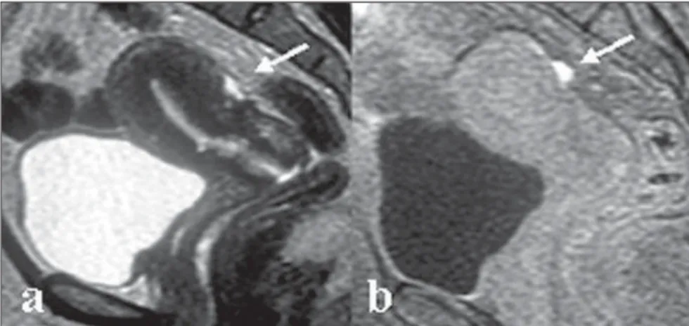
MRI may be a limited method in cases where the endometriosis presents with small infiltrative implants. Especially in cases where the lesions are visualized as
whitish foci at laparoscopy, MRI may be negative. However, in some patients hyperintense foci may be characterized on fat-suppressed T1-weighted sequences, representing small areas of hemor-rhage(4,13,14)(Figure 1).
At MRI, deep, solid lesions affecting the posterior cul-de-sac (Figure 2) present low to intermediate intensity signal, with fur-ther hyperintense foci on fat-suppressed (blood) T1-weighted images, hyperintense
signal on T2-weighted images with vari-able contrast-enhancement after intrave-nous gadolinium injection. Hypersignal foci on T1-weighted images result from ectatic endometrial glands with hemor-rhagic content surrounded by fibrotic tis-sue. The extensive fibrosis component (Figure 3) generally characterized in the histological study of these lesions is re-sponsible by the variable post-contrast-en-hancement(4,7). Some foci of endometriosis
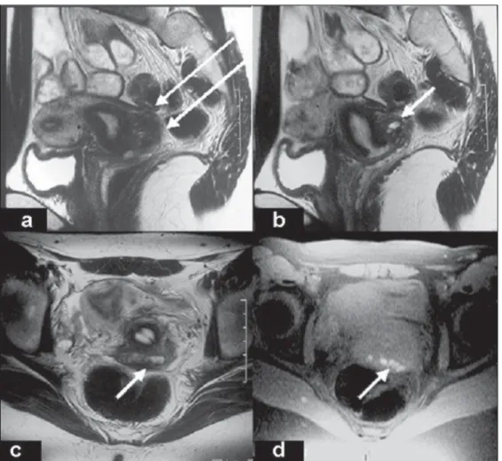
Figure 2. Axial, T2-weighted image (a), T1-weighted image with fat suppression (b) and T1-weighted image (d) show adhesion in the posterior cul-de-sac confirmed by laparoscopy (c). MRI shows postero-medially displaced ovaries with undefined cleavage plane between the uterine and rectosigmoid serosas, determining posterior cul-de-sac obliteration indicative of tethering process (long arrows). Additional find-ings: small endometriomas in the deep portion of both ovaries (short arrows).
Figure 4. T2-weighted, coronal (a), sagittal (b) and axial (d) planes, and axial, fat-suppressed T1-weighted images demonstrating vesical en-dometriosis. A 32-year-old patient with pelvic pain during urination. MRI demonstrates deep vesical en-dometriosis in the vesicouterine, retrocervical septa, and upper portion of the rectovaginal septum. Hypoin-tense nodular thickening on T2-weighted sequences, with partially defined margins, affecting the right, posterolateral part of the bladder and the corresponding vesicovaginal sep-tum (long arrows); additionally, inter-mingled hemorrhagic foci (arrow heads) can be observed. In addition, hypointense nodular thickening is observed on T2-weighted sequences of the retrocervical region and right utero-sacral ligament, extending to-wards the vaginal fornix and upper portion of the rectovaginal septum (short arrows).
Figure 3. Axial, T2-weighted image (a), fat-suppressed T1-weighted im-age (b), and sagittal T2-weighted (c), and fat-suppressed T1-weighted (d) images show involvement of the utero-sacral ligament. MRI demon-strates isointense nodular lesion with spiculate margins on T1-weighted sequences, and hypointense lesion adjacent to the right utero-sacral liga-ment on T2-weighted sequences (ar-rows), with hyperintense foci on fat-suppressed T1-weighted sequences, indicating hemorrhage.
localized in the Douglas cul-de-sac (Figure 4) may present with abundant glandular component with a mild fibrotic reaction. In these cases, high signal intensity is
ob-served on T1-weighted images, and vari-able signal intensity on t2-weighted im-ages, with the solid glandular component demonstrating variable
contrast-enhance-ment after gadolinium injection(4,8,14). In
Figure 5. T2-weighted, sagittal (a) and coronal (b) images, and axial, fat-suppressed T1-weighted image (c) demonstrate ureteral endometriosis. MRI shows a hypointense, nodular lesion on T2-weighted sequences, with ill-defined margins (short arrows), and intermingled foci of bleeding (arrow head) extraperitoneally localized in the left pelvis, involving the pelvic portion of the ipsilateral ureter, determining upstream dilatation (long arrows).
as bilateral or asymmetrical thickening and nodularity within these ligaments should be taken into consideration(4,8,14).
MRI may demonstrate focal or diffuse vesical involvement by endometriosis (Fig-ure 6). The majority of patients present focal areas of parietal thickening with eventual hypersignal foci on fat-suppressed
T1-weighted images. Invasion of the mu-cosa is not frequent in cases of vesical en-dometriosis and MRI can demonstrate al-terations even in cases of asymptomatic patients with normal cystoscopy(4,8,13).
MRI sensitivity in the diagnosis of en-dometriosis with rectal involvement is rela-tively low, because of artifacts associated
strated by MRI as a parietal thickening with hypointense signal on T2-weighted images. Hyperintense hemorrhagic foci may be identified on fat-suppressed T1-weighted images(4,7,8,13).
CONCLUSION
In patients with deep pelvic en-dometriosis, clinical and sonographic re-sults may be normal or poorly elucidative, difficulting the diagnosis determination. In these cases, MRI is essential for an accu-rate differential diagnosis. MRI can evalu-ate areas otherwise inaccessible by laparoscopy, identifying and evaluating the extent of lesions in the sub-peritoneal re-gion and in the presence of dense adhe-sions. Because of its multiplanar capacity and excellent tissue characterization, MRI plays an essential role in the preoperative evaluation of patients with deep pelvic endometriosis.
Acknowledgements
The authors thank Dr. Cláudio Crispi, for kindly letting the author have the ma-terial utilized in the present study.
REFERENCES
1. Craig V. Endometriosis of the urinary tract. Urol Clin North Am. 2002;29:625–35.
2. Brandão A. Endometriose. In: Brandão A, Wer-ner Jr H, Daltro P, editores. Ressonância magné-tica em obstetrícia e ginecologia. Rio de Janeiro: Revinter; 2003. p.133–50.
3. Kuligowska E, Deeds L, Lu K. Pelvic pain: over-looked and underdiagnosed gynecologic condi-tions. Radiographics. 2005;25:3–20.
4. Frate CD, Girometti R, Pittinno M, et al. Deep ret-roperitoneal pelvic endometriosis: MR imaging appearance with laparoscopic correlation. Radiographics. 2006;26:1705–18.
5. Zacharia TT, O’Neill MJ. Prevalence and distri-bution of adnexal findings suggesting endo-metriosis in patients with MR diagnosis of adenomyosis. Br J Radiol. 2006;104:303–7. 6. Crosignani P, Olive D, Bergqvist A, et al.
Advances in the management of endometriosis: an update for clinicians. Hum Reprod. 2006;12: 179–89.
7. Bazot M, Darai E, Hourani R, et al. Deep pelvic endometriosis: MR imaging for diagnosis and prediction of extension of disease. Radiology. 2004;232:379–89.
8. Woodward PJ, Sohaey R, Mezzetti Jr TP. Endometriosis: radiologic-pathologic correlation. Radiographics. 2001;21:193–216.
9. Olive DL, Schwartz LB. Endometriosis. N Engl J Med. 1993;328:1759–69.
10. Wykes CB, Clark TJ, Khan KS. Accuracy of laparoscopy in the diagnosis of endometriosis: a systematic quantitative review. Br J Obstet Gynaecol. 2004;111:1204–12.
11. Francisco VV, D’Ippolito G, Silva GPA, et al. Prevalência de artefatos em exames de ressonân-cia magnética do abdome utilizando a seqüênressonân-cia GRASE: comparável com as melhores seqüências rápidas? Radiol Bras. 2005;38:323–8.
12. Bezerra MRL, Soares AFF, Faintuch S, et al. Iden-tificação das estruturas músculo-ligamentares do assoalho pélvico na ressonância magnética. Ra-diol Bras. 2001;34:323–6.
13. Kinkel K, Chapron C, Balleyguier C, et al. Mag-netic resonance imaging characteristics of deep endometriosis. Hum Reprod. 1999;14:1080–6. 14. Kataoka ML, Togashi K, Yamaoka T, et al.