Quality in cytopathology: an analysis of
the internal quality monitoring indicators
of the Instituto Nacional de Câncer
Qualidade em citopatologia: análise de indicadores de monitoramento
interno de qualidade do Instituto Nacional de Câncer
Mario Lucio C. Araujo Jr;Daniela A. Santana; Lívia B. Almeida; Shirley B. S. Quintana; Gloria Regina F. Silva; Rachel C. S. P. Fonseca
Instituto Nacional de Câncer (INCA).
First submission on 16/09/14; last submission on 20/02/15; accepted for publication on 24/03/15; published on 20/04/15
ABSTRACT
Introduction: Quality control programs are required to ensure the effectiveness of Pap smear, which still remain a key strategy for control
of cervical cancer worldwide. Objective: This study was based on the retrospective and quantitative analysis of the post-analytical phase indicators from the internal quality monitoring (IQM) program for cytopathology laboratories, such as: positivity rate, atypical squamous cell (ASC)/satisfactory exams ratio, ASC/abnormal test results ratio, ASC/squamous intraepithelial lesions (SIL) ratio, percentage of tests
compatible with high-grade squamous intraepithelial lesion (HSIL), and total of false negative. Materials and methods: The information
was extracted from the computerized system of the Section for Integrated Technology in Cytopathology (Seção Integrada de Tecnologia em Citopatologia [SITEC]), a reference institution for cancer cytopathology, from July 2013 to June 2014. From a total of 156,888 Pap smears, 157,454 were considered satisfactory for indicator analysis and 566 were excluded because they were considered unsatisfactory and/or rejected for analysis. The data was organized in tables using Microsoft Excel 2010 software, and categorized as indicators. Results: The averages for the indicators were: 7.2% for positivity rate, 56.9 for ASC/abnormal test ratio, 4.1 for ASC/satisfactory tests ratio, 1.4 for
ASC/SIL ratio, 0.6% percentage for tests compatible with HSIL, and 2.1% for false-negative rate. Conclusion: The results show that an
Internal Quality Monitoring Program is essencial to ensure quality for cytopathology laboratories, and a randomized review of at least 10% of the negative exams, as recommended by the Brazilian Ministry of Health/Instituto Nacional de Câncer (INCA), since is an effective method, especially for large laboratories.
Key words: cytopathology; Pap smear; cervix; quality indicators.
INTRODUCTION
Cervical cancer appears as a major public health problem and is the fourth most common cancer among women(1). The
conventional cervical smear test (Pap smear) is the main strategy of cervical cancer screening programs worldwide. To ensure its effectiveness the structuring of a quality control system is required(2-5).
On December 30, 2013 entered into force on Ordinance N. 3388, which redeines the National Qualiication in Cytopathology (Qualiicação Nacional em Citopatologia
[QualiCito]) for cervical cancer prevention(6), under the
Health Care Attention for People with Chronic Diseases (Rede de Atenção à Saúde das Pessoas com Doenças Crônicas). QualiCito consists in establishing standards and evaluation the quality of Pap smear of the cervix by monitoring the performance of public and private laboratories service providers to the Uniied Health System (Sistema Único de Saúde [SUS]). Its execution occurs through internal quality monitoring (IQM), external quality monitoring (EQM) and compliance with the criteria set out for quality assessment
and hiring laboratories(6).
The Section for Integrated Technology in Cytopathology
(Seção Integrada de Tecnologia em Citopatologia [SITEC]) belongs to the Pathology Division of the Instituto Nacional de Câncer José Alencar Gomes da Silva (INCA) and implemented the IQM as a tool to develop the diagnosis, improve the technical procedures, and conduct continuing education. By implementing the quality indicators, we can assess the overall performance of the laboratory, performing individual statistical analysis of professionals, as well as provide the data obtained for conducting internal and/or external audit(3, 5).
This paper presents the quality indicators of post-analytical phase of IQM in SITEC/INCA, which include the positivity rate, the percentage of tests compatible with atypical squamous cells (ASC) among satisfactory tests, the ASC percentage among abnormal tests, the ASC/squamous intraepithelial lesion (SIL) ratio, the percentage of tests compatible with high-grade squamous intraepithelial lesion (HSIL) in addition to the percentage of negative false exams.
OBJECTIVE
To assess the main quality indicators and the percentage of false negative tests by IQM of SITEC/INCA.
MATERIALS AND METHODS
This retrospective and quantitative study, consists in the analysis of the indicators of post-analytical phase of IQM,
proposed in QualiCito(6) and in Quality Management Manual
for Cytopathology Laboratories(3), whose selected variables are
described in their respective formulas as follows:
1) positivity rate
nº of abnormal tests in a particular location and year × 100 total of satisfactory tests
2) percentage of tests compatible with ASC among satisfactory tests nº of tests with ASC-US and ASC-H × 100
total of satisfactory tests
ASC-US: atypical squamous cells of undetermined significance; ASC-H: atypical squamous cell, cannot exclude a high-grade squamous intraepithelial lesion.
3) percentage of tests compatible with ASC among abnormal tests nº of tests with ASC-US and ASC-H × 100
total of abnormal tests
4) ASC/SIL ratio
nº tests compatible with ASC-US e ASC-H nº of tests with LSIL and HSIL
LSIL: low-grade squamous intraepithelial lesion.
5) percentage of tests compatible with HSIL nº of tests with HSIL × 100
total of satisfactory tests
6) percentage of false negative tests. False negative are the smears that were classiied as negative by the routine screening tests, but which were considered abnormal by the Cytopathology Quality team of SITEC/INCA, in random review of at least 10% of exams. These tests are adressed to cytopathologists to conirm the diagnosis.
The information in this study was taken from the computerized system of SITEC/INCA, a reference institution for cancer cytopathology, including the period from July 2013 to June 2014. From a total of 157,454 cervical smear tests using the conventional method, 156,888 were considered satisfactory for the indicators analysis, and 566 were excluded because they were classiied as unsatisfactory and/or rejected.
In SITEC/INCA’s irst cervical smear test reading is performed by cytotechnologists. Senior cytotechnologists compose the quality control team and they review at least 10% of cases classiied as negative and those with information of clinical risk. All positive cases are addressed to the team of pathologists for emit the inal report.
All data obtained for analysis were organized in tables using Microsoft Excel 2010 software, in which they were categorized into. The study was approved by the Research Ethics Committee (Comitê de Ética em Pesquisa [CEP]) of INCA, CAAE 32632314.6.0000.5274.
RESULTS
Table 1 shows the monthly percentage of quality indicators
present average of 4.1%; the ASC/SIL ratio remained between 1.1-1.9; and the percentage of exams compatible with HSIL showed variation between 0.4%-0.8% during the study period.
The average of each indicator for the assessed period is shown in Figure 1.
The false negative rate varies between 1.6% and 2.5% in the period, with an average of 2.1%, as shown in Figure 2.
DISCUSSION
Through this study it was observed that the positivity rate (7.2%) of SITEC/INCA in the period remained within the standard recommended by the QualiCito(6) and the Quality
Management Manual for Cytopathology Laboratory(3), which is
equal to or greater than 3% and between 3%-10%, respectively, as shown in Table 2.
Between July 2013 and June 2014 the positivity rate ranged from 3.8% to 9.2%. It is not possible to assert whether the variations among the analyzed months occur by chance or if there is seasonality, since this study is limited to evaluate the
data for only one year. The extension of evaluation of these data
for other years can help to respond these hypotheses.
FIGURE 1 − SITEC average July 2013 to June 2014
SITEC: Integrated Section of Technology in Cytopathology (Seção Integrada de Tecnologia em Citopatologia); ASC: atypical squamous cell; SIL: squamous intraepithelial lesion; HSIL: high-grade squamous intraepithelial lesion.
FIGURE 2 − Total and percentage of false negative in the period of July 2013 to June 2014, average 2.1%
FN: false negative.
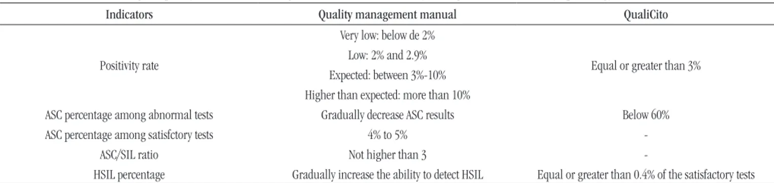
TABLE 2 − Percentage of quality indicators according to QualiCito and the Quality Management Manual for cytopathology laboratories (INCA, 2012)
Indicators Quality management manual QualiCito
Positivity rate
Very low: below de 2%
Equal or greater than 3% Low: 2% and 2.9%
Expected: between 3%-10% Higher than expected: more than 10%
ASC percentage among abnormal tests Gradually decrease ASC results Below 60%
ASC percentage among satisfctory tests 4% to 5%
-ASC/SIL ratio Not higher than 3
-HSIL percentage Gradually increase the ability to detect HSIL Equal or greater than 0.4% of the satisfactory tests
QualiCito: National Qualification in Cytopathology in preventing cervical cancer; INCA: Instituto Nacional de Câncer; ASC: atypical squamous cell; SIL: squamous intraepithelial lesion; HSIL: high-grade squamous intraepithelial lesion.
TABLE 1 − Quality Indicators from SITEC/INCA during the period from July 2013 to June 2014
Indicators 2013Jul 2013Aug 2013Sept 2013Oct 2013Nov 2013Dec 2014Jan 2014Feb 2014 Mar 2014Apr 2014May 2014Jun Mean of period
Positivity rate 3.8 7.6 6.4 8.0 5.8 8.3 9.2 6.2 8.7 6.5 8.2 7.4 7.2
ASC percentage among abnormal test results 51.9 56.8 58.9 53.8 51.4 59.8 56.3 63.7 58.6 58.0 55.3 58.0 56.9
Percentage of ASC compatible test
results among satisfactory tests 2.0 4.3 3.8 4.3 3.0 5.0 5.2 4.0 5.1 3.8 4.6 4.3 4.1
ASC/SIL ratio 1.1 1.4 1.5 1.2 1.1 1.6 1.4 1.9 1.5 1.5 1.3 1.5 1.4
Percentage of tests results compatible with HSIL 0.4 0.5 0.4 0.7 0.5 0.6 0.8 0.4 0.7 0.5 0.6 0.7 0.6
SITEC: Integrated Section of Technology in Cytopathology (Seção Integrada de Tecnologia em Citopatologia); INCA: Instituto Nacional de Câncer; ASC: atypical squamous cell; SIL: squamous intraepithelial lesion; HSIL: high-grade squamous intraepithelial lesion.
60.0
50.0
40.0
30.0
20.0
10.0
0.0 72
Positivity
ASC percentage among abnormal
tests results Percentage of
tests compatible
with ASC and
fully satisfactory
ASC/SIL ratio Percentage of
tests compatible
with HSIL
4.1
1.4 0.6 56.9
1.6% 2.0% 2.4% 1.8% 2.4% 2.5%
80 70 60 50 40 30 20 10 0
67 54
72
66
32
76
FN Percentage Total FN
The positivity rate expresses the prevalence of cellular changes in the tests and the sensitivity of the screening process to detect lesions in the examined population. When this index is below the recommended, it may indicate that positive tests were not identiied, increasing the rate of false negative results(7).
The literature suggests that countries like the United
States(8), Norway(9) and the United Kingdom(10) show the
positivity rate of 6.8%, 4.9% e 6.4%, respectively. These countries with effective results in cytological screening has been decreased the incidence and mortality rates from cervical cancer. It is important to mention that laboratories need to be alert to keep the positivity rate within the recommended intervals, as the conirmation of this index higher than 3% is one of the parameters discussed in the QualiCito(6) to contract
and/or contract renewal with laboratories providing services to the SUS.
The mean percentage of exams compatible with ASC among the satisfactory in SITEC/INCA in the period was 4.1%, the expected value was between 4%-5% of tests, according to the Quality Management Manual(3). The atypical cells of
undetermined signiicance were considered as suspicious cases where the presence of cellular changes are insuficient for the diagnosis of squamous intraepithelial lesions, have their diagnosis variable in relation to the SIL. SIL criteria are clear and well-deined, while the diagnosis compatible with ASC is more variable and less reproducible than the diagnosis of low- and high-grade SIL, meaning that the agreement between the surveyors tend to be higher for SIL than for ASC(3).
The mean of ASC percentage among abnormal tests in SITEC/INCA in the period was 56.9%, and the expected value was lower than 60% according to QualiCito(6). The Quality
Management Manual(3) recommends that there should be a
progressive reduction of this rate. High ASC indices indicate problems in the sample, in the laboratory examination or in both. This indicator comprises an indirect assessment of quality, but does not allow an independent analysis of the quality of the process. The increase of this index is harmful for women as well as the health care network because it leads to an increase in the number of tests intended to repetition(3).
The ASC/SIL ratio, in accordance with the Quality
Management Manual(3), must not exceed three times the
SIL rate. Thus, it is possible to identify the low professional performance in the screening. In SITEC/INCA, the average of this indicator was 1.4%, which is within the established limit. According to Türkmen(11), ASC/SIL ratio may be lower in
high-risk populations and higher in low-high-risk populations. The 1.4%
ASC/SIL ratio from SITEC/INCA is compared to those found in the study of Renshaw(12), which corresponds to1.5%, of Chebib(13)
whose average is 1.15%, and of Catteau(14),which was 1.9%.
The percentage of exams compatible with HSIL in SITEC/ INCA ranged between 0.4%-0.8% in the period, which is consistent with QualiCito(6) that establishes the indice superior
a 0.4% index, and the Manual of Quality Management(3)
recommends gradually increase the ability to detect HSIL. The HSIL percentage for all satisfactory tests reported in the literature was 0.5% for the United States (15), 0.6% in Canada(16),
1.1% in the United Kingdom(10), and 1.14% in Norway(9).
The high-grade lesions represent those which genuinely are precursors of cervical cancer with potential for progression, and its detection is the major goal of secondary prevention of cervical cancer(3).
The cervical Pap smear has been criticized because its sensitivity rate is not so high. In the United States, for example, it corresponds to approximately 79% in conventional cytology(17).
The false negative exams are mainly related to collecting, screening, and interpretation errors of cytophatological
diagnosis(2-4, 6). The review of cases for quality control is an
effective method to minimize the number of negative false
diagnosis(17, 18).
There are several methods of review to monitor the quality of cervical screening, to the discretion of each laboratory to choose the method that better meets its proile. The review methods recommended by the Quality Management
Manual(3) and QualiCito(6) include: analysis of cytohistological
correlation, retrospective review of the tests, random review of 10%, review of smears selected based on clinical criteria for risk, quick review of 100% of negative smears, fast prescreening of all smears, among others(3). The choice of IQM method in
the laboratory must be made by evaluating the volume of
examination and the quantitative professionals, in order to not exceed the workload(1, 9, 13).
In SITEC/INCA, the IQM is carried out using the method of random review of at least 10% of negative smears. If the team has found any signiicant cytomorphological change that changes the diagnostic procedure, the reviewer sends the case with a probable diagnosis to the cytopathologist to the inal report.
Analysis of our study results show that the random review method of at least 10% of negative smears is appropriate, especially for laboratories with a large number of Pap smears.
smears unfeasible, requiring impractical time and greater quantitative of professionals, not available at the institution.
Using the random review of at least 10% of negative cases, we detected during the analysis period an average of 2.1% of
false negative tests. Bonilha et al.(19), using the same method
obtained a rate of 2.4%. Currens et al.(20) detected 0.18% false
negative cases with the random review of at least 10% by Pap smear, but in liquid-based cytology method.
Through IQM it is possible to assess the causes of false negative exams and to plan actions of continuing education with the professionals. It takes place during the SITEC weekly meeting with the Cytopathology Quality team to discuss scientiic articles related to the topic and to propose improvement strategies
of data on the quality of preventive gynecological examinations performed at INCA. In addition, the good practice in IQM gives the laboratory greater credibility with their customers.
CONCLUSION
IQM strategy is essential to ensure quality in cytopathology. We believe that for large laboratories, the random review of at least 10% of exams considered negative, is effective to identify issues that must be addressed in continuing education, in order to obtain reliable indicators that meet the regulations established by Qualicito and by the references in the national and international literature.
RESUMO
Introdução: A estruturação de um sistema de controle de qualidade é necessária para garantir a efetividade do exame preventivo
ginecológico, que permanece como principal estratégia para controle do câncer do colo do útero no mundo. Objetivo: Análise
retrospectiva e quantitativa dos indicadores da fase pós-analítica do monitoramento interno de qualidade (MIQ) para laboratórios de citopatologia, como: índice de positividade, razão atipias escamosas de significado indeterminado (ASC)/exames satisfatórios, razão ASC/exames alterados, razão entre ASC/lesões intraepiteliais escamosas (SIL), percentual de exames compatíveis com lesão intraepitelial
escamosa de alto grau (HSIL), além do total de falso negativos. Material e método: As informações foram extraídas do sistema
informatizado da Seção Integrada de Tecnologia em Citopatologia (SITEC), instituição de referência em citopatologia oncológica, no período de julho de 2013 até junho de 2014. De um total de 157.454 exames citopatológicos no período, 156.888 foram considerados satisfatórios para análise dos indicadores e 566 foram excluídos por serem classificados como insatisfatórios e/ou rejeitados. Todos os dados obtidos para análise foram organizados em tabelas utilizando-se o programa Microsoft Excel 2010, nas quais foram
categorizados em indicadores. Resultados: A média obtida dos indicadores foi de 7,2% para o índice de positividade, 56,9 para a
razão de ASC/exames alterados, 4,1 para a razão ASC/exames satisfatórios, 1,4 para ASC/SIL, 0,6% para exames compatíveis com
HSIL e 2,1% de exames falso negativos. Conclusão: Os resultados demonstram que o MIQ é fundamental para manter a qualidade
dos laboratórios de citopatologia, e a revisão aleatória de pelo menos 10% dos exames considerados negativos, conforme orientação do Ministério da Saúde/Instituto Nacional de Câncer (INCA), é um método eficaz principalmente em laboratórios de grande porte.
Unitermos: citopatologia; colpocitológico; colo uterino; indicadores da qualidade.
REFERENCES
1. Ministério da Saúde. Brasil. Instituto Nacional de Câncer José Alencar Gomes da Silva. Coordenação-Geral de Prevenção e Vigilância. Estimativa 2014: incidência de câncer no Brasil. Rio de Janeiro; 2014.
2. Arcuri RA, Cunha KCF, Alves EC, et al. Controle interno da qualidade em citopatologia ginecológica: um estudo de 48.355 casos. J Bras Patol Med Lab. 2002; 38(2): 141-7.
3. Ministério da Saúde. Brasil. Instituto Nacional de Câncer José Alencar Gomes da Silva. Coordenação-Geral de Prevenção e Vigilância. Divisão de
Detecção Precoce e Apoio à Organização de Rede. Manual de gestão da qualidade para laboratório de citopatologia. Rio de Janeiro: Inca; 2012. 4. Confortini M, Di Stefano C, Biggeri A, et al. Daily peer review of abnormal cervical smears in the assessment of individual practice as an additional method of internal quality control. Cytopathology. 2014. DOI:10.1111/cyt.12195.
5. Siegl EJ, Miller JW, Khan K, Harris SE. Quality assurance through quality improvement and professional development in the national breast and cervical cancer early detection program. Cancer. 2014; 120 Suppl 16: 2584-90.
câncer do colo do útero (QualiCito), no âmbito da Rede de Atenção à Saúde das Pessoas com Doenças Crônicas. Diário Oficial [da República Federativa do Brasil]. Brasília (DF); 2013 December 13. Seção 1, p. 42.
7. Bortolon PC, Silva MAF, Corrêa FM, et al. Avaliação da qualidade dos laboratórios de citopatologia do colo do útero no Brasil. Rev Bras Cancer. 2012; 58(3): 435-44.
8. Davey DD, Neal MH, Wilbur DC, Colgan TJ, Stver PE, Mody DR. Bethesda 2001 implementation and reporting rates: 2003 practices of participants in the College of American Pathologists Interlaboratory Comparison Program in Cervicovaginal Cytology. Arch Pathol Lab Med. 2004; 128(11): 1224-9.
9. Nygård JF, Skare GB, Thoresen SØ. The cervical cancer screening pro-gramme in Norway, 1992-2000: changes in Pap smear coverage and incidence of cervical cancer. J Med Screen, London. 2002; 9(2): 86-91. 10. NHS Cancer Screening Programmes. NHS Cervical Screening Pro-gramme Statistical Bulletin (England 2010-11). [Internet]. Available at: http://www.can cerscreening.nhs.uk/cervical/statistics.html.
11. Türkmen IÇ, Bassüllü N, Korkmaz P, et al. Patients with epithelial cell abnormality in PAP smears: correlation of results with follow-up smears and cervical biopsies. Türk Patoloji Derg. 2013; 29(3): 179-84. 12. Renshaw AA, Auger M, Birdsong G, et al. ASC/SIL ratio for cytotechnologists: a survey of its utility in clinical practice. Diagn Cytopathol. 2009; 38(3): 180-3.
13. Chebib I, Rao RA, Wilbur DC, Tambouret RH. Using the ASC/SIL ratio, human papillomavirus, and interobserver variability to assess and
MAILING ADDRESS
Mario Lucio Cordeiro Araujo Junior
Rua Cordeiro da Graça, 156; Santo Cristo; CEP: 20220-400; Rio de Janeiro-RJ, Brazil; e-mail: [email protected]
monitor cytopathology fellow training performance. Cancer Cytopahol. 2013; 121(11): 638-43.
14. Catteau X, Simon P, Noël JC. Evaluation of the oncogenic human papillomavirus DNA test with liquid-based cytology in primary cervical cancer screening and the importance of the ASC/SIL ratio: a Belgian study. ISRN Obstetrics and Gynecology. 2014; 2014: ID 536495, 5 p. 15. Eversole GM, Moriarty AT, Schwartz MR, et al. Practices of participants in the college of american patholo gists interlaboratory comparison program in cervicovaginal cytology, 2006. Arch Pathol Lab Med. 2010; 134(3): 331-5.
16. BC Cancer Agency. Cervical cancer screening program. 2009 Annual report. [Internet]. Vancouver, 2010. 30 p. Available at: http://www. bccancer.bc.ca/NR/rdonlyres/A6E3D1EC93C44B66A7E8B025721184B2/4 4207/2009CCSP_Annual_ReportFINALFeb1910.pdf.
17. Renshaw AA, Elsheikh TM. Predicting screening sensitivity from workload in gynecologic cytology: a review. Diagn Cytopathol. 2010; 39(11): 832-6.
18. Tarkkanen J, Geagea A, Nieminen P, Anttila A. Quality improvement project in cervical cancer screening: practical measures for monitoring laboratory performance. Acta Obstet Gynecol Scand. 2003; 82(1): 82-8. 19. Bonilha JL, Valença CFM, Micelli JP, Zanovelo EM, Silva J, Cury PM. Controle da qualidade em colpocitologia: visão rápida com campo marcado. J Bras Patol Med Lab. 2006; 42(6): 441-8.