CLINICAL AND BIOCHEMICAL MANIFESTATIONS PRODUCED BY SCORPION (Hemiscorpius lepturus) VENOM IN EXPERIMENTAL ANIMALS
ZARE MIRAKABBADI A. (1), ZOLFAGHARIAN H. (1), HEDAYAT A. (1), JALALI A. (2)
(1) Department of Venomous Animals and Antisera Production, Razi Vaccine and Serum Research Institute, Hesarak, Karaj, Iran; (2) Department of Pharmacology and Toxicology, School of Pharmacy, Jundishapur University, Ahvaz, Iran.
ABSTRACT: Several studies have been published about the clinical and biochemical manifestations produced by the venom of scorpions of the Buthidae family, but very few reports have indicated the manifestations induced by the venom of the Scorpionidae family. Hemiscorpius lepturus is an important scorpion species present in the south and southwestern part of Iran, causing morbidity and mortality in children and adults. For the present study, H. lepturus venom was extracted by electric shock and subcutaneously injected (6.3mg/kg) into a group of six rabbits. Blood collection was carried out before and three hours after venom injection for determination of osmotic fragility and levels of blood sugar, alanine aminotransferase (ALT), aspartate aminotransferase (AST), lactate dehydrogenase (LDH), creatine phosphokinase (CPK) and alkaline phosphatase (ALP). In vitro studies were also carried out to verify the osmotic fragility of red blood cells (RBCs) exposed to venom concentrations ranging from 0–90μg/2ml blood. Results showed the extreme effect of this venom on the lysis of RBCs both in vitro and in vivo. Venom injection caused significant (p>0.001) increase in ALT, AST, LDH and blood sugar levels. There was also an increase in CPK, and ALP levels after venom injection; however, it was not statistically significant. All animals died four hours after having received the venom. The current study revealed that the neurological effect of H. lepturus venom is similar to that of scorpions of the Buthidae family. However, they differ in RBCs lysis, which was highly significant when induced by H. lepturus venom, probably due to the presence of a type of phospholipase in this venom. Further studies are needed to provide a clearer view of the mechanism of action of H. lepturus venom.
KEY WORDS: Hemiscorpius lepturus, scorpions, osmotic fragility, Iran, biochemical changes.
CONFLICTS OF INTEREST: There is no conflict.
CORRESPONDENCE TO:
INTRODUCTION
Venomous scorpions pose a great health hazard to children and adults in the
southwestern part of Iran (1, 14), where the most common scorpion genus is
Hemiscorpius. Although H. lepturus belongs to the Scorpionidae family and its venom
gland is very small, it can cause death to both children and adults (1, 5, 14). Many
studies have reported manifestations caused by venoms of scorpions of the Buthidae
family (3, 10, 17, 18) but very few have indicated the signs and symptoms as well as
the serum biochemical changes induced by H. lepturus venom (14).
The most significant manifestation in envenomation by H. lepturus is hemolysis.
Approximately 40% of the stung patients need hemodialysis and, sometimes, packed
cells treatment (14). The mechanism of death caused by H. lepturus envenomation is
thought to be quite different from that caused by scorpions of the Buthidae family;
clinicians in the south of Iran believe that toxin distribution rate of H. lepturus venom
is very slow (14). Thus, the current study aimed at investigating cellular and
biochemical alterations caused by H. lepturus envenomation and at verifying whether
its mechanism of manifestations is quite different from or similar to that of scorpions
of the Buthidae family.
MATERIALS AND METHODS
Five thousand scorpions (H. lepturus) were collected from the southwestern part of
Iran and identified by experts at Razi Vaccine and Serum Research Institute, Iran.
Venom was milked by electric shock, lyophilized and preserved until used. The
venom LD50 was determined in mice (18–20g) by the method of Reed and Muench
(15).
Six male healthy rabbits of 1.5±0.2kg were selected for this experiment. The venom
(6.3mg/kg of body weight) was subcutaneously injected. Blood sampling was carried
out via a polyethylene cannula (G21) inserted into the marginal ear vein of each
overnight-fasted animal before and three hours after venom injection. Part of the
collected blood was poured into heparinized tubes for osmotic fragility tests and part
of it underwent serum separation for analysis of blood sugar, ALT, AST, LDH, CPK
In vivo osmotic fragility test was carried out with whole blood collected before and
three hours after venom injection; blood samples were exposed to various NaCl
concentrations (from 0 to 0.9%) and incubated for 30min at 37 °C (13).
For in vitro osmotic fragility test, 2ml of normal whole blood was incubated at various
concentrations of H. lepturus venom (1, 10, 30, 60 and 90μg) for 30min at 37°C;
then, samples were exposed to NaCl concentrations ranging from 0 to 0.9% for
30min at 37 °C (13, 16).
RESULTS
The venom LD50, determined in mice, was 126 μg/mouse. After venom injection
(6.3mg/kg), animals were observed for signs and symptoms during the whole
experiment. Within 20–35min after venom administration, all animals started to
present signs of envenomation like edema at the injection site, increased heartbeat
rate, salivation and lacrimation, and altered breathing rhythm. Although urination and
change in the urine color from light yellow to red were expected, no such alterations
were observed in the experimentally envenomed rabbits during the experiment.
Edema at the injection site was measured and characterized by a 1–2cm swollen red
patch. In two rabbits, the swollen red patch was extended to 8–10cm. Within two
hours after injection, all animals showed wheezing and coughing. No hematuria or
dysentery was observed in envenomed rabbits. Two hours after venom injection, all
animals showed muscle contraction and paralysis. They could not stand on their feet.
Death occurred as a consequence of increased heartbeat rate, difficult breathing and
complete feet paralysis, similarly to cyanide poisoning.
Figure 1 shows the osmotic fragility of RBCs, normal and exposed to various
concentrations of H. lepturus venom. Hemolysis rate increased with increasing
concentrations of venom. The figure clearly shows a significant (p<0.001) shift to the
right as the venom concentration increased. Changes in the osmotic fragility of RBCs
exposed to 1μg venom were insignificant, compared with control.
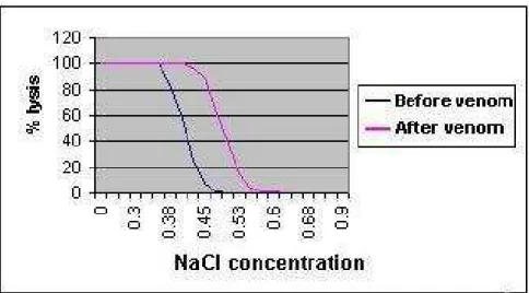
Figure 2 shows the comparison of osmotic fragility of RBCs from rabbits’ blood
samples before and after venom injection. The figure clearly shows a significant shift
to the right, which indicates an increased osmotic fragility of RBCs exposed to H.
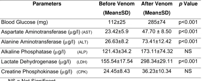
Table 1 shows the levels of serum enzymes ALT, AST, LDH, CPK and ALP as well
as blood sugar. There was a highly significant (p<0.001) increase in the
concentration of LDH, ALT and AST in the serum of rabbits three hours after venom
injection. Although there was also an increase in CPK and ALP levels after venom
injection, it was not significant when compared with the levels of these enzymes
before injection. Blood sugar also significantly (p<0.001) increased after H. lepturus
venom administration
Figure 1. In vitro osmotic fragility of rabbits’ red blood cells incubated at increasing
concentrations of NaCl and exposed to various concentrations of H. lepturus venom.
Figure 2. In vivo osmotic fragility of rabbits’ red blood cells incubated at increasing
Table 1. Comparison of various biochemical parameters before and three hours after
venom injection into experimental rabbits.
Parameters Before Venom
(Mean±SD)
After Venom
(Mean±SD)
p Value
Blood Glucose (mg) 112±25 285±74 p<0.001
Aspartate Aminotransferase (μg/l) (AST) 23.42±5.9 47.70 ± 8.50 p<0.001 Alanine Aminotransferase (μg/l) (ALT) 26.63±8.2 73.41±12.42 p<0.001 Alkaline Phosphatase (μg/l) (ALP) 121.43±34.2 173.11±74.32 NS Lactate Dehydrogenase (μg/l) (LDH) 155.54±17.54 298.34±29.11 p<0.001 Creatine Phosphokinase (μg/l) (CPK) 24.45±8.43 36.23±10.34 NS
NS = Not Significant
DISCUSSION
Hemiscorpius lepturus is a dangerous scorpion from the south and southwestern part
of Iran that causes mortality and morbidity in both children and adults (1, 14).
Although many scientific reports have clarified the complex mechanism of action of
venoms of scorpions of the Buthidae family (3, 4, 10, 16), scarce studies showed the
mechanism involved in envenomation by scorpions of the Scorpionidae family,
especially H. lepturus (14).
Clinical reports have indicated a high hemolysis rate in patients stung by H. lepturus
(14). In the southwest of Iran, hemolysis due to envenomation by such scorpion is so
common that when a patient is suspected of having been stung by a scorpion, the
first clinical test is to check the presence of hemoglobin in the patient’s urine. An
interesting fact in H. lepturus sting is the absence of local pain or the presence of
very mild pain at the sting site (14). Many times, absence of local pain can delay
treatment.
In the current study, local signs manifested as a local reddish edema of about 2–3cm
around the injection site between 15 and 20min after venom administration. Systemic
signs and symptoms (increased heart rate and breathing) were observed 20–35min
following injection. However, the grading of clinical symptoms was not established in
lepturus venom is very slow, compared with that of venoms of the Buthidae family;
thus, they propose the removal of the local tissue after scorpion sting. The
manifestation of systemic signs and symptoms occurred from 30 to 35min after
venom injection, which is not significantly different from the time of emergence of
systemic signs and symptoms in cases of envenomation by other scorpions (10, 17).
This indicates that the distribution rate of neurotoxins is similar in envenomation by
all scorpions including those of the Scorpionidae family. All the animals died within 3–
4h after having received the venom. This was also similar to the time scorpions of the
Buthidae family needed to kill rabbits (18). Thus, removing the skin at the sting site
seems to be of no use and may even complicate the treatment.
Increased osmotic fragility of RBCs exposed to scorpion (Mesobuthus tamulus)
venom, both in vitro and in vivo, was previously reported (11). In the present study,
when RBCs were exposed in vitro to various concentrations of H. lepturus venom,
there was a highly significant (p<0.001) increase in osmotic fragility. This is an
indicator of the venom direct action on RBCs. In vivo results of osmotic fragility also
confirmed the increased osmotic fragility of RBCs, which may be due to the presence
of an enzyme, like phospholipase A2, in the venom of H. lepturus and which may be
the cause of hematuria in patients stung by this scorpion species. Whether the action
of venom on other tissues is similar to that on RBCs is to be clarified in future studies.
Determination of blood sugar levels in the serum of rabbits showed a highly
significant increase compared with the levels before venom administration. The
levels of CPK, ALT, AST and LDH enzymes also increased after venom injection. All
these changes were similar to those observed in experimental animals receiving
venom of scorpions of the Buthidae family (18, 19). Clinical manifestations of
scorpion envenomation appear to be secondary to the activation of both sympathetic
and parasympathetic autonomic nervous system (3, 4, 10). Such manifestations
reveal that scorpion envenomation causes an autonomic storm (6, 7, 9). In the
present study, significant hyperglycemia was noticed after H. lepturus venom
administration, which is quite compatible to the results of other studies (2, 8, 12, 19).
The proposed mechanism for this effect included: peripheral and central stimulation
of the adrenergic system; activation of β-receptors with catecholamine and serotonin
secretion; blockage of insulin secretion; and insulin resistance (3, 16, 19). The
reports (12, 17). Aspartate aminotransferase, as a transferase enzyme, is distributed
to all parts of the body but mostly concentrates in the liver and the heart; therefore,
the increase in its levels may be attributed to myocardial infarction or hepatic failure.
Alanine transferase, acting similarly to AST, is considered a liver-specific enzyme. It
increases more and remains longer than AST during hepatic failure or inflammation.
Therefore, the increase in AST and ALT levels may be due to a direct action of the
venom on the liver and the heart.
Thus, according to the obtained results, the signs and symptoms of envenomation by
H. lepturus occur through two mechanisms: directly, through the effect of venom
components on various cells, including RBCs, causing hemolysis; and indirectly,
through the effect of neurotoxins enhancing the release of catecholamines with
consequent stimulation of the somatic and autonomic nervous system.
REFERENCES
1 CHOMAILI B. Scorpionism and its manifestation in Iran. In: Congress of Iranian
Association of Pediatric Physicians, I, Ahvaz, Iran, 1993, p 104. Abstracts.
2 EL-ASMAR MF., SOLIMAN SF., ISMAIL M., OSMAN OH. Glycemic effect of
venom from the scorpion Buthus minax (L.Koch). Toxicon, 1974, 12, 249-51.
3 GUERON M., LLIA R. And SOFER S. The cardiovascular system after scorpion
envenomation: A review. J. Toxicol. Clin. Toxicol., 1992, 30, 245-58.
4 ISMAIL M. The treatment of the scorpion envenoming syndrome: The Saudi
experience with serotherapy. Toxicon, 1994, 32, 1019-26.
5 ISMAIL M. The scorpion envenoming syndrome. Toxicon, 1995, 33, 825-58.
6 ISMAIL M., ABD-ELSALAM MA., MORAD AM. Do changes in body temperature
following envenomation by the scorpion Leiurus quinquestriatus influence the course
of toxicity? Toxicon, 1990, 28, 1265-84.
7 MAZZEI DE DAVILA CA., DAVILA DF., DONIS JH., BELLABARBA GA.,
VILLARREAL V., BARBOZA JS. Sympathetic nervous system activation, antivenin
administration and cardiovascular manifestation of scorpion envenomation. Toxicon,
2002, 40, 1339-46.
8 MURTHY KRK., BILLIMORIA FR., KHOPKAR M., DAVE KN. Acute hyperglycemia
and hyperkalaemia in acute myocarditis produced by scorpion (Buthus tamulus)
9 MURTHY KRK., VAKIL AE., YEOLEKAR KE. Insulin administration reverses the
metabolic and electrocardiographic changes in acute myocarditis induced by Indian
red scorpion (Buthus tamulus) venom in experimental dogs. Indian Heart J., 1990,
42, 35-42.
10 MURTHY KRK., ZARE MA. Effect of Indian red scorpion (Mesobuthus tamulus
concanesis, Pocock) venom on thyroxine and triiodothyronine in experimental acute
myocarditis and its reversal by species antivenom. Indian J. Exp. Biol., 1998, 36,
16-21.
11 MURTHY KRK., ZARE MA. The use of antivenom reverses haematological and
osmotic fragility changes of erythrocytes caused by Indian red scorpion. Effect of
Indian red scorpion Mesobuthus tamulus concanesis Pocock in experimental
envenoming. J. Venom. Anim. Toxins, 2001, 7, 113-38.
12 OMRAN MA., ABDEL-RAHMAN MS. Effect of scorpion Leiurus quinquestriatus (H
and E) venom on the clinical chemistry parameters of the rat. Toxicol. Lett., 1992, 61,
99-109.
13 PARAPART AK., LORENZ PB., PARPART ER., GREGG JR., CHASE AM. The
osmotic resistance (fragility) of human red cells. J.Clin.Invest., 1947, 26, 636.
14 RADMANESH M. Clinical study of Hemiscorpion lepturus in Iran. J. Trop. Med.
Hyg., 1990, 93, 327-32.
15 REED LH., MUENCH H. A simple method for estimating 50 percent end-points.
Am. J. Hyg., 1938, 27, 493.
16 ZARE MA. Scorpion venom poisoning: reversal of biochemical hormonal and
pathophysiological disturbances by antivenom and insulin therapy. Bombay:
University of Bombay, 1992. 229p. [Thesis – PhD]
17 ZARE MA., JALALI A., JAHROMI EA., VATANPUR H., AKBARY A. Biochemical
changes and manifestation of envenomation produced by Odonthobuthus doriae
venom in rabbits. J. Venom. Anim. Toxins incl.Trop. Dis., 2006, 12, 67-77.
18 ZARE MA., MURTHY KRK., HAGHNAZARI L. Scorpion venom poisoning in
experimental animals. Arch. Inst. Razi, 1994, 44/45, 67-72.
19 ZARE MA., MURTHY KRK., JAFAR MB. Insulin as an effective drug in scorpion