Minimally Invasive Surgery for Intracochlear
Schwannoma Removal and Simultaneous
Cochlear Implantation
Ricardo Ferreira Bento
1Eloisa Maria Mello Santiago Gebrim
2Ana Tereza de Matos Magalhães
1Larissa Vilela Pereira
1Anna Carolina de Oliveira Fonseca
11Department of Otolaryngology, School of Medicine, Universidade de
São Paulo, São Paulo, SP, Brazil
2Department of Radiology, School of Medicine, Universidade de São
Paulo, São Paulo, SP, Brazil
Int Arch Otorhinolaryngol 2016;20:271–274.
Address for correspondence Ricardo Ferreira Bento, MD, PhD, Department of Otorhinolaryngology, HCFMUSP, Rua Dr. Ovídio Pires de Campos, 225 - Cerqueira César, São Paulo SP 05403-010, Brazil (e-mail: [email protected]).
Introduction
Minimally invasive surgeries are more common in today’s clinical practice. The cochlea is a particularly differentiated organ with delicate microscopic architecture that is easily damaged during surgical procedures. Access to the cochlea through the internal ear was taboo for surgeons until the stapedotomy approach was introduced by Shea. With the
advent of cochlear implant (CI) surgery, the barrier to cochlear surgery was broken. CI surgery has been performed for more than 30 years and is the standard procedure for restoring hearing in patients with severe-to-profound hearing loss. CI indications have expanded to allow for the restoration of moderate-to-severe hearing loss. For this purpose, preservation of the cochlear archi-tecture is essential.1
Keywords
►
cochlea
►
intracochlear
schwannoma
►
hearing loss
►
hearing rehabilitation
►
cochlear implantation
Abstract
Introduction
Hearing preservation has not yet been reported in patients undergoing
resection of intracochlear schwannomas. This study describes a minimally invasive
procedure for intracochlear schwannoma resection with simultaneous cochlear
implan-tation that resulted in good hearing.
Objective
This study aims to describe a minimally invasive procedure for intracochlear
schwannoma resection with simultaneous cochlear implantation.
Data Synthesis
The technique described in this study was developed for a 55-year-old
male with a 20-year history of bilateral progressive hearing loss and tinnitus that had a
mass in the left apical turn of the cochlea measuring 0.3 cm. Surgery accessed the apical
turn of the cochlea. We performed mastoidectomy and posterior tympanotomy and
removed incus and tensor tympani muscle to expose the cochlear apex. The tumor was
identi
fi
ed and completely resected. After the cochlea was anatomically preserved, it was
implanted with a straight electrode via round window insertion. The histopathological
examination con
fi
rmed intracochlear schwannoma. Speech perception test revealed
100% speech recognition with closed sentences and the average audiometric threshold
(500 to 2000 Hz) was 23 dB.
Conclusion
Our technique led to rehabilitation of the patient and improved hearing
without damaging the intracochlear structure, making it possible to perform CI in the
same procedure with good results.
received
October 19, 2015
accepted
February 12, 2016
published online
April 7, 2016
DOI http://dx.doi.org/ 10.1055/s-0036-1581091.
ISSN 1809-9777.
Copyright © 2016 by Thieme Publicações Ltda, Rio de Janeiro, Brazil
THIEME
Von Ilberg et al2 first described a cochlear surgery that preserved residual hearing. Substantial progress has been made in cochlear surgical techniques, including the develop-ment of atraumatic cochleostomy and round window electrode insertion and the use of intraoperative lubricant solutions, which improved the ability to slide the electrode into the cochlea. Additionally, new implants are continuously being developed, and the improvements in electrode design have facilitated atraumatic surgery with the aim of preserving residual hearing.1 Skarzynski et al3 and Bento et al4 have demonstrated that residual hearing can be preserved during CI surgery.
Progress in cochlear implant surgical techniques and tech-nology have brought a new interest in bimodal stimulation. Now, specific frequencies can be stimulated to improve cochlear tonotopy. Electroacoustic implants allow for bimodal stimula-tion. High frequencies are electrically stimulated, and low frequencies are acoustically amplified. The bimodal stimulation results in less noise and improved musical perception.5
Intralabyrinthine schwannomas (ILS) are tumors that pri-marily arise from within the membranous labyrinth (the co-chlea, vestibule, and semi-circular canals). To date, only 137 cases have been reported.6Salzman et al presented an anatomically based classification system for ILS, including intravestibular, intracochlear, vestibulocochlear, transmodiolar, transmacular, and transotic. Intracochlear schwannomas (ICS) are the domi-nant ILS type according to this classification system. The treat-ment of ICS depends on tumor size and growth.6The degree of hearing loss, the presence of vestibular symptoms, and the pathologic diagnosis must also be considered.7
Hearing preservation has not yet been reported in patients undergoing resection of intracochlear schwannomas. Anacu-sis often occurs as a result of the natural history of the tumor or after surgical removal.8
This study describes a minimally invasive procedure for intracochlear schwannoma resection with simultaneous cochlear implantation that resulted in good hearing.
To our knowledge, we are thefirst to report an ICS with a CI during the same surgery procedure and that is our report’s main contribution. Kronenberg et al do report an ICS with a CI, but their ICS was incidentally discovered during surgery and the CI performed three years later, whereas ours was previously diag-nosed and planned to perform during the same surgery. The results of the patient̀s auditory speech perception were fairly good. The score was 100% in closed set and 30% in open set presentation of topic-related, everyday sentences.9
Review of a Particular Subject
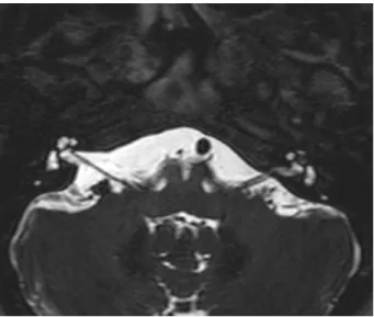
The technique described in this study was developed for a patient with a 20-year history of bilateral progressive hearing loss and tinnitus. The patient was an otolaryngol-ogist and was hindered from working in his chosen profes-sion due to hearing loss and incapacitating vertigo. His hearing had gradually deteriorated on the left side, result-ing in profound hearresult-ing loss. The patient also developed vertigo. Magnetic resonance imaging (MRI) with gadolini-um revealed a mass in the left apical turn of the cochlea measuring 0.3 cm (►Figs. 1,2, and3).
His hearing had also deteriorated on the right side, and his hearing aid (HA) was still providing some benefit to the right ear (in contrast to the left side)►Fig. 4. demonstrates his
audiometry with bilateral sensorineural hearing loss, severe at the right side and profound at the left side.
Fig. 1 Pre-gadolinium T1.
Fig. 2 Post-gadolinium T1, enhanced nodular mass in the left apical turn of the cochlea measuring 0.3 cm.
Fig. 3 T2, Low signal mass in the left apical turn of the cochlea.
International Archives of Otorhinolaryngology Vol. 20 No. 3/2016
This surgery accessed the apical turn of the cochlea. We performed mastoidectomy and posterior tympanotomy, removing the incus and tensor timpani muscle to expose the cochlear apex. The cochlear wall was burred with a small diamond burr. A small window in the apical turn was opened, and the membranous labyrinth was exposed.
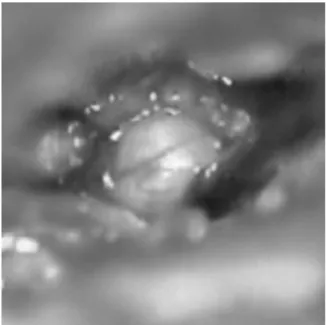
The opening was enlarged with a delicate curette. The tumor was identified, pulled out with a delicate hook, and completely resected (►Figs. 5and6).
The small window in the apical turn was sealed with bone wax. After the cochlea was anatomically preserved, it was
implanted with a straight electrode (Nucleus CI422) via round window insertion. Computed tomography scans indicated complete electrode insertion into the cochlea. The histopath-ological examination confirmed intracochlear schwannoma. We performed tumor resection to treat the patient’s vertigo, and performed simultaneous CI implantation to restore his hearing. Immediately after surgery, the patient’s vertigo and dizziness resolved and the tinnitus improved. A speech processor was activated four weeks after implanta-tion. All electrodes (22/22) were used for electric stimulation, and neural response telemetry indicated that all electrodes were functional. After three months of speech processor use, an auditory speech perception test revealed 100% speech recognition with closed sentences, and the average
Fig. 5 Apical turn of cochlea exposed with tumor inside. Fig. 6 Apical turn of the cochlea exposed following tumor removal. Fig. 4 Audiometry with bilateral sensorineural hearing loss, severe at the right side and profound at the left side.
audiometric threshold (500 to 2000 Hz) was 23 dB. The patient continues to use a HA in the contralateral ear and his speech perception with open sentences is 70% using only the HA. However, with bimodal stimulation (HAþCI), the
score is 100%, which indicates that CI improved this patient’s speech recognition.
Radiological examinations during thefive follow-up years did not indicate tumor growth.
Discussion
With the recent improvements in imaging techniques, an increasing number of reports have described intralabyrin-thine schwannomas.10
MRI with gadolinium is the best diagnostic tool to identify this disease. Intracochlear schwannomas may have slightly higher signal intensities than normal intralabyrinthine fluid on unenhanced T1-weighted images. On T2-weighted images, the schwannomas appear as hypointense lesions with sharp borders, and the fluid has a high signal. After gadolinium administration, the schwannomas present as strongly enhanced, sharply circumscribed lesions on T1-weighted images.11
Asymmetric sensorineural hearing loss is almost invariably present in patients with intracochlear schwannoma. Tinnitus, ear fullness, and vertigo are also common. Intracochlear schwan-nomas are difficult to diagnose, and diagnosis is often delayed because the presenting symptoms overlap with other otologic diseases (particularly Ménière’s disease). Sometimes, patients present with mixed hearing loss and the conductive component can be secondary to stapes movement interference caused by tumor pressure on the vestibular surface of the stapes footplate or secondary to endolymphatic hydrops.12,13
Other lesions can mimic ILS on contrast-enhanced MRIs, including labyrinthitis (typically of viral etiology), labyrin-thitis ossificans, hemorrhage, and lipoma.6
Conclusion
Treatment should preserve hearing and rehabilitate the patient. This minimally invasive procedure allows for simul-taneous tumor removal and cochlear implantation with good audiological results. To the best of our knowledge, this report is thefirst description of simultaneous intracochlear schwan-noma resection and cochlear implantation.
With this technique, we accessed the apical turn of the cochlea (with minimal damage to other structures) and removed the tumor. Thus, the intracochlear structure was preserved, which facilitated successful cochlear implantation and hearing restoration.
References
1 Bento RF. Tratado de Implante Coclear e Próteses Auditivas
Implantáveis. 1st ed. Rio de Janeiro, Brazil: Thieme Publicações Ltda; 2014
2 von Ilberg C, Kiefer J, Tillein J, et al. Electric-acoustic stimulation of the auditory system. New technology for severe hearing loss. ORL J Otorhinolaryngol Relat Spec 1999;61(6):334–340
3 Skarzyński H, Lorens A, D’Haese P, et al. Preservation of residual
hearing in children and post-lingually deafened adults after cochlear implantation: an initial study. ORL J Otorhinolaryngol Relat Spec 2002;64(4):247–253
4 Bento RF, Brito Neto R, Castilho AM, et al. Resultados auditivos com
o implante coclear multicanal em pacientes submetidos a cirurgia no Hospital das Clínicas da Faculdade de Medicina da Universidade de São Paulo. Braz J Otorhinolaryngol 2004;70:632–637
5 Kiefer J, Pok M, Adunka O, et al. Combined electric and acoustic
stimulation of the auditory system: results of a clinical study. Audiol Neurootol 2005;10(3):134–144
6 Salzman KL, Childs AM, Davidson HC, Kennedy RJ, Shelton C,
Harnsberger HR. Intralabyrinthine schwannomas: imaging
diag-nosis and classification. AJNR Am J Neuroradiol 2012;33(1):
104–109
7 Miller ME, Moriarty JM, Linetsky M, Lai C, Ishiyama A.
Intra-cochlear schwannoma presenting as diffuse Intra-cochlear enhance-ment: diagnostic challenges of a rare cause of deafness. Ir J Med Sci 2012;181(1):131–134
8 Jiang ZY, Kutz JW Jr, Roland PS, Isaacson B. Intracochlear
schwan-nomas confined to the otic capsule. Otol Neurotol 2011;32(7):
1175–1179
9 Kronenberg J, Horowitz Z, Hildesheimer M. Intracochlear
schwan-noma and cochlear implantation. Ann Otol Rhinol Laryngol 1999; 108(7 Pt 1):659–660
10 Neff BA, Willcox TO Jr, Sataloff RT. Intralabyrinthine
schwanno-mas. Otol Neurotol 2003;24(2):299–307
11 Magliulo G, Colicchio G, Romana AF, Stasolla A. Intracochlear
schwannoma. Skull Base 2010;20(2):115–118
12 Green JD Jr, McKenzie JD. Diagnosis and management of
intra-labyrinthine schwannomas. Laryngoscope 1999;109(10):
1626–1631
13 Kennedy RJ, Shelton C, Salzman KL, Davidson HC, Harnsberger
HR. Intralabyrinthine schwannomas: diagnosis, management,
and a new classification system. Otol Neurotol 2004;25(2):
160–167
International Archives of Otorhinolaryngology Vol. 20 No. 3/2016