Transpterygoid Approach to a Dermoid Cyst in
Pterygopalatine Fossa
Alexandre Beraldo Ordones
1Marco Aurélio Fornazieri
1Fábio de Rezende Pinna
1Thiago Freire Pinto Bezerra
1Richard Louis Voegels
1Luiz Ubirajara Sennes
11Department of Otolaryngology, The School of Medicine, The
University of São Paulo, São Paulo/SP, Brazil
Int Arch Otorhinolaryngol 2014;18:83–86.
Address for correspondence Marco Aurélio Fornazieri, MD, Dr. Eneas de Carvalho Aguiar Street, 255/6thfloor—6167, Cerqueira Cesar/SP, Brazil (e-mail: [email protected]).
Introduction
Dermoid cysts are congenital defects involving pluripotent stem cells arising from an ectopic site. This disease presents a slightly higher predominance in males, and most patients are diagnosed within thefirst fourth decades of life.1Although up to one third of dermoid cysts are found at birth, most of them are diagnosed in the second and third decades.2
Almost 7% of all dermoids are located in the head and neck.3 They are frequently found in the region of the lateral part of the eyebrow, in the periorbital region, and in the midline nasal region. In the neck they usually occur in the submental region,
above the hyoid and always in the midline.3 This tumor accounts for fewer than 5% of all intracranial masses.2
Tumors arising from the sinonasal region usually present late as their symptoms are often banal and may be overlooked by patients and their clinicians. The recent onset of unilateral nasal symptoms, without improvement with medical thera-py, and orbital and neurologic symptoms should be investi-gated with imaging studies. Computed tomography (CT) and magnetic resonance imaging (MRI) are used to characterize tumors of this region.4
Endoscopic sinus surgery is a well-established technique for the treatment of sinus diseases, including chronic sinusitis
Keywords
►
dermoid cyst
►
epidermal cyst
►
head and neck
neoplasms
►
skull base
►
nasal cavity
►
pterygopalatine fossa
Abstract
Objective
To describe a case of dermoid cyst arising from the pterygopalatine fossa
and review the literature.
Methods
We report a case of a 23-year-old man who suffered a car accident 2 years
before otolaryngologic attendance. He had one episode of generalized tonic-clonic
seizure and developed a reduction of visual acuity of the left side after the accident.
Neurologic investigation was performed and magnetic resonance imaging revealed an
incidental
fi
nding of a heterogeneous ovoid lesion in the pterygopalatine fossa,
hyperintense on T2-weighted imaging.
Results
Endoscopic sinus surgery with transpterygoid approach was performed. The
ovoid lesion was noted in the pterygopalatine fossa. Puncture for intraoperative
evaluation showed a transparent thick
fl
uid. Surprisingly, hair and sebaceous glands
were found inside the cyst capsule. The cyst was excised completely. Histologic
examination revealed a dermoid cyst. The patient currently has no evidence of
recurrence at 1 year postoperatively.
Conclusion
This unique case is a rare report of a dermoid cyst incidentally diagnosed.
An endoscopic transnasal transpterygoid approach may be performed to treat
success-fully this kind of lesion. Although rare, it should be considered in the differential
diagnosis of expansive lesions in the pterygopalatine fossa, including schwannoma,
angio
fi
broma, esthesioneuroblastoma, osteochondroma, cholesterol granuloma,
hem-angioma, lymphoma, and osteoma.
received
May 15, 2013
accepted
June 27, 2013
DOI http://dx.doi.org/ 10.1055/s-0033-1353370.
ISSN 1809-9777.
Copyright © 2014 by Thieme Publicações Ltda, Rio de Janeiro, Brazil
THIEME
and nasal neoplasms. Endoscopic sinus surgery to approach nasal neoplasms is limited, as the tumor can be too exten-sive.5In this report, we discuss diagnosis, differential diagno-sis, and possible approaches to lesions arising from the pterygopalatine fossa.
Case Report
A 23-year-old man was admitted to a tertiary care center after a car accident, in which he sustained a pelvic/femoral frac-ture. There was no evidence of any associated injury, and a conversion to tracheostomy after long-term intubation was performed. He also presented afirst episode of generalized tonic-clonic seizure. In addition, he presented reduced visual acuity in the left side.
The patient was referred for brain MRI; a heterogeneous ovoid lesion hyperintense in T2-weighted imaging in the pterygopalatine fossa was discovered. As a matter of fact, this lesion was indenting the middle fossa and was closely related to the maxillary artery, sphenopalatine ganglia, and maxillary posterior wall. The patient was referred to the
Otolaryngology Department of Clinics Hospital–University of São Paulo.
Prior to surgery, a wide range of differential cell counts were obtained. Laboratory workup, including a complete blood count with differentials and determination of liver enzymes and electrolytes, showed no abnormal findings. Brain CT showed a heterogeneous lobulated lesion in the left masticator space (infratemporal region), 33, 52 cm
wide, medial to lateral pterygoid muscle, with erosion of the inferior wall of foramen rotundum (►Fig. 1). We repeated
brain MRI, which showed a lobulated extra-axial formation in the left masticator space, between lateral and medial ptery-goid muscles, with fat heterogeneous tissue, hypointense in T2-weighted image, gadolinium-enhanced in T1-weighted image (►Fig. 2). There were small hyperintense lesions in
T1-weighted images in the suprasellar cistern and in the sylvian fissure, suggesting previous dermoid rupture (►Fig. 2B).
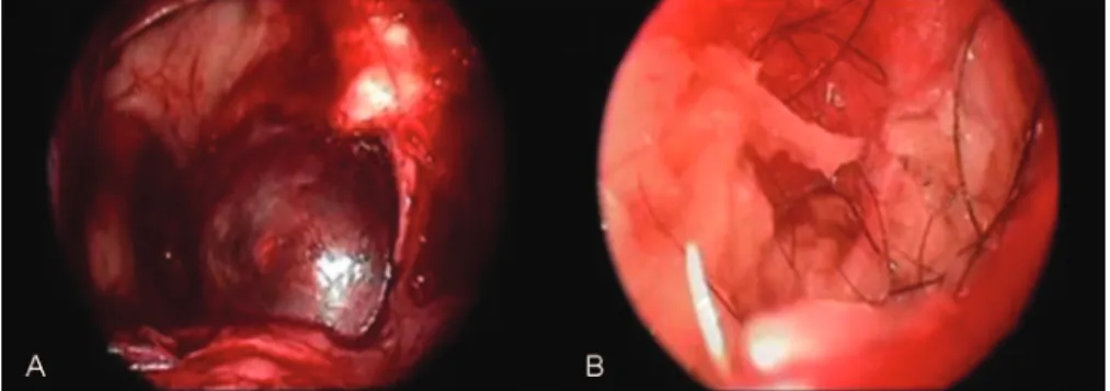
A transpterygoid transnasal endoscopic approach and resection of the lesion was performed. The puncture of the lesion showed initially a transparent thick liquid. Unexpectedly, hair and sebaceous glands were found inside the cyst capsule (►Fig. 3). The cyst was excised
completely.
Histopathologic examination confirmed the diagnosis of a dermoid cyst. The patient’s postoperative course was unremarkable.
Discussion
Benign and malignant tumors can arise from any of the structures within the infratemporal fossa and paraphar-yngeal space.6Dermoid inclusion cysts are benign tumors and are mainly unilocular and expand slowly, enlarging over years or decades, by the accumulation of cutaneous products. They may show lipid content, derived from sebaceous secretions, and secretions of apocrine sweat glands and hair.2
The causes of dermoids include failure of surface ectoderm to separate from underlying structures and sequestration of
Fig. 1 Computed tomography showing a heterogeneous lobulated lesion in the infratemporal region.
Fig. 2 Magnetic resonance imaging (MRI) of the lesion with heterogenous content in the inferior region. (A, B) T1-weighted coronal MRI. (C) T1-weighted axial MRI showing invasion of the infratemporal fossa.
International Archives of Otorhinolaryngology Vol. 18 No. 1/2014
surface ectoderm at lines of epithelial fusion during embry-onic development. Most congenital dermoid cysts probably arise due to an embryologic accident during early stages of development, between the third and fifth weeks of gestation.2,3
Clinical examination of the fossa pterygoid is difficult because it is deep lying and not easily accessible. Lesions are often discovered only at a late stage either because there are no clinical signs or, if they do occur, they are so common as to be overlooked.1,6,7In our case, the diagnosis was incidental. Tumors that arise in pterygopalatine fossa are usually asymp-tomatic. CT and MRI provide precise imaging information and may be necessary for making the diagnosis. In addition, imaging data can help to differentiate infection from tumor lesions and primary tumors from secondary tumors.1,7 Al-though they are slow-growing, dermoid inclusions cysts produce pressure changes on surrounding structures that are visible at radiograph study.
In a case series, Yu et al evaluated 86 patients with tumoral lesions of pterygopalatine and infratemporal spaces.1 Most of the lesions (81%) originated in oral and maxillofacial regions other than pterygopalatine and infra-temporal spaces and extended to this region. Only one case of teratoma was found. The most frequent diseases reported among the other cases were squamous cells carcinoma, adenoid cystic carcinoma, inflammatory disease, and hemangioma.
Differential diagnosis in cases of tumoral lesions in ptery-goid fossa should be performed (►Table 1).1,8–13
Treatment of dermoid cysts consists of complete surgical excision of the lesion, avoiding recurrence.6Rupture of der-moid cysts can produce severe chemical meningitis, usually attributed to the irritating effects of the cholesterol in the cellular debris.2 In our case, brain MRI showed signs of previous rupture, which may account for the patient’s epilepsy.
Uppal et al excised a dermoid cyst in the infratemporal fossa, extending inferiorly to the parapharyngeal space, using a lateral approach to that region.6In our case, we performed a minimally invasive treatment, using an endoscopic trans-nasal transpterygoid approach with a wide access to the lesion. It was possible to proceed with complete excision of the tumor, without intraoperative complications.
The clinical diagnosis and management of these lesions can be challenging because of the relative inaccessibility of the region, which contains the maxillary artery, the maxil-lary nerve, and the pterygopalatine ganglia, with its branches.
Surgical advantages of the endoscopic endonasal approach in comparison with traditional transcranial approaches in-clude a more direct midline exposure, decreased brain pa-renchyma injury, and lack of neurovascular structure manipulation. From the patient’s perspective, decreased sur-gery time, decreased length of stay, increased patient com-fort, and lack of external incision are advantages of the endoscopic endonasal approach.
Although rare, dermoid cysts should be considered in the differential diagnosis of expansive lesions in the pterygopa-latine fossa and can be excised by a transnasal transpterygoid endoscopic approach.
Fig. 3 Intraoperative endoscopic visualization of the dermoid cyst.
Table 1 Differential diagnosis of lesions in pterygoid region
Meningoencephalocele Malignant lymphoma
Maxillary sinus carcinoma Squamous cell carcinoma
Olfactory neuroblastoma Inflammatory disease
Osteochondroma Adenoid cystic carcinoma
Cholesterol granuloma Adenocarcinoma
Myoepithelial carcinoma Hemangioma
Ameloblastoma Leiomyosarcoma
Malignantfibrohistiocytoma Undifferentiated carcinoma
Giant-cell tumor Chondrosarcoma
Keratocyst Osteoblastoma
Mixed tumor Malignant myoepithelioma
Mucoepidermoid carcinoma Liposarcoma
Hemangiopericytoma Hemangioendothelioma
Undifferentiated sarcoma Malignant mixed tumor
Lymphangioma Neurofibroma
Rhabdomyosarcoma
Author Contributions
All authors participated in the medical treatment of the patient, including postoperative care. In addition, they performed data collection and analysis and the writing of the paper.
References
1 Yu Q, Wang P, Shi H, Luo J, Sun D. The lesions of the pterygopalatine and infratemporal spaces: computed tomography evaluation. Oral Surg Oral Med Oral Pathol Oral Radiol Endod 1998;85:742–751 2 Smirniotopoulos JG, Chiechi MV. Teratomas, dermoids, and
epi-dermoids of the head and neck. Radiographics 1995;15:1437–1455 3 Vrabec JT, Schwaber MK. Dermoid tumor of the middle ear: case
report and literature review. Am J Otol 1992;13:580–581 4 European Position Paper on Endoscopic Management of Tumors
of the Nose, Paranasal Sinuses and Skull Base. Rhinology 2010 (Suppl 22):1–143
5 Snyderman CH, Pant H, Carrau RL, Prevedello D, Gardner P, Kassam AB. What are the limits of endoscopic sinus surgery? The expanded
endonasal approach to the skull base. Keio J Med 2009;58: 152–160
6 Uppal HS, D’Souza AR, De R, Irving RM. Dermoid cyst of the infratemporal fossa. J Laryngol Otol 2002;116:150–152
7 Faye N, Lafitte F, Williams M, et al. The masticator space: from anatomy to pathology. J Neuroradiol 2009;36:121–130
8 Nishikawa T, Ishida H, Nibu K. A rare spontaneous temporal meningoencephalocele with dehiscence into the pterygoid fossa. Auris Nasus Larynx 2004;31:429–431
9 Souza RP, et al. Maxillary sinus carcinoma: an analysis of ten cases. Radiol Bras 2006;39:397–400
10 Seccia V, Lenzi R, Casani AP, Muscatello L. Ectopic olfactory neuroblastoma arising in the pterygopalatine fossa. Otolaryngol Head Neck Surg 2010;142:460–461
11 Wu W, Hu X, Lei D. Giant osteochondroma derived from pterygoid process of sphenoid. Int J Oral Maxillofac Surg 2007;36:959–962 12 Weiland DA, Aygun N. An unusual presentation of a cholesterol granuloma in a pneumatized pterygoid process of the sphenoid sinus. Otolaryngol Head Neck Surg 2007;136:153–154
13 Ghosh A, Saha S, Saha VP, Sadhu A, Chattopadhyay S. Infratemporal fossa myoepithelial carcinoma—a rare case report. Oral Maxillofac Surg 2009;13:59–62
International Archives of Otorhinolaryngology Vol. 18 No. 1/2014