Neurodegeneration with
brain iron accumulation
A case report
Daniel Nassif1,João Santos Pereira1,2, Mariana Spitz1,2, Cláudia Capitão1, Alessandra Faria1
ABSTRACT. Pantothenate kinase-associated neurodegeneration (PKAN) is an autosomal recessive disorder caused by mutation in the PANK2 gene. It is characterized by abnormal brain iron accumulation, mainly in the globus pallidus. PKAN
is included in a group of disorders known as neurodegeneration with brain iron accumulation (NBIA). We report a case of atypical PKAN with its most characteristic presentation, exhibiting marked psychiatric symptoms, speech disorder and focal dystonia. Brain MRI has great diagnostic importance in this group of disorders and, in this case, disclosed the eye-of-the-tiger sign. Genetic testing confirmed the diagnosis.
Key words: PKAN, NBIA, PANK2.
NEURODEGENERAÇÃO COM ACÚMULO CEREBRAL DE FERRO: RELATO DE CASO
RESUMO. Neurodegeneração associada à pantotenato-quinase (PKAN) é uma entidade autossômica recessiva causada pela mutação do gene PANK2. Caracteriza-se por depósito cerebral anormal de ferro, particularmente nos globos pálidos.
PKAN faz parte de um grupo de desordens conhecidas como neurodegeneração com acúmulo cerebral de ferro (NBIA). Relatamos um caso de PKAN atípica com sua apresentação mais característica, sendo evidentes sintomas psiquiátricos marcados, distúrbio da fala e distonia focal. A ressonância magnética de crânio possui grande importância diagnóstica neste grupo de desordens, e neste caso, demonstrou o sinal do olho de tigre. O teste genético confirmou o diagnóstico.
Palavras-chave: PKAN, NBIA, PANK2.
INTRODUCTION
N
eurodegeneration with brain iron accu-mulation (NBIA) syndromes are neuro-degenerative disorders whose main feature is the abnormal accumulation of iron, predomi-nantly in the globus pallidus. Clinical indings are heterogeneous, but typically include pro-gressive extrapyramidal disorder, with variable involvement of the pyramidal, peripheral ner-vous, autonomic, cognitive, psychiatric, visual and cerebellar systems.1,2Pantothenate kinase-associated neuro-degeneration (PKAN) represents the most prevalent form of NBIA, accounting for 50% of cases. his is caused by mutation in the
PANK2 gene.3 PKAN is subdivided into the
classic form, with early onset and rapid evolu-tion, and the atypical form, with later onset and slower progression.4
he use of brain magnetic resonance imag-ing to disclose the accumulation of brain iron is important for conirming the diagnosis and also facilitates diferential diagnosis among NBIAs. In PKAN, the presence of the eye-of-the-tiger sign is a deining characteristic.5
Genetic testing is available for most of the NBIA-associated syndromes, including PKAN.1
Currently, however, only symptomatic treatment of these disorders is possbile.1-3
We report a case of atypical PKAN with typical radiological indings and genetic con-irmation of the entity.
This study was conducted at the Movement Disorders Sector, Neurology Service, Pedro Ernesto University Hospital, State University of Rio de Janeiro, Rio de Janeiro RJ, Brazil.
1Movement Disorders Sector, Neurology Service, Pedro Ernesto University Hospital, State University of Rio de Janeiro, Rio de Janeiro RJ, Brazil. 2Post Graduate
Stricto Sensu Program in Medical Sciences, School of Medical Sciences, State University of Rio de Janeiro, Rio de Janeiro RJ, Brazil.
João Santos Pereira. Setor de Distúrbios do Movimento / Neurologia / HUPE/UERJ – Av. 28 de Setembro, 77 / 2º andar – 20550-031 Rio de Janeiro RJ – Brasil. E-mail: [email protected]
Disclosure: The authors report no conflicts of interest.
Received January 27, 2016. Accepted in final form March 28, 2016.
CASE REPORT
A 21-year-old female student presented with a four-year history of irritability, learning diiculties with decline in academic performance, and progressive emotional lability. he neurological examination revealed the presence of dysarthria, focal dystonia involving the left hand, and facial tics, as well as the presence of hyperrelexia and spasticity of the lower limbs, with lexor plantar relex. Tests for strength, coordination and sensitivity were unremarkable. No parkinsonism, chorea or myoclonus was evident. he neuro-ophthal-mologic exam, including fundoscopy was normal. he patient had 10 years of education and a total score of 23 on the Mini-Mental State Exam (MMSE). She scored a total of 39 points on the Beck Depression Inventory (BDI), indicating marked depressive symp-toms. here was no history of neurological disease or consanguinity.
he patient was submitted to subtests from the WAIS III battery (Wechsler Adult intelligence Scale – 3rd
edi-tion) and to speciic tests for assessing attention, mem-ory, language, praxis, perception and executive functions, and proved cooperative and communicative during the exam. he results indicated a worse performance on processing speed, attention switching, long-term mem-ory, verbal reasoning, abstraction, verbal luency, plan-ning and perceptual organization (including strategy in constructional praxis). Inhibitory control was mildly impaired whereas working memory was within normal limits but with luctuating performance on speciic tasks. he phonoaudiological assessment revealed mild dysar-thria, with impairment of phonation and articulation, in addition to sporadic gagging.
Figure 1. Blood smear shows numerous acanthocytes.
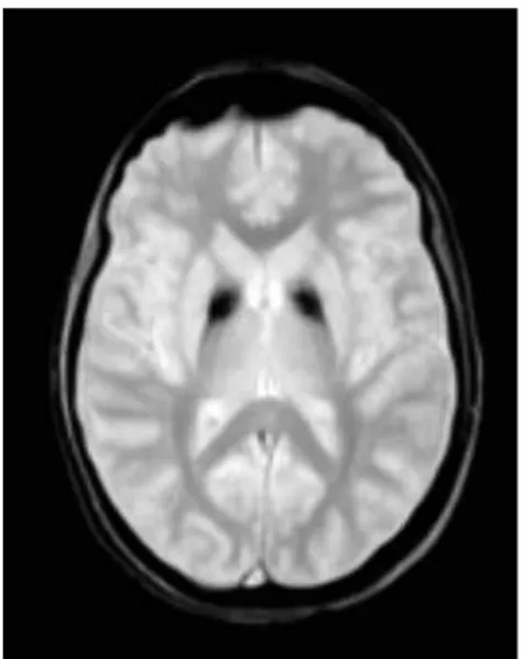
Figure 2. T2-weighted magnetic resonance imaging showing symmetrical central hyperintensity surrounded by hypointense signal in globus pallidus, giving “eye of tiger” appearance.
Figure 3. T2* shows low signal in corresponding areas from iron deposition.
Laboratory tests, including full blood count, bio-chemical evaluation, renal and liver function tests were normal. Sera copper and ceruloplasmin concentrations, as well as urinary copper level, were also normal. Blood smear showed a large number of acanthocytes (Figure 1).
A T2 weighted brain magnetic resonance imaging demonstrated symmetrical central hyperintensity sur-rounded by hypointense signal in globus pallidus, consis-tent with the “eye-of-the-tiger” sign. T2* demonstrated low signal in corresponding areas from iron deposition (Figures 2 and 3).
Given the suspected diagnosis, genetic testing was performed and the molecular analysis of itron 5 of the
PANK2 gene revealed the presence of the c.1537-3C>G
pantothenate-kinase protein, resulting in a non-functional protein. his inding conirms the diagnosis of pantothenate kinase-associated neurodegeneration (PKAN).
DISCUSSION
Hallervorden-Spatz syndrome was irst described in 1922 by two German neuropathologists, Julius Haller-vorden and Hugo Spatz, whose studies were based on pathological specimens obtained under Nazi euthanasia programs in individuals with physical and intellectual disabilities.6,7 For decades, despite the clinical
hetero-geneity, the majority of patients with pathological or radiological evidence of brain iron accumulation were diagnosed as having Hallervorden-Spatz, although they probably had other NBIA syndromes.8
he irst subtype of the Hallervorden-Spatz syn-drome, identiied by the mutation in the PANK2 gene
and speciic radiological and clinical indings, was denominated pantothenate kinase-associated neurode-generation or PKAN.9
In 2003, a new nomenclature for the syndrome was proposed prompted by the anti-ethical activities of the neuropathologists who originally described the syn-drome and due to the recognition of genetic mutations associated with the clinical syndrome, particularly the
PANK2 gene. his led to introduction of the terms
neu-rodegeneration with brain iron accumulation (NBIA) and pantothenate kinase-associated neurodegeneration
(PKAN) to denote patients with suspected or proven mutation in the PANK2 gene.4,8,10
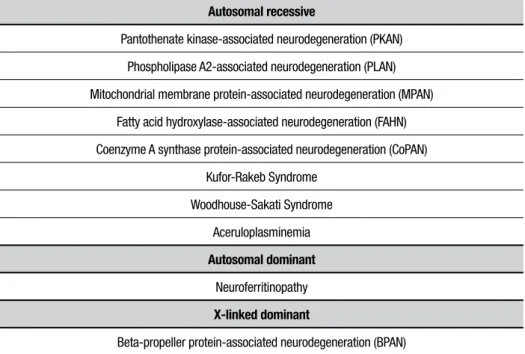
A number of genes associated with NBIA have been identiied, including PANK2, PLA2G6, FA2H, C19orf12, ATP13A2, CP, and FTL. he terminology of the syn-dromes associated with these genes follows a system in which the irst letters refer to the physiopathogenic molecular base involved and the last letters to “associated neurodegeneration”, for example, pantothenate kinase-associated neurodegeneration or PKAN, as depicted in Table 1.1,3
PKAN accounts for 50% of NBIA cases.3 It is a rare
autosomal recessive disorder associated with mutation in the PANK2 gene located on chromosome 20p13 and encodes pantothenate kinase, a key regulating enzyme of coenzyme-A synthesis.1,11
PKAN is subdivided into two main types, based on age of onset, symptoms and progression: (1) classic (2) atypical.4 Some cases have overlapping features3 and
oth-ers do not it this subdivision.11
he onset of classic PKAN generally occurs at around three years of age and before six years in 90% of cases. It typically presents with gait diiculties.2,12 Patients
pres-ent pyramidal and extrapyramidal signs with marked dystonia.2 he dystonia generally dominates the clinical
picture and begins assymetrically.12 Severe tongue
pro-trusion dystonia can occur.13 he presence of dystonic
opisthotonus or oromandibular dystonia suggests
inclu-Table 1. NBIA subtypes described to date with acronym and mode of inheritance.
Autosomal recessive
Pantothenate kinase-associated neurodegeneration (PKAN)
Phospholipase A2-associated neurodegeneration (PLAN)
Mitochondrial membrane protein-associated neurodegeneration (MPAN)
Fatty acid hydroxylase-associated neurodegeneration (FAHN)
Coenzyme A synthase protein-associated neurodegeneration (CoPAN)
Kufor-Rakeb Syndrome
Woodhouse-Sakati Syndrome
Aceruloplasminemia
Autosomal dominant
Neuroferritinopathy
X-linked dominant
Beta-propeller protein-associated neurodegeneration (BPAN)
sion of PKAN as a diferential diagnosis.14 he
neuro-ophthalmologic exam can reveal pigmentary retinopa-thy and saccade and pupil abnormalities.15 Some patients
have acanthocytes in peripheral blood.16
he progression of classic PKAN is not linear, exhib-iting periods of marked worsening. Bulbar compromise can lead to nutritional and respiratory problems.3 Loss
of walking ability occurs within 10-15 years of disease onset.4 Some patients evolve to death within the irst
decade of disease onset whilst others reach adulthood.3
Atypical PKAN is more heterogeneous than the clas-sic form, emerging in the second or third decade of life with slower evolution.3,4,8 Psychiatric symptoms and
speech disorders are characteristic,11 and were found in
the present case. Psychiatric symptoms include depres-sion, emotional lability, impulsivity, aggressivity and tourettism with motor and verbal tics.17 Speech disorders
include dysarthria, hypophonia, spasmodic dysphonia and palilalia. Pigmentary retinopathy is rare.3,4
Unlike the classic form, motor involvement is less marked, with loss of walking at 15-40 years of disease.4,17
Dystonia tends to dominate the clinical picture in ado-lescents, while patients over 20 years of age tend to pres-ent parkinsonism as the predominant clinical symptom. Pyramidal signs can occur. Motor involvement may not be signiicant in the initial stage of the disease.3
Brain magnetic resonance imaging is an important diagnostic tool. he characteristic inding is the eye-of-the-tiger sign, characterized by bilateral hypointensity of the globus pallidus with a central area of hyperinten-sity on T2-weighted images. Hypointenhyperinten-sity in substantia nigra may be seen in some patients. Areas of hypointen-sity at sites of iron deposits are visible on T2*.18 he area
of hypointensity correlates pathologically with abnormal iron deposition, while central hyperintense signal occurs due to neuronal loss with gliosis.19
As the disease progresses, the area of hypointensity can dominate the radiological presentation. It is believed
that the central hyperintense area can disappear.8,18
here are controversial reports in the literature on the correlation of mutation in the PANK2 gene and the
pres-ence of the eye-of-the-tiger sign.4,20 his same sign has
been described in other pathologies, such as neurofer-ritinopathy, corticobasal degeneration, multiple system atrophy18 and in a case report with syndromic diagnosis
of early onset parkinsonism, with response to dopami-nergic therapy.21
he management of PKAN remains symptomatic, as is the case for other NBIAs. Dystonia can be controlled with the use of benzodiazepines, anticholinergics and botulinum toxin. Baclofen can be used for relieving spasticity.1,3 Stereotactic surgical modalities such as
thalamotomy22 and palidotomy23 and deep brain
stimu-lation of the internal globus pallidus,24 can help control
symptoms but do not arrest disease progression.1
Despite the few options, the beneit of using iron che-lates, such as deferiprone, is questionable.1,3
It can be concluded that, although rare, neurode-generation with brain iron accumulation syndromes should be included as part of the diferential diagnosis in patients with progressive extrapyramidal syndrome, particularly when brain iron deposits are depicted on magnetic resonance imaging. Multidisciplinary rehabili-tation programs can promote satisfactory conditions in these patients and contribute toward improving their quality of life.
Author contribution. Daniel Nassif: design, analysis of data,
intellectual contribution to the writing of the manu-script; João Santos Pereira: design, analysis of data, intellectual contribution to the writing of the manu-script; Mariana Spitz: design, analysis of data, intel-lectual contribution to the writing of the manuscript; Claudia Capitão: analysis of data, Alessandra Faria: analysis of data.
REFERENCES
1. Schneider SA. Neurodegeneration with Brain Iron Accumulation. Curr Neurol Neurosci Rep 2016;16:9. DOI 10.1007/s11910-015-0608-3. 2. Schneider SA, Hardy J,Bhatia KP. Syndromes of Neurodegeneration with
Brain Iron Accumulation (NBIA): an Update on Clinical Presentations, Histological and Genetic Underpinnings, and Treatment Considerations. Mov Disord. 2012;27(1):42-53.
3. Hogarth P. Neurodegeneration with Brain Iron Accumulation: Diagnosis and Management. J Mov Disord 2015;8(1):1-13
4. Hayflick SJ, Westaway SK, Levinson B, et al. Genetic, clinical, and radiographic delineation of Hallervorden-Spatz syndrome. N Engl J Med. 2003;348:33-40.
5. Kruer MC, Boddaert N, Schneider SA, et al. Neuroimaging features of neurode generation with brain iron accumulation. AJNR Am J Neu roradiol 2012;33:407-414.
6. Van Craenenbroeck A, Gebruers M, Martin J-J, Cras P. Hallervorden- Spatz disease: historical case presentation in the spotlight of nosological evolution. Mov Disord. 2010;25:2486-2492.
7. Shevell M. Racial hygiene, active euthanasia, and Julius Hallervorden. Neurology 1992;42:2214-2219.
8. Gregory A, Hayflick SJ. Neurodegeneration with brain iron accumulation. Folia Neuropathol. 2005;43:286-296.
9. Zhou B, Westaway SK, Levinson B, Johnson MA, Gitschier J,Hayflick SJ. A novel pantothenate kinase gene (PANK2) is defective in Hallervorden-Spatz syndrome. Nat Genet 2001;28:345-349.
10. Zeidman LA, Pandey DK. Declining use of the Hallervor den-Spatz disease eponym in the last two decades. J Child Neurol 2012;27: 1310-1315.
geno-type of pantothenate kinase-associated neurodegeneration. Neurology 2005;64:1810.
12. Hayflick SJ. Neurodegeneration with brain iron accumulation: from genes to pathogenesis. Semin Pediatr Neurol. 2006;13:182-185.
13. Schneider SA, Aggarwal A, Bhatt M, et al. Severe tongue protrusion dystonia: clinical syndromes and possible treatment. Neurology. 2006; 67:940-943.
14. Stamelou M, Lai SC, Aggarwal A, et. al. Dystonic Opisthotonus: A “Red Flag” for Neurodegeneration With Brain Iron Accumulation Syndromes? Mov Disord 2013;28:1325-1329.
15. Egan RA, Weleber RG, Hogarth P, et al. Neuro-ophthalmologic and electroretinographic findings in pantothenate kinase associated neuro-degeneration (formerly Hallervorden-Spatz syndrome). Am J Ophthalmol. 2005;140:267-274.
16. Gregory A, Hayflick SJ. Pantothenate Kinase-Associated Neurodegen-eration. In: Pagon RA, Adam MP, Ardinger HH, Bird TD, Dolan CR, Fong CT, et al., editors. GeneReviews(R) [Internet]. Seattle (WA): University of Washington; 1993-2014.
17. Gregory A, Polster BJ, Hayflick SJ. Clinical and genetic delineation of neurodegeneration with brain iron accumulation. J Med Genet. 2009;46:73-80.
18. Amaral LL, Gaddikeri S, Chapman PR, et al. Neurodegeneration with brain Iron accumulation: clinicoradiological approach to diagnosis. J Neuroimaging: Off J Am Soc Neuroimaging. 2015;25:539-551. 19. Guillerman RP. The eye-of-the-tiger sign. Radiology 2000;217:895-
896.
20. Hartig MB, Hortnagel K, Garavaglia B, et al. Genotypic and phenotypic spectrum of PANK2 mutations in patients with neurodegeneration with brain iron accumulation. Ann Neurol 2006;59:248-256.
21. Barbosa ER, Bittar MR, Bacheschi LA, Comerlatti LR, Scaff M. Parkin-sonismo precoce associado a lesões palidais de tipo “eye-of-the-tiger”. Arq Neuropsiquiatr 1995;53(2):294-297.
22. Tsukamoto H, Inui K, Taniike M, et al. A case of Hallervorden- Spatz disease: progressive and intractable dystonia controlled by bilateral thala-motomy. Brain Dev. 1992;14:269-272.
23. Balas I, Kovacs N, Hollody K. Staged bilateral stereotactic pallido-thalamotomy for life-threatening dystonia in a child with Hallervorden-Spatz disease. Movement Disorders: Off J Movement Disorder Soc. 2006;21:82-85.