TINU syndrome: review of the literature and case report
Sindrome TINU: revisão de literatura à propósito de um caso
Authors
Marcelo A. Pinheiro 1
Matheus B. C. Rocha 1
Beatriz Oliveira Neri 2
Isabelle Oliveira Parahyba 1
Luis A.R. Moura 3
Claudia Maria Costa de Oliveira 1,2
Marcos Kubrusly 1,2
1 UniChristus.
2 Universidade Federal do
Ceará.
3 Universidade Federal de São
Paulo.
Submitted on: 06/16/2015. Approved on: 09/28/2015.
Correspondence to:
Claudia Maria Costa de Oliveira. Hospital Universitário Walter Cantideo-UFC Centro Universitário UniChristus.
Rua Professor Jacinto Botelho, nº
500/500, Fortaleza, Ceará, Brazil.
Introdução: A síndrome nefrite tubuloin-tersticial e uveíte (síndrome TINU) é uma entidade clínica incomum e a maioria dos pacientes são adolescentes e mulheres jovens. O caso relatado a seguir refere-se a uma paciente idosa com manifesta-ções oftalmológicas que antecederam às renais, sendo provavelmente o primeiro caso descrito no Brasil. Relato de Caso: Paciente feminina, 60 anos, procurou at-endimento médico por queixa de "olhos vermelhos". Três meses após o primeiro episódio do quadro ocular, a paciente cur-sou com sintomas sistêmicos e disfunção renal. A biópsia renal evidenciou nefrite tubulointersticial com sinais de atividade. Discussão: A fisiopatologia da síndrome TINU permanece pouco entendida, pro-vavelmente envolvendo a imunidade ce-lular e humoral. Essa síndrome pode ser diferenciada de condições sistêmicas asso-ciadas à uveíte e à nefrite, sendo necessária a exclusão de outras doenças antes de se confirmar seu diagnóstico, especialmente na presença de achados oftalmológicos. Conclusão: A suspeição clínica e o con-hecimento do manejo desta patologia por nefrologistas, internistas e oftalmologistas são mandatórios no tratamento do paci-ente com Síndrome TINU.
R
ESUMOPalavras-chave: doenças autoimunes; lesão renal aguda; nefrite intersticial; uveíte.
Introduction: Tubulointerstitial nephritis and uveitis syndrome (TINU Syndrome) is an uncommon clinical entity, and the majority of patients are adolescents and young women. The case reported refers to an elderly patient with ophthalmolo-gic symptoms detected earlier than kidney manifestations, being probably the first case described in Brazil. Case Report: Fe-male patient, 60 years-old, sought medi-cal attention for complaints of "red eye". Three months after the first episode of eye manifestation, the patient presented with systemic symptoms and renal dysfunction. Renal biopsy showed tubulointerstitial ne-phritis with signs of activity. Discussion:
The pathophysiology of TINU Syndrome remains poorly understood, probably in-volving both cellular and humoral immu-nity. This syndrome can be differentiated from systemic conditions associated with nephritis and uveitis, and prior exclusion of other diseases is necessary to confirm diagnosis, especially in the presence of ophthalmologic findings. Conclusion:
The clinical suspicion and the knowled-ge of the manaknowled-gement of the disease by nephrologists, internists and ophthalmo-logists is mandatory in the treatment of patients with TINU Syndrome.
A
BSTRACTKeywords: acute kidney injury; autoim-mune diseases; interstitial; nephritis; uveitis.
I
NTRODUCTIONTubulointerstitial nephritis (TIN) is a significant cause of kidney failure in children and adults, and it is characterized by renal histological and functional abnormalities.1
entity,2-5 first described in 1975 by Dobrin
et al.,6receiving the name tubulointerstitial
nephritis and uveitis syndrome (TINU syndrome).7 TINU is characterized by
favorable acute tubulointerstitial nephritis and recurrent chronic uveitis.8
women.10 Its etiology is unknown, with several
possible triggers that can start it, such as infections and drug use.2,11
Diagnosis is mainly clinical, proving the presence of TIN and uveitis with no evidence of other systemic or infectious diseases. Symptoms are mostly nonspecific, including fever, headache, fatigue and anorexia,1,12,13 causing the clinical
diagnosis to be difficult and often delaying treatment onset.1
The case described below was found in an elderly patient, with pre-nephritis ophthalmic manifestations, and it appears to be the first report of its kind from Brazil.
C
ASE REPORTFemale patient, 60 years old, with a past of systemic hypertension, sought medical treatment in September 2010, complaining of a “Red Eye” (Figure 1) of sudden onset, with pain on palpation and eye movement, associated with mild bilateral eyelid edema. She was diagnosed with non-bacterial conjunctivitis. Corticosteroid-based eye drops were prescribed for seven days, with symptom resolution. Seven days after interrupting the use of eye drops, the patient developed a new ophthalmic crisis, to which the prior approach was maintained, and symptoms subsided.
Thirty days after the condition had started, the patient presented with low-intensity pain in her upper left quadrant, continuous, not ventilator-dependent, for 15 days, progressing to severe pain. A chest X-ray revealed mild pulmonary infiltrates in the left lung base. It was suggested the possibility of atelectasis secondary to pain or pneumonic disease, treated with clarithromycin for 7 days and 2 days with dexamethasone/indomethacin, to which she responded with clinical and radiological improvement. Thirty days after pain resolution, the patient had an episode similar to her initial ophthalmic condition. Three months after the first episode of her eye disease, she presented with systemic symptoms and lost 5 kg (7% of body weight) in one month without changes to her blood pressure.
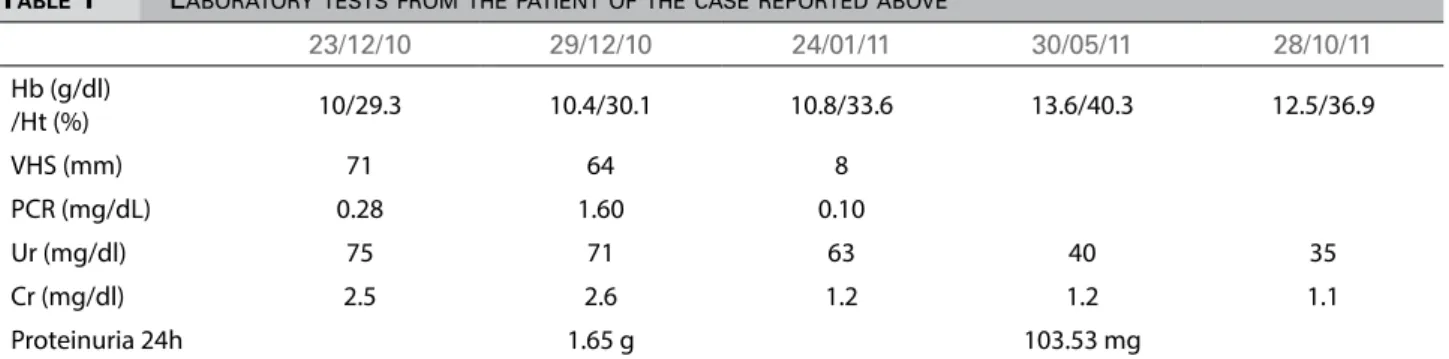
Laboratory tests showed azotemia - creatinine: 2.5 mg/dL and urea: 75 mg/dl, while her baseline creatinine was 0.9 mg/dl. Urinalysis with urine glucose ++, without other changes; 24-hour proteinuria: 1,65g; Renal ultrasound: kidneys of normal size, slight increase in cortical echogenicity and good cortex-medulla differentiation. The other tests carried out are shown on Table 1. After admission, she was submitted to pulse therapy with methylprednisolone 500 mg for 3 days followed by prednisone 1 mg/kg/day for 1 month, and the medication was weaned during for 4 months, with progressive improvement in her tests. In January 2011 (post-pulse therapy), she was submitted to a renal biopsy, which showed glomeruli within normal limits (global sclerosis: 2/30), tubular epithelial degenerative changes with spotted atrophy and mild diffuse interstitial fibrosis and tubulointerstitial nephritis with signs of inflammatory activity. Direct immunofluorescence revealed no glomerular deposits and tubular cylinders with IgM and IgA (Figure 2).
Three months after discontinuing the immunosuppressive medication, she had a new episode of anterior uveitis, and was started on corticosteroid eye drops and ketorolac. As a side effect, she had an increase in intraocular pressure, and was started on brinzolamide/timolol and brimonidine tartrate. We tried to wean her off the corticosteroid eye drops at times, but always with worsening of uveitis.
TABLE 1 LABORATORYTESTSFROMTHEPATIENTOFTHECASEREPORTEDABOVE
23/12/10 29/12/10 24/01/11 30/05/11 28/10/11
Hb (g/dl)
/Ht (%) 10/29.3 10.4/30.1 10.8/33.6 13.6/40.3 12.5/36.9
VHS (mm) 71 64 8
PCR (mg/dL) 0.28 1.60 0.10
Ur (mg/dl) 75 71 63 40 35
Cr (mg/dl) 2.5 2.6 1.2 1.2 1.1
Proteinuria 24h 1.65 g 103.53 mg
Figure 2. Kidney biopsy. A. Interstitial inlammation consisting of lymphoid cells that dissociate the tubular epithelium. (PAS - 400x); Renal biopsy B. Interstitial broadening by ibrosis and lymphocytic inlammatory iniltrate (Masson trichrome - 400x).
Currently, after 52 months of follow up, the patient remains in use of cyclosporine ophthalmic emulsion because of the failure in interrupting the medicine. Urinalysis is within the normal range, and urea and creatinine with values of 36 mg/dL and 1.0 mg/dL, respectively.
D
ISCUSSIONThe TINU syndrome has an approximate prevalence of 3.5 cases/million people and an incidence of 0.2 cases/ million/year,3,7 probably being underdiagnosed;14,15
without racial or ethnic predominance among the cases reported.16
There is a higher incidence among women,2,9 in
the ratio of 3: 1 and, according to recent studies, the prevalence in men has increased.3
The disease manifests earlier in men, ranging from
from 10-74 years of age.2,7,10 There are few cases
described in the literature involving the elderly.7,9
The main manifestations are nonspecific and associated with the renal disease, including, in decreasing order, fever, weight loss, fatigue and malaise, anorexia, abdominal pain, among others.1,2,7,9,15-17
Uveitis may precede (21%), happen concurrently (15%) or succeed (65%) nephritis,9,16,18 with eye
symptoms starting, in average, one month after the systemic effects. The main ocular disease is anterior uveitis, which occurs in 80% of patients, and the most common symptoms are pain and redness.2
it is the result of an autoimmune process that involves cellular and humoral immunity.8 The renal and ocular
injuries in the clinical course of TINU may suggest a common antigen to these two structures that could be the target of a crossed autoimmune reaction.19
This syndrome can be differentiated from systemic conditions associated with uveitis and nephritis. TIN has a variety of etiologies and it would be no different with TINU syndrome. The role of infections and non-infectious causes is speculated in the immune response, leading to the development of the syndrome.1,2,8,9,11,20,21
Kim et al. consider that the main causes of
concomitant nephritis and uveitis, which can be mistaken for the TINU syndrome are: sarcoidosis, Sjogren’s syndrome,21 lupus, Wegener’s granulomatosis,
Behcet’s disease,22 and it is imperative to order tests to
rule these out.
In 2001, Mandeville et al.2 published the TINU
syndrome diagnostic criteria, requiring the presence of acute interstitial nephritis and uveitis, with no other systemic disease as a cause, in addition to completing the 3 following criteria: 1. abnormal kidney function; 2. abnormal urinalysis; and 3. Systemic disease lasting ≥ 2 weeks.
Laboratory tests in the evaluation of TINU syndrome are not typical. Moreover, the ANCA, antinuclear antibody, anticardiolipin antibodies, anti-DNA antibody and rheumatoid factor may be positive and may be hypocomplementemia, and polyclonal hypergammaglobulinemia and circulating immune complexes.2,23,24
The patient had mild anemia, elevated inflammatory markers, azotemia and increased proteinuria. C4 was high (60.8 mg/dL), and C3 dosage was normal (154 mg/dL). C-ANCA, P-ANCA and FAN were negative.
Histological evidence of TIN should be sought for the definitive diagnosis of the syndrome. However, an invasive procedure such as a kidney biopsy may not be suitable for all patients, and should be considered in a case-by-case basis.3
TIN may resolve spontaneously, but the treatment of patients with progressive renal failure include the use of corticosteroids. The nephropathy becomes chronic in 11% of cases and only 5% of patients require dialysis.18 In this case, after pulse therapy, the patient
experienced a reduction in urea and creatinine levels. Topical steroids associated with cycloplegic agents are commonly recommended as a treatment option for anterior uveitis.16,25 If refractory,
one should consider using oral steroids15 or
immunosuppressive agents.5,9,15,25 Reports indicate
that in 14% of the cases the symptoms persist for more than 3 months5,9 and uveitis recurrence
occurs in about 50% of patients.7,26 In a series of
33 cases, the median value for disease persistence was 7 months, ranging from 1 to 147 months.27
Although this patient of ours manifested the TINU syndrome at 60 years of age, she developed a favorable renal function outcome, presenting azotemic values within the normal range even after 52 months of follow-up. Due to the increase of intraocular pressure with use of topical corticosteroids, we decided to use cyclosporine eye drops, making continuous use without possibilities of discontinuing it, as it always resulted in uveitis recurrence.
C
ONCLUSIONThis tubulointerstitial nephritis and uveitis case makes us suggest that this rare syndrome should be considered in the differential diagnosis of unex-plained tubulointerstitial nephritis, especially in the presence of ocular disorders, even in older patients. There is evidence suggesting that TINU is underdiag-nosed, especially in young patients who have mild and asymptomatic kidney disease. It is necessary to disclose this information among nephrologists, inter-nists and ophthalmologists, because their interaction is paramount in the treatment of patients with the TINU syndrome.
R
EFERENCES1. Jahnukainen T, Ala-Houhala M, Karikoski R, Kataja J, Saarela V, Nuutinen M. Clinical outcome and occurrence of uveitis in children with idiopathic tubulointerstitial nephritis. Pediatr Ne-phrol 2011;26:291-9. DOI:http://dx.doi.org/10.1007/s00467-010-1698-4
2. Mandeville JT, Levinson RD, Holland GN. The tubuloin-terstitial nephritis and uveitis syndrome. Surv Ophthalmol 2001;46:195-208. PMID: 11738428 DOI: http://dx.doi. org/10.1016/S0039-6257(01)00261-2
3. Helms E, Servilla KS, Hartshorne MF, Harris A, Nichols MJ, Tzamaloukas AH. Tubulointerstitial nephritis and uvei-tis syndrome: use of gallium scintigraphy in its diagnosis and treatment. Int Urol Nephrol 2005;37:119-22. PMID: 16132773DOI: http://dx.doi.org/10.1007/s11255-004-2356-1 4. Eyileten T, Dede F, Yenicesu M, Celebi G, Vural A. Idiopa-thic tubulointerstitial nephritis and uveitis-'TINU syndrome'. Nephrol Dial Transplant 2008;23:771-2. DOI: http://dx.doi. org/10.1093/ndt/gfm624
6. Dobrin RS, Vernier RL, Fish AL. Acute eosinophilic inters-titial nephritis and renal failure with bone marrow-lymph node granulomas and anterior uveitis. A new syndrome. Am J Med 1975;59:325-33. PMID: 1163543 DOI:http://dx.doi. org/10.1016/0002-9343(75)90390-3
7. Weinstein O, Tovbin D, Rogachev B, Basok A, Vorobiov M, Kratz A, et al. Clinical manifestations of adult tubulointerstitial nephri-tis and uveinephri-tis (TINU) syndrome. Int Ophthalmol 2010;30:621-8. DOI:http://dx.doi.org/10.1007/s10792-010-9369-9
8. Timmermans SA, Huitema JJ, Wirtz JJ. Keep an eye out for tubulo-interstitial nephritis. Neth J Med 2013;71:523-5. 9. Han JM, Lee YJ, Woo SJ. A case of tubulointerstitial nephritis
and uveitis syndrome in an elderly patient. Korean J Ophthal-mol 2012;26:398-401. DOI: http://dx.doi.org/10.3341/ kjo.2012.26.5.398
10. Abed L, Merouani A, Haddad E, Benoit G, Oligny LL, Sartelet H. Presence of autoantibodies against tubular and uveal cells in a patient with tubulointerstitial nephritis and uveitis (TINU) syndrome. Nephrol Dial Transplant 2008;23:1452-5. DOI: http://dx.doi.org/10.1093/ndt/gfm890
11. Fried T. Acute interstitial nephritis. Why do the kidneys sud-denly fail? Postgrad Med 1993;93:105-6.
12. Clarkson MR, Giblin L, O'Connell FP, O'Kelly P, Walshe JJ, Conlon P, et al. Acute interstitial nephritis: clinical features and response to corticosteroid therapy. Nephrol Dial Transplant 2004;19:2778-83. DOI:http://dx.doi.org/10.1093/ndt/gfh485 13. Baker RJ, Pusey CD. The changing profile of acute
tubuloin-terstitial nephritis. Nephrol Dial Transplant 2004;19:8-11. DOI: http://dx.doi.org/10.1093/ndt/gfg464
14. Tan Y, Yu F, Qu Z, Su T, Xing GQ, Wu LH, et al. Modi-fied C-reactive protein might be a target autoantigen of TINU syndrome. Clin J Am Soc Nephrol 2011;6:93-100. DOI: http:// dx.doi.org/10.2215/CJN.09051209
15. Thomassen VH, Ring T, Thaarup J, Baggesen K. Tubuloin-terstitial nephritis and uveitis (TINU) syndrome: a case report and review of the literature. Acta Ophthalmol 2009;87:676-9. PMID: 18700885 DOI:http://dx.doi.org/10.1111/j.1755-3768.2008.01302.x
16. Parameswaran S, Mittal N, Joshi K, Rathi M, Kohli HS, Jha V, et al. Tubulointerstitial nephritis with uveitis syndrome: A case report and review of literature. Indian J Nephrol 2010;20:103-5. DOI: http://dx.doi.org/10.4103/0971-4062010;20:103-5.65307
17. Onyekpe I, Shenoy M, Denley H, Riad H, Webb NJ. Re-current tubulointerstitial nephritis and uveitis syndrome in a renal transplant recipient. Nephrol Dial Transplant 2011;26:3060-2. DOI: http://dx.doi.org/10.1093/ndt/gfr352 18. Hinkle DM, Foster CS. Tubulointerstitial nephritis and uvei-tis syndrome. Int Ophthalmol Clin 2008;48:9-13. DOI:http:// dx.doi.org/10.1097/IIO.0b013e3181694db9
19. Khammassi N, Mohsen D, Ounissi M, Tougourti MN, Ha-mza M. TINU syndrome and lymphoid interstitial alveoli-tis: a new observation. Tunis Med 2011;89:210-2. PMID: 21308636
20. Alon US. Tubulointerstitial nephritis. In: Avner ED, Harmon WE, Niaudet P, Yoshikawa N, eds. Pediatric Nephrology. 6th ed. Berlin and Heidelberg: Springer-Verlag; 2009. p.1081-98. 21. Kim DH, Jang YS, Do WS, Oh DM, Kim SH, Kwak EK, et al.
A case of tubulointerstitial nephritis and uveitis syndrome in an old age female. Korean J Nephrol 2010;29:250-5. 22. Lee G, Ashfaq A. Tubulointerstitial nephritis and uveitis
(TINU syndrome). 2014 [cited 2014 Ago 3]. Available from: http://www.uptodate.com/contents/tubulointerstitial-nephri-tis-and-uveitis-tinu-syndrome?source=search_result&search =tinu&selectedtitle=1~6#h14
23. Goda C, Kotake S, Ichiishi A, Namba K, Kitaichi N, Ohno S. Clinical features in tubulointerstitial nephritis and uveitis (TINU) syndrome. Am J Ophthalmol 2005;140:637-41. PMID: 16226516 DOI: http://dx.doi.org/10.1016/j.ajo.2005.04.019 24. Paladini A, Venturoli V, Mosconi G, Zambianchi L, Serra L,
Valletta E. Tubulointerstitial nephritis and uveitis syndrome in a twelve-year-old girl. Case Rep Pediatr 2013;2013:652043. DOI: http://dx.doi.org/10.1155/2013/652043
25. Gion N, Stavrou P, Foster CS. Immunomodulatory therapy for chronic tubulointerstitial nephritis-associated uveitis. Am J Ophthalmol 2000;129:764-8. PMID: 10926986 DOI: http:// dx.doi.org/10.1016/S0002-9394(00)00482-7
26. Levinson RD, Park MS, Rikkers SM, Reed EF, Smith JR, Martin TM, et al. Strong associations between specific HLA-DQ and HLA-DR alleles and the tubulointerstitial nephritis and uveitis syndrome. Invest Ophthalmol Vis Sci 2003;44:653-7. PMID: 12556395 DOI: http://dx.doi.org/10.1167/iovs.02-0376 27. Mackensen F, Billing H. Tubulointerstitial nephritis and