Letters to the Editor
Radiol Bras. 2016 Mai/Jun;49(3):199–204
203
accompanied by at least two of the minor criteria (not necessarily simultaneously), including recurrent genital ulcers, ocular lesions, skin lesions, and a positive pathergy test(12), none of which were observed in our patient. Other causes of pulmonary artery aneu-rysms are trauma, infection, pulmonary hypertension, and Marfan syndrome(8–11).
There is no standard treatment for Hughes-Stovin syndrome, the most widely used treatment option being immunosuppres-sion therapy involving a combination of glucocorticoids and cy-clophosphamide, which has the potential to stabilize aneurysms or even promote regression in some cases(11). The use of antico-agulants is controversial because of the risk of fatal hemoptysis, being allowed only in selected cases and provided jointly adminis-tered with immunosuppression therapy(7–11). Other possible treat-ments include surgical resection and arterial embolization, which are used in most cases in which there is massive hemoptysis(11).
REFERENCES
1. Batista MN, Barreto MM, Cavaguti RF, et al. Pulmonary artery sar-coma mimicking chronic pulmonary thromboembolism. Radiol Bras. 2015;48:333–4.
2. Yamanari MGI, Mansur MCD, Kay FU, et al. Bullet embolism of pul-monary artery: a case report. Radiol Bras. 2014;47:128–30. 3. Pessanha LB, Melo AMF, Braga FS, et al. Acute post-tonsillectomy
negative pressure pulmonary edema. Radiol Bras. 2015;48:197–8. 4. Francisco FAF, Rodrigues RS, Barreto MM, et al. Can chest
high-reso-Bruno Niemeyer de Freitas Ribeiro1, Renato Niemeyer
Ribeiro1, Gláucia Zanetti2, Edson Marchiori2
1. Instituto Estadual do Cérebro Paulo Niemeyer, Rio de Janeiro, RJ, Brazil. 2. Universidade Federal do Rio de Janeiro (UFRJ), Rio de Janeiro, RJ, Brazil. Mailing address: Dr. Edson Marchiori. Rua Tho-maz Cameron, 438, Valparaíso. Petrópolis, RJ, Brazil, 25685-120. E-mail: [email protected].
lution computed tomography findings diagnose pulmonary alveolar microlithiasis? Radiol Bras. 2015;48:205–10.
5. Koenigkam Santos M. Diagnosis of pulmonary alveolar microlithiasis [Editorial]. Radiol Bras. 2015;48(5):ix–x.
6. Fernandes GL, Teixeira AA, Antón AGS, et al. Churg-Strauss syndrome: a case report. Radiol Bras. 2014;47:259–61.
7. Cruz VA, Muniz YA, Silva Torres PPT, et al. Síndrome de Hughes-Sto-vin. Rev Bras Reumatol. 2009;49:747–52.
8. Chung MP, Yi CA, Lee HY, et al. Imaging of pulmonary vasculitis. Radiology. 2010;255:322–41.
9. El Aoud S, Frikha F, Snoussi M, et al. Moderate hemoptysis caused by Hughes-Stovin syndrome. Clin Pract. 2014;4:647.
10. Silva R, Escobar A, Vega R, et. al. Síndrome Hughes-Stovin: caso clínico. Rev Med Chile. 2013;141:922–6.
11. Khalid U, Saleem T. Hughes-Stovin syndrome. Orphanet J Rare Dis. 2011;6:15.
12. Belczak SQ, Aun R, Valentim L, et. al. Tratamento endovascular de aneurismas da aorta em pacientes com doença de Behçet: relato de dois casos. J Vasc Bras. 2010;9:89–94.
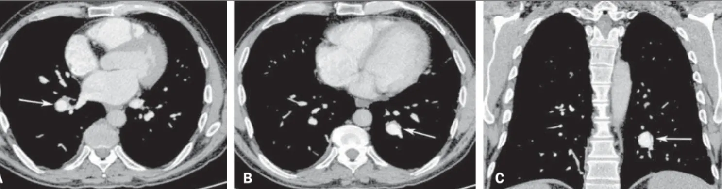
Figure 1. Contrast-enhanced computed tomography of the chest, with axial slices (A,B) and coronal slices (C), showing aneurysms in branches of the pulmonary arteries (arrows).
A
B
C
http://dx.doi.org/10.1590/0100-3984.2015.0048
Differential diagnosis of anterior sacral meningocele during the evaluation of post-hysterectomy pelvic collections
Dear Editor,
Here, we present the case of a 34-year-old woman who suf-fered postoperative pain and fever after subtotal abdominal hys-terectomy. Conventional radiography of the pelvis showed unilat-eral sacral curvature (Figure 1A). Computerized tomography (CT) performed on the second postoperative day revealed a dense locu-lated collection, interspersed with small air bubbles, in the pelvic cavity and a cyst-like formation with hypodense liquid content in the presacral space, communicating with the spinal canal, dislo-cating the rectum to the right (Figure 1B). A diagnosis of anterior sacral meningocele (ASM) was made, and the surgical team was informed of its coexistence with the postoperative pelvic collections. A new procedure was carried out to drain the collections, care being taken to avoid the sacculation caused by the ASM, which was vis-ible and palpable. Magnetic resonance imaging (MRI) was car-ried out in order to monitor the postoperative drainage and to char-acterize the malformation in greater detail (Figure 1C).
Various conditions related to anomalies in central nervous system development have been reported in Brazil(1–3). ASM is a rare form of spinal dysraphism, in which the meningeal sac herni-ates into the presacral space(4,5). It accounts for approximately 5% of all retrorectal masses and is more prevalent in women(6).
ASM can occur in isolation or in association with other con-genital abnormalities, such as urocon-genital malformations, anorectal malformations, lipoma, teratoma, epidermoid tumor, and dermoid cyst(7,8). Due to its occult nature, it is generally diagnosed in the second or third decades of life. It can be asymptomatic or present with nonspecific symptoms, such as constipation, urological symp-toms, and, in rare cases, neurological symptoms(9). The diagnos-tic investigation can include conventional radiography, ultrasound, CT, and MRI.
Letters to the Editor
Radiol Bras. 2016 Mai/Jun;49(3):199–204
204
can reveal herniation of the meningeal sac. MRI is the test of choice for evaluating ASM, because it creates high contrast between soft tissues, which makes it able to detect any commu-nication between the ASM and the subarachnoid space, and provides detailed information about other related abnormalities that might be present(4). However, when a communication with the subarachnoid space is narrow, MRI can fail to show it. In such cases, myelography with intrathecal injection of contrast can be necessary(12).
The differential diagnosis of ASM includes cystic lesions lo-cated in the presacral region(7,13): tumors of the gastrointestinal or genitourinary tract; epidermoid or dermoid cysts; aneurysmal bone cyst; hamartoma; hydatid cyst; lipoma; lymphangioma; perineural cyst; rectal duplication cyst; gynecologic tumors; ter-atoma; or teratocarcinoma. The most important means of estab-lishing the definitive diagnosis is detecting communication be-tween the cystic lesion and the subarachnoid space(11).
In the case in question, making the diagnosis of ASM was particularly important because the patient underwent laparotomy to drain the hemorrhagic collections in the pelvis. During that procedure, an unwarranted, inadvertent intervention in the men-ingocele could have had disastrous consequences.
REFERENCES
1. Simão MN, Helms CA, Richardson WJ. Magnetic resonance imaging findings of disc-related epidural cysts in nonsurgical and postoperative microdiscectomy patients. Radiol Bras. 2012;45:205–9.
2. Barros ML, Fernandes DA, Melo EV, et al. Central nervous system mal-formations and associated defects diagnosed by obstetric ultrasonogra-phy. Radiol Bras. 2013;45:309–14.
3. Holanda MMA, Rocha AB, Santos RHP, et al. Basal sphenoethmoidal
encephalocele in association with midline cleft lip and palate: case re-port. Radiol Bras. 2011;44:399–400.
4. Villarejo F, Scavone C, Blazquez MG, et al. Anterior sacral meningo-cele: review of the literature. Surg Neurol. 1983;19:57–71. 5. Sharma V, Mohanty S, Singh DR. Uncommon craniospinal dysraphism.
Ann Acad Med Singapore. 1996;25:602–8.
6. Beyazal M. An asymptomatic large anterior sacral meningocele in a patient with a history of gestation: a case report with radiological find-ings. Case Rep Radiol. 2013;2013:842620.
7. Shedid D, Roger EP, Benzel EC. Presacral meningocele: diagnosis and treatment. Semin Spine Surg. 2006;18:161–7.
8. McGregor C, Katz S, Harpham M. Management of a parturient with an anterior sacral meningocele. Int J Obstet Anesth. 2013;22:64–7. 9. Mohta A, Das S, Jindal R. Anterior sacral meningocele presenting as
constipation. J Pediatr Neurosci. 2011;6:40–3.
10. Kovalcik PJ, Burke JB. Anterior sacral meningocele and the scimitar sign. Report of a case. Dis Colon Rectum. 1988;31:806–7. 11. Naidich TP, Fernbach SK, McLone DG, et al. John Caffey Award.
Sonography of the caudal spine and back: congenital anomalies in chil-dren. AJR Am J Roentgenol. 1984;142:1229–42.
12. Manson F, Comalli-Dillon K, Moriaux A. Anterior sacral meningocele: management in gynecological practice. Ultrasound Obstet Gynecol. 2007;30:893–6.
13. Hemama M, Lasseini A, Rifi L, et al. A sacral hydatid cyst mimicking an anterior sacral meningocele. J Neurosurg Pediatr. 2011;8:526–9.
Ronaldo Garcia Rondina1, Richard Volpato1, Luiz Felipe Alves
Guerra1, Diego Lima Nava Martins1, Laís Bastos Pessanha1
1. Universidade Federal do Espírito Santo (UFES), Vitória, ES, Brazil. Mailing address: Dr. Ronaldo Garcia Rondina. Rua Júlio César de Oliveira Serrano, 135, Bl. 3, ap. 302, Mata da Praia. Vitória, ES, Brazil, 29065-720. E-mail: [email protected].
Figure 1. A: Conventional radiography showing unilateral sacral curvature (scimitar sacrum). Detail: Three-dimensional CT reconstruction for better characterization of the findings. B: CT with reformatting in the sagittal plane, showing a cyst-like formation with hypodense liquid content on the presacral space, apparently commu-nicating with the spinal canal, dislocating the rectum to the right. C: MRI with sagittal slices in a T2-weighted sequence, showing morphological and structural alteration with sacral dysraphism, in which the dural sac is insinuated toward the presacral space, with homogeneous cerebrospinal fluid contents. It is also possible to observe the tethered spinal cord and conus medullaris at the L3 level.