F
IELD USE OF THE
DOT-ELISA TEST FOR VISCERAL
LEISHMANIASIS IN HONDURAS
Bryce C. WuZton;2 MichaeZ G. Pappas;” MameZ Sierra, Ji. ;’ Rzlta Haikowski:2 Peter Tackson; and
Ram&v
cust02~03 ’
I
NTRODUCTION
Serologic tests are universally accepted as an indispensable tool for di- agnosing visceral leishmaniasis at health care facilities. Additionally, serodiagnosis is the most valuable single procedure in any surveillance program, since it per- mits rapid screening of large populations in remote areas without clinical evalua- tion, yields incidence data for the popu-
lation, and provides an efficient means of early case detection (1).
Several types of tests with a generally acceptable degree of reliability have been used as an aid in the diagnosis of visceral leishmaniasis. These include complement furation, both with a non- specific acid-fast bacterial antigen (2)
This article will also be published in Spanish in the Bo- /et&~ de h 0$&a Sanitam Panamericana, 1986, Department of Immunology, Walter Reed Army Insti- tute of Research, Washington, D.C. 20307, USA. The views of the authors do not purport to reflect the posi- tion of the U.S. Department of the Army or the U.S. Department of Defense.
’ Ministry of Public Health, Tegucigalpa, Honduras.
and with a homologous parasite antigen (3); direct agglutination (4); and indirect fluorescent antibody (5). Recently, the
enzyme-linked immunosorbent assay
(ELISA) utilizing soluble antigen has also
been found effective (G, 7).
However, efficient utilization of serodiagnostic tests in endemic areas is often difficult because the tests must be performed in a central laboratory, while the patients live in remote areas with poor means of transport and communi- cation. Consequently, serologic methods are generally available only for those pa- tients who come to a major hospital. In many cases this delays diagnosis for a hazardously long time, because the pa- tient is not seen at a medical facility with adequate diagnostic capabilities until he is gravely ill. The development of sero-
logic tests using dried blood specimens s
on filter paper has provided some im- -,
provement in this respect, particularly s
with regard to seroepidemiologic studies, 2
since it has simplified the taking and .$
processing of specimens and has elimi- nated the need for refrigeration during
2
transport. However, extremely few pri- ?I
mary health care facilities have this diag-
nostic service available from a central lab- 3
oratory.
A test that could be per- formed at the village level without any laboratory facilities whatever, a true “field test,” would be of inestimable value for improved patient care and for epidemiologic surveillance in endemic areas. At present, even when serodiag- nostic services are available, the delays encountered between taking the speci- men and obtaining a test result are con- siderable; and when a positive result is obtained from a patient who lives in a re- mote village that can be reached only by several hours travel on foot or horseback, the responsible physician or health au- thority is then faced with the problem of locating the patient and getting him to a medical facility that can confirm the di- agnosis, which causes still further delays. So if testing could be done at the time the specimen was originally taken, the test would be of much more value; and, in the case of positive reactions, arrange- ments for overcoming transport or simi- lar problems could be made immedi- ately, so as to get the patient to a health care facility for confirmatory biopsies and, if necessary, immediate treatment.
Such a field test should have
the following characteristics: (1) It
should require only a small volume of blood or serum (an amount obtainable without venipuncture). (2) It should be inexpensive. (3) It should utilize materi- als and equipment that are not easily \3
5
damaged or broken and that are easily transportable on foot or horseback. (4) It -
s should require no electricity or facilities
% that may be unavailable at villages in de-
.s veloping countries. (5) It should not re-
9, quire closely controlled temperatures.
Q ;i (6) It should be read visually. (7) It
: should produce results within a few
3
hours.
The Dot-ELISA technique of Pappas et al. (S-9) is a micro-ELISA utiliz-
148 ing a suspension of Leishmzniu donovani
promastigotes dotted onto nitrocellulose filter discs as antigen. It is visually read, inexpensive, and portable; in addition, its sensitivity and specificity are equiva- lent to the standard ELISA and superior to the CF test (9). Because this test seemed to meet the essential requirements and to be adaptable for field use, it was se- lected for a preliminary trial in a known endemic area in Honduras (1 O-l 1).
The isolated rural localities where the work was performed are repre- sentative of those where the disease is en- countered in the New World. The work thus constitutes a realistic trial under dif- ficult conditions such as are likely to be encountered in other endemic areas.
M
ATERIALS
AND METHODS
The Localities and Subjects Tested
Previously diagnosed visceral leishmaniasis patients who could be lo- cated provided a basis for selecting locali- ties and subjects for testing. The 305 per- sons tested were classified into four groups as follows:Group I consisted of nine former visceral leishmaniasis patients between four and eight years of age, who had two to seven years previously been parasitologically diag- nosed and treated with meglumine antimo- niate (Glucantime@).
one mother), who constituted a high-expo- sure group.
Gr0.Q III included a total of 244 children, ranging from six months to eight years of age, who resided in these and nearby localities and who were selected by random sampling done at clinics, schools, and homes. Grozlp IV consisted of subjects with other hemoflagellate infections, includ- ing three children with parasitologically diag-
nosed cases of extensive and long-standing cutaneous leishmaniasis involving multiple
disseminated lesions and four subjects with
serologically diagnosed Chagas’ disease.
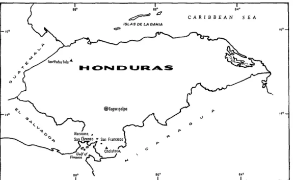
Most of the localities involved were in the Southwestern section of the country, in a known endemic area (Figure 1). Six were in the municipality of San Francisco de Coray and three were in the municipality of Choluteca. Various small localities in the municipalities of Na- caome and San Lorenzo were also repre- sented; and a few samples were also
taken in Colonia Estados Unidos in the Capital (Tegucigalpa), where one former patient infected in San Francisco de Coray was residing.
Blood Samples
Finger-prick blood samples
(see photograph) were collected in
0.1 ml capillary tubes, after which the open end was sealed. with plasticine modeling clay and the tube was immedi- ately shaken down to remove any air bubbles. The specimens were kept at the ambient temperature for one to four hours to hasten coagulation and clot re- traction, and were then stored on wet ice until processed.
FIGURE 1. A map of Honduras showing the location of the study areas.
I I I
880 840
CARIBBEAN SEA
One of the authors (Or. Sierra) taking a capillary blood specimen from a young child. Capillary blood specimens obtained by finger-prick are much easier to obtain from young children than are venous bbod specimens.
To extract the serum, the tube
was nicked with a file and broken off at
the bottom, just above the plasticine
plug. The clot usually adhered to the
plug and could be extracted intact while
the tube was held nearly horizontal, leav-
ing essentially clear serum in the tube.
Any red cells remaining did not interfere
with the test.
The sera were diluted with
phosphate-buffered
saline containing
1%
bovine serum albumin. The plastic
tip of a micropipette was inserted into
the end of the capillary tube, and
.Olml
of serum was removed. This was placed
in .15 ml of diluent in a microtiter plate
well (to achieve an initial
1:16 dilution),
and
.05ml of this latter solution was
transferred to a test well containing an
antigen disc (see below) and an equal
volume of diluent to provide a final dilu-
tion of
1: 32for the screening test.
Antigen Preparation
Dot-ELISA antigen prepara-
tion has been described in detail else-
where (8,. Antigen was prepared at Wal-
ter Reed from L. donovani (WR 311)
promastigotes, which were dotted on ni-
trocellulose filter paper in
1~1 volumes
(2.5x
10’parasites). These “dots” were
antigen discs were cut out with a punch as needed in the field.
The Test Procedure
The Dot-ELISA was performed in standard $6well flat-bottom microti- ter plates, essentially in the manner de- scribed by Pappas et al. (8), the only sig- nificant modification being the use of pH 7.4 phosphate-buffered saline (PBS)
in place of triethanolamine-buffered sa-
line (TESS). The PBS was transported as a
10X concentrate to conserve space and weight, and was diluted with distilled water in Honduras. Regarding storage in the field, the antigen, bovine serum al- bumin, control sera, conjugate, and sub- strate were kept in an insulated container and were protected against high temper- atures with wet ice or other coolants. However, the recommended tempera- ture of 4 “C was seldom maintained be- cause of high ambient temperatures that at times reached 30-34’ C. The tests were conducted at ambient temperatures that reached 2%30°C.
The shaking of the microtiter plates during the various reactions was accomplished manually by moving the plates with a rotary motion in small cir- cles on the surface of a table, instead of with the mechanized shaker described in the original account of the procedure. Antigen discs were cut from the nitrocel- lulose papers and were placed in the wells of the microtiter plates just before each test run.
The test procedure can be summarized as follows:
I) Blocking: Add .075 ml of 5%
BSA~ in PBS to each well containing an antigen disc. Shake the microtiter plate for one minute, incu- bate for 30 minutes, and aspirate off.
’ Bovine serum albumin, fraction V. Sigma Chemical Co., St. Louis, Missouri, USA.
2) Antibody reaction: Place 0.1 ml of test serum (diluted 1:32 in PBS-l% BSA) in well, shake for one minute, incubate for 30 minutes, as- pirate off.
3) Washing: Wash discs by adding 0.1 ml of .05 % NP405 in PBS. Shake for one min- ute and aspirate. Repeat twice, then allow the so- lution to stand for 10 minutes on the third wash. 4) Con+gate reaction: Add .05 ml of affinity-purified horseradish peroxidase-conju- gated goat antihuman IgG (gamma chain-spe- cific),” optimally diluted 1:lOO in PBS-l% BSA to
each well. Shake for one minute, incubate for 30 minutes, and aspirate. Wash three times, as above.
j) Substrate development: Add .05 ml of rhe precipitable substrate 4ClN’ to each test well, shake for one minute, incubate for 30 minutes, and aspirate. Wash two times in PBS
(without BSA) and a third time in distilled water. Aspirate and air-dry.
Microvolumes of sera and other reagents were measured with an adjustable-volume pipette8 with plastic disposable tips. However, the volumes of the wash solutions were not critical, and were not precisely measured; they were delivered into the wells with a Pasteur pi- pette, estimating the 0.1 ml required for washing. All the solutions were with- drawn from the wells with a Pasteur pi- pette connected to a 20 ml syringe by a length of quarter-inch diameter rubber surgical tubing, an arrangement that fa- cilitated aspiration of the wells in rapid succession.
’ Nonidet P-40 (a nonionic surfactant), Calbiochem- Behring Corp., La Jolla, California, USA.
’ Kirkegaard and Perry Laboratories, Inc., Gaithersburg,
Maryland, USA.
’ 4-chloro-1-naphtol, a peroxide-chromogenic precipita- ble substrate (Kirkegaard and Perry Laboratories), was prepared by adding equal volumes of substrate solution and then diluting 1:3 in PBS.
’ Gilson pipetman (.OOl ml - 0.2 ml).
RE
SULTS
Positive reactions appeared as blue-purple dots on the white nitrocellu- lose discs. In some instances the dots be- came more distinct as the filter-paper dried. There were virtually no equivocal reactions, the dots being either present or absent. Positive control sera were posi- tive in all the runs, while negative con- trol sera and reagent controls were unre- active.
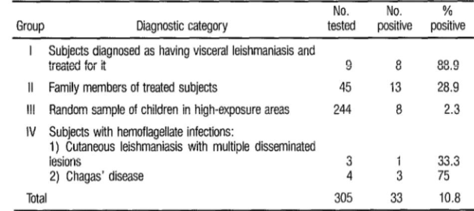
There were significant differ- ences in the rates of positivity of the four
groups screened (Table 1). In Group I,
eight of nine (88.9%) of the parasitolo- gically diagnosed visceral leishmaniasis patients reacted positively at the screen- ing dilution of 1:32. And in Group II, the family members with presumably in-
creased exposure to infection, 13 of 45
(28.9 % ) reacted positively. In contrast, only 8 of 244 (2.3 % ) of the apparently healthy children from the general popu- lation in the area reacted positively.
A noteworthy number of pos- itive responses were also obtained from the subjects with related protozoan in- fections (Group IV). That is, three out of four subjects with serologically diag- nosed cases of Chagas’ disease and one out of three children with extensive long- standing disseminated lesions of cutane- ous leishmaniasis also yielded a positive response.
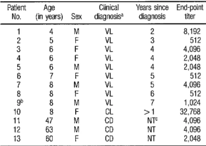
Sera from the visceral leish- maniasis patients in Group I, as well as sera from the cutaneous leishmaniasis and Chagas’ disease patients in Group IV that responded positively, were then fully titrated to determine their end- points (Table 2). All of the nine previ- ously diagnosed and treated visceral leishmaniasis patients in Group I were reactive at titers that ranged from 1: 5 12
to 1:8,192. The one Group I serum that
was inexplicably nonreactive in the
screening test yielded an end-point titer of 1: 1,024 when titrated. The reciprocal titers exhibited by sera of Group IV pa- tients with other hemoflagellate infec- tions ranged from 2,048 to 32,768. These latter titers were surprisingly high, especially since all had yielded titers of only 5 12 with homologous species anti- gen.
TABLE 1. Results of Dot-ELBA testing of the four groups screened far visceral leishmaniasis in Honduras.
No. No. % Group Diagnostic category tested positive positive
I Subjects diagnosed as having visceral leishmaniasis and
treated for it 9 8 88.9 II Family members of treated subjects 45 13 28.9 Ill Random sample of children in high-exposure areas 244 8 2.3 IV Subjects with hemoflagellate infections:
1) Cutaneous leishmaniasis with multiple disseminated
lesions 3 1 33.3
2) Chagas’ disease 4 3 75
TABLE 2. End-point titers of sera from Group I and Group IV subjects that gave a positive screening test reaction.
Patient Age Clinical Years since End-point No. (in years) Sex diagnosisa diagnosis titer
1 4 M VL 2 8,192
2 5 VL 3 512
3 4
: F VL 4 4,096 F VL 4 2,048 5 6 M VL 4 2,048 6 7 F VL 5 512 7 8 M VL ; 4.096
8 8 VL 512
gb 8 ; VL 7 1,024 10 8 F >I 32,768 11 47 M c”o’ NTC 4,096 12 63 M CD NT 4,096 13 60 F CD NT 2,048
d VL=visceral leshmaniasls: CL=cutaneous leishmanlasls; CD=Chagas’ disease b This subject yielded a negative reactjon upon mitral Dotwsn screening at a dllutlon of 1 32 c NT=Not treated.
D
ISCUSSION
AND CONCLUSIONS
The localities where this trial was carried out appear representative of those where visceral leishmaniasis is found in the New World, and the condi- tions of the trial constituted a realistic testing under difficult conditions likely to be encountered in other endemic areas. The antigen discs and reagents proved to be stable over extended pe- riods of time, and high ambient temper- atures (up to 28°C) prevailing during the assay did not appear to adversely affect the results.
The degree of nonspecificity of the test, as indicated by the positive reactions seen in sera from one subject with multiple disseminated lesions of cu- taneous leishmaniasis and from three of four subjects with Chagas’ disease, is probably about the same as for any sero- logic test utilizing promastigotes as anti- gen. This specificity could possibly be
improved by use of amastigotes as anti- gen, an improvement that has been demonstrated in the indirect fluorescent antibody test for cutaneous leishmaniasis (13). However, attaining some improved specificity this way would not be of great practical importance, since the purpose of serologic screening is to select individ- uals for definitive clinical evaluation. In the case of both cutaneous leishmaniasis and Chagas’ disease, early diagnosis is beneficial, and the differential diagnosis is not difficult. Hence, from the point of view of primary medical care in develop- ing countries, the incidental diagnosis of Chagas’ disease should be regarded as a serendipitous advantage rather than a drawback.
second antibody, which has been shown to reduce the number and titers of false positive reactions due to related proto- zoan diseases and interfering hyperglob- ulinemia (primarily IgM) observed in populations residing in the tropics (9). It is not possible at this time to determine whether the positive reactions in Group III represented clinically inapparent in- fections with visceral leishmaniasis, or were due to subclinical Chagas’ disease or other related protozoan infections.
The nine Group I subjects (all with past visceral leishmaniasis infections that had been treated two to seven years before) had titers that were considerably lower than those encountered in active disease cases-the latter sometimes rang ing up to 1:524,288 (8). These lower ti- ters probably resulted from declines in antibody levels after treatment, although the temporal response of antibodies to effective treatment is not yet known. The possibility that such long-lasting persis- tence of antibody indicates treatment failure cannot be ruled out.
A definitive diagnosis of vis- ceral leishmaniasis can only be made by demonstration of the parasites, either by direct examination or by culture, usually of biopsy material from the bone marrow or spleen. Although splenic biopsy is now accepted as a safe and reliable proce- dure for diagnosis and assessing treat- ment efficacy (14), it is not entirely with-
% out risk and normally should not be
2 done outside a medical facility. It also re-
-
s quires skilled, medically qualified opera-
% tors, and should be performed only
x when there is a strong clinical suspicion
‘4 u of infection. %
Q provides a means of screening that per- Within this context, serology mits this relatively time-consuming and
154
expensive biopsy procedure to be limited to those patients with a high probability of infection. Additionally, serodiagnosis is undoubtedly the most valuable single procedure in any program for surveil- lance of visceral leishmaniasis, since it permits screening of populations in re- mote areas with coverage of a large num- ber of subjects in a short time without need of clinical evaluation.
The Dot-ELISA technique ap- pears to be a practical and economical one for rapid screening at the village level. Only .05 ml of patient serum is re- quired, an amount easily acquired by finger-prick. The test is cheap, costing less than five cents (US) per test well, and can be performed by personnel without a high level of technical training. All the material required can be transported eas- ily on foot or horseback, and no electric- ity or facilities not readily available at the village level are required. We believe that this test can be of great value in areas en- demic for visceral leishmaniasis anywhere in the world.
A
CKNOWLEDGMENTS
S
UMMARY
The dot enzyme-linked im-
munosorbent assay (Dot-ELISA), a rapid, visually read microtechnique for serodi- agnosis of human visceral leishmaniasis, was field-tested in a known endemic area in the Republic of Honduras. Of 305 in- dividuals screened at a serum dilution of
1: 32, positive reactions were observed in eight of nine parasitologically diagnosed visceral leishmaniasis patients who had received treatment, 13 of 45 family members of patients (a high-risk group), and eight of 244 randomly selected chil- dren in the endemic area. Cross-reactions were observed in one of three children with parasitologically confirmed cutane- ous leishmaniasis and three of four adults serologically positive for Chagas’ disease. End-point titrations performed on the visceral leishmaniasis sera gave reciprocal
titers ranging from 5 12 to 8,192, which are lower than those usually encountered in untreated active cases.
This test does not require electricity, and all materials are easily transportable on foot. It is rapid, simple to perform, and inexpensive, yet sensi- tive and relatively specific under field conditions. It could prove to be a valu- able tool for primary health care facilities
and for epidemiologic surveys in the
many endemic areas where no serologic
testing capability currently exists.
l&E
FERENCES
1 Lobel, H. O., and I. G. Kagan. Seroepide-miology of parasitic diseases. Annu Rev Micro- biol32:329-347, 1378.
Pellegrino, J.? 2. Brener, and U. M. Santos. Complement fuation test in kala-azar using Mycobuctehm bzctyrzkm antigen. J Pa7aritol 44645, 1948.
Hockmeyer, W. T., B. W. Wellde, C. L. Sabwa, D. H. Smith, P. H. Rees, and P A. A. Kager. A complement fmation test for visceral leishmaniasis using homologous parasite anti- gen. Ann pop Med Parasitol 781489-493, 1984.
Allain, D., and I. G. Kagan. A direct aggluti- nation test for leishmaniasis. Am J Fop Med Hyg 241232-236, 1975.
Duxbury, R. E., and E. H. Sadun. Fluorescent antibody test for serodiagnosis of visceral leishmaniasis. Am J Fop Med Hyg 13:525- 529, 1964.
6 Hommel, M., W. Peters, J. Ranque, M. Ouihci. and G. Lanotte. The micro-ELISA tech- Gque in the serodiagnosis of visceral leishma- niasis. Ann Trap Mea’ Parasitol 72:213-218, 1978.
Edrissian, Gh.H., and P Darabian. A compar- ison of enzyme-linked immunosorbent assay and indirect fluorescent antibody test in the sero-diagnosis of cutaneous and visceral leishmaniasis in Iran. Tram R SOG ??op Med Hyg 73~289-292, 1979.
Pappas, M. G., R. Hajkowski, and W. T. Hockmever. Dot enzvme-linked immunosor- bent assay (Dot-ELISAj: a micro technique for the rapid diagnosis of visceral leishmaniasis. J ImmzlnolMethodr 64:205-214, 1983. 9 Pappas, M. G., R. Hajkowski, and W. T.
Hockmeyer. Standardization of the dot en- zyme-linked immunosorbent assay (Dot-ELISA) for human visceral leishmaniasis. Am J Fop MedHyg 33:1105-1111, 1984.
10 Pappas. M. G., R. Haikowski, L. T. Cannon Sr.: and W. T. Hockmeyer. Dot enzyme-linked immunosorbent assay (Dot-ELISA): Compari- son with standard ELISA and complement fua- tion assays for the diagnosis of human visceral leishmaniasis. Vet Parado/ 14:239-249, 1984. Navin, T. R., M. Sierra, R. Custodio: F. Steurer, C. H. Porter, and T. Ruebush. Epide- miologic study of visceral leishmaniasis in Honduras. Am J i?op Med Hyg 34:1069- 1075, 1985.
13 Walton, B. C., W. H. Brooks, and I. Arjona. Serodiagnosis of American leishmaniasis by
14 Rees, F? H., P A. A. Kager, B. T Welide, and the indirect fluorescent antibody test. Am J
W. T. Hockmeyer. The response of Kenyan ??op MedHyg 21:296-299, 1972.