RESUMO.- [Avaliação do eletrocardiograma em cães com leishmaniose visceral.] Devido à identificação prévia de miocardite e arritmias tanto em seres humanos quanto em cães portadores de leishmaniose, 105 cães com sorologia positiva para tal enfermidade tiveram seus eletrocardio -gramas avaliados quanto à presença de distúrbios do ritmo e alterações nas ondas eletrocardiográficas. Poucas varia -ções expressivas foram observadas, exceto parada sinusal, bloqueio de ramo direito e complexos atriais prematuros em 14,3%, 4,8%, e 4,8% dos pacientes estudados, respecti -vamente. Ademais, a análise do ECG demonstrou alterações sugestivas de sobrecarga do átrio e do ventrículo esquer -dos e hipóxia do miocárdio. Embora o comprometimento cardíaco em cães com leishmaniose já tenha sido demons -trado anteriormente, apenas uma pequena parte dos cães avaliados apresentou alteração no eletrocardiograma, não sendo possível correlacionar com a ocorrência de miocar -dite nesta pesquisa.
TERMOS DE INDEXAÇÃO:Leishmaniose visceral, eletrocardiogra -fia, arritmias, caninos, protozoário, eletrofisiologia.
Assessment of the electrocardiogram in dogs with visceral
leishmaniasis
1Marlos G. Sousa2*, Roberta Carareto2, Jeanna G. Silva2 and Juliana Oliveira2
ABSTRACT.- Sousa M.G., Carareto R., Silva J.G. & Oliveira J. 2013. Assessment of the electro-cardiogram in dogs with visceral leishmaniasis.Pesquisa Veterinária Brasileira 33(5):643-647. Escola de Medicina Veterinária e Zootecnia, Universidade Federal do Tocantins, Rodov.
BR-153 Km 112, Araguaína, TO, 77800-970, Brazil, E-mail: [email protected]
As myocarditis and arrhythmias have been shown to occur in both human beings and dogs with leishmaniasis, electrocardiograms of 105 dogs serologically positive for this di -sease were assessed for rhythm disturbances and changes in ECG waves. A few expressive alterations were seen, including sinus arrest, right bundle branch block, and atrial prema -ture beats in 14.3%, 4.8%, and 4.8% of the studied subjects, respectively. Also, the analysis of ECG waves showed changes suggestive of left atrium and ventricle enlargements, and myocardial hypoxia in some animals. Although cardiac compromise has been previously reported in dogs with leishmaniasis, only a small subset of dogs showed any alteration in the electrocardiogram, which cannot support the occurrence of myocarditis in this inves -tigation.
INDEX TERMS: Visceral leishmaniasis, electrocardiography, arrhythmias, canines, protozoan, elec -trophysiology.
1 Received on October 2, 2012.
Accepted for publication on March 18, 2013.
2 Escola de Medicina Veterinária e Zootecnia, Universidade Federal do
Tocantins (UFT), Rodov. BR-153 Km 112, Campus EMVZ/UFT, Araguaína, TO 77800-970, Brazil. *Corresponding author: [email protected]
INTRODUCTION
Visceral leishmaniasis is a generalized infection of the mononuclear phagocyte system, caused by the protozoan
Leishmania chagasi. It is known to have a high viscerotro -pism and its transmission results from bites of phleboto -mine sand flies containing the infective promastigote form. Although different modalities of the disease have been do -cumented, the infection is presented clinically by irregular fever, anemia, hepatosplenomegaly, intestinal disorders, hemorrhages, weight loss, alopecia or hypotrichosis, and several other associated signs (Cimerman & Cimerman 2001).
Because of the multisystemic nature of leishmaniasis, the parasite may infect several corporal tissues in human beings, dogs, and many other animals. One of the most important features of visceral leishmaniasis in humans remains on the compromise of organs related to the mo -nonuclear phagocyte system where the parasite multiplies, leading to the organ enlargement in response to infection (Cimerman & Cimerman 2003).
-re enough, p-resent edema, leukocyte infiltration, focal ne -crosis, and proliferation of Anitschkow myocytes. All these alterations were once named as “reactional myocardium” (De-Morais et al. 1988, Puerto-Alonso et al. 2006). In acute form, myocarditis generally results in an unexplained de -velopment of arrhythmias or cardiac failure after a recent episode of infectious disease. Its diagnosis may be equivo -cal because of the absence of clini-cal or clincopathologi-cal findings specific for myocarditis (Kittleson & Kienle 1998). When myocarditis is present, several unspecific changes on the electrocardiogram (ECG) are generally documented, including, but not limited to, changes in the ST-segment, in the amplitude of T wave and QRS complex and the occur -rence of atrioventricular conduction disturbances. Besides that, signs of hypokinesia of the myocardial walls, altered myocardium echogenicity, or pericardial effusion can be detected in the echocardiographic examination (De-Morais et al. 1988, Kittleson & Kienle 1998, Torrent et al. 2005).
Because of the increasing number of dogs with leish -maniasis and prolonged pauses and/or evidence of ar -rhythmias on auscultation, this study was aimed to assess cardiac rhythm and the ECG parameters in dogs with con -firmed visceral leishmaniasis.
MATERIALS AND METHODS
One-hundred-five mature dogs of either sex (55 males and 50 females), with mean weight of 9.8 kg (2.6 to 31.3 kg), serologi -cally positive for visceral leishmaniasis by indirect immunofluo -rescence and Elisa with title higher than 1:40, were prospectively included in the study. All animals presented at least three of the following clinical signs suggestive of leishmaniasis: hypotrichosis, periocular alopecia, weight loss, onycogriphosis, skin lesions, and either hyporexia or anorexia, besides lymphadenomegaly and/or hepatosplenomegaly detected at physical examination.
Standard 6-lead computer-based ECGs (leads I, II, III, aVR, aVL, aVF) were recorded for three minutes in all dogs positioned in ri -ght lateral recumbency with a speed of 50 mm/s, calibrated for 10 mm/mV. Electrocardiograms were evaluated for cardiac rhythm, P wave duration (Pms) and amplitude (PmV), PR and QT intervals duration, QRS complex duration, R wave amplitude, and heart rate (HR). Also, ST segment was assessed regarding its level, as well as the T wave regarding its polarity (Tpol) and amplitude (TmV). Lead II was chosen to assess such parameters, whereas the mean elec -trical axis (MEA) was calculated through the net QRS amplitude of leads I and III.The duration of both P wave and QRS complex was compared with the normal range proposed by Wolf et al. (2000), while the comparison of the remaining parameters was accompli -shed against the reference table of Tilley & Burtnick (2004).
Mean and standard deviations were calculated for the para -metric data, while the non-para-metric results were grouped by percentage of occurrence. Chi-square test was applied to contin -gency tables created to check for differences in the occurrence of arrhythmias according to gender (males vs. females) and body weight (up to 5 kg, from 5.1 to 15 kg, and greater than 15.1 kg). An unpaired T test was calculated to investigate differences in the parametric results between males and females, as well as an one --way analysis of variance for the several variables according to body weight. The software Prism for Windows (Prism v.5.04, Gra -phpad Software, San Diego, CA, USA)3 was used for all analyses,
and significance was set at P < 0.05.
RESULTS
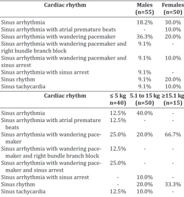
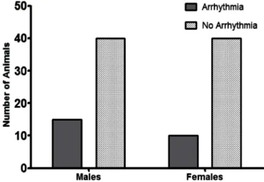
Several breeds were represented in the study, including Pinscher (6.7%), English Cocker Spaniel (2.8%), Boxer and German Shepherd (1.9% each), Dalmation, Lhasa-Apso, and Rottweiller (0.9% each), but the majority of the dogs were mixed-breed animals (82.9%). Regarding gender and body weight, 52.4% were male and 47.6% were female, whereas 38.1% weighed less than 5 kg, 47.6% weighed between 5.1 and 15 kg, and the minority (14.3%) weighed over 15.1 kg. The distribution of rhythms and conduction disturbances in dogs with confirmed visceral leishmaniasis is represen -ted on Table 1. Some important findings are shown in Fi -gure 1. For the purpose of creating a contingency table to allow chi-square analyses to be performed, only abnormal rhythms were considered as arrhythmias, which included atrial premature beats, bundle branch blocks, and sinus ar -rest. The remainder of rhythms documented in this inves -tigation was considered as non-arrhythmia. Therefore, sta -tistical significance was not attained by chi-square for the occurrence of arrhythmias according to gender (Figure 2, P = 0.3822). Sinus arrhythmia with wandering pacemaker was more prevalent in males, and females more frequently presented sinus arrhythmia. However, differences regar
-3 Prism v.5.04, Graphpad Software, San Diego, CA, USA.
Table 1. Percent distribution of cardiac rhythms and conduction disturbances identified in dogs with confirmed
visceral leishmaniasis
Cardiac rhythm (n=105) 28.6% Sinus arrhythmia with wandering pacemaker 23.7% Sinus arrhythmia
14.3% Sinus rhythm
9.5% Sinus arrhythmia with wandering pacemaker and sinus arrest 9.5% Sinus tachycardia
4.8% Sinus arrhythmia with wandering pacemaker and right bundle branch block
4.8% Sinus arrhythmia with atrial premature beats 4.8% Sinus arrhythmia with sinus arrest
Cardiac rhythm Males Females
(n=55) (n=50)
Sinus arrhythmia 18.2% 30.0% Sinus arrhythmia with atrial premature beats - 10.0% Sinus arrhythmia with wandering pacemaker 36.3% 20.0% Sinus arrhythmia with wandering pacemaker and 9.1% right bundle branch block
Sinus arrhythmia with wandering pacemaker and 9.1% 10.0% sinus arrest
Sinus arrhythmia with sinus arrest 9.1% Sinus rhythm 9.1% 20.0% Sinus tachycardia 9.1% 10.0%
Cardiac rhythm ≤ 5 kg 5.1 to 15 kg ≥15.1 kg
n=40) (n=50) (n=15)
Sinus arrhythmia 12.5% 40.0% -Sinus arrhythmia with atrial premature 12.5% - beats
Sinus arrhythmia with wandering pace- 25.0% 20.0% 66.7%
maker
Sinus arrhythmia with wandering pace- 12.5% - maker and right bundle branch block
Sinus arrhythmia with wandering pace- 25.0% - maker and sinus arrest
-ding cardiac rhythms were found when taking body weight into account, with chi-square test demonstrating statistical significance (Figure 3, P < 0.0001) in this setting. Arrhyth -mias and conduction disturbances were significantly more frequent in dogs weighting less than 5 kg.
Increased Pms was documented in 33.3% of the dogs, whereas 26.7% showed increased duration of the QRS complex. Also, spiked T waves and ST segment shifts were demonstrated in approximately 19.0% and 9.5%,of the stu -died animals, respectively. Mean and standard deviations of the numeric parameters are listed on Table 2. The com -parison of these parameters considering gender showed a significant difference (P = 0.0497) for QT interval only.
When body weight was considered, comparison was also significantly different (P = 0.0259) for R wave amplitude. The distribution of the non-parametric data is shown on Table 3.
Fig.1. Representative ECG findings in dogs with visceral leish -maniasis. (a) Sinus arrhythmia; (b) and (c) sinus arrest. The first marked RR interval has 440 msec, with the next one being more than two-fold longer at 947 msec. Wandering pa -cemaker is also present – notice the varying amplitude of P waves; (d) incomplete right bundle branch block; (e) atrial premature beat (arrow). Also seen are the spiked T waves (All ECG tracings are shown in lead II, 50 mm/s, 1cm = 1mV).
Fig.2. Occurrence of arrhythmias according to gender in dogs with visceral leishmaniasis. Chi-square test (P=0.3822) attained no statistical significance when only atrial premature beats, bundle branch blocks, and sinus arrest were considered as ar -rhythmias or conduction disturbances.
Fig.3. Occurrence of arrhythmias according to body weight in dogs with visceral leishmaniasis. Statistical significance was recorded for chi-square test (P<0.0001) when comparing the occurrence of atrial premature beats, bundle branch blocks, and sinus arrest grouped as arrhythmias or conductions dis -turbances versus the remainder of cardiac rhythms.
Table 2. Mean and standard deviation of the ECG parametric parameters recorded in dogs with confirmed visceral leishmaniasis
Parametera All dogs (n=105) Males (n=55) Females (n=50) ≤ 5 Kg (n=40) 5.1 to 15 Kg (n=50) ≥ 15.1 kg (n=15)
Pms 46.9 ± 5.1 46.9 ± 6.1 46.9 ± 4.0 49.1 ± 3.4 44.2 ± 5.5 50.0 ± 3.0
PmV 0.20 ± 0.06 0.18 ± 0.05 0.22 ± 0.06 0.21 ± 0.08 0.20 ± 0.04 0.20 ± 0.07
PR 87.7 ± 12.8 89.7 ± 15.6 85.8 ± 9.3 85.9 ± 14.4 86.8 ± 11.4 96.7 ± 14.2 QRS 58.6 ± 10.7 60.3 ± 14.4 56.8 ± 4.1 59.2 ± 17.0 56.7 ± 3.6 63.3 ± 3.5
R 1.02 ± 0.46 1.13 ± 0.37 0.90 ± 0.53 0.70 ± 0.30C 1.19 ± 0.47D 1.32 ± 0.26D
QT 198.8 ± 21.9 207.6 ± 25.7A 189.0 ± 11.7B 195.9 ± 22.2 198.0 ± 21.1 209.0 ± 30.0
HR 111.7 ± 30.6 101.9 ± 28.3 122.4 ± 30.7 114.9 ± 39.3 113.7 ± 26.0 96.3 ± 22.4
a P
ms: duration of P wave (msec); PmV = amplitude of P wave (mV); PR = duration of PR interval (msec); QRS = duration of QRS com
-plex (msec); R = amplitude of R wave (mV); QT = duration of QT interval (msec); HR = heart rate (bpm). A,B Significant difference
DISCUSSION
The majority of the dogs enrolled in this study presented sinus arrhythmia with wandering pacemaker, which is con -sidered a normal cardiac rhythm for canines (Tilley & Bur -tnick 2004). However, an expressive amount of dogs were shown to have sinus arrest, which may explain the pauses frequently auscultated in these animals (Ohara 2007), but is uncommonly associated with myocarditis (Woolley et al. 2007). Paroxysmal sinus arrest may be a normal finding in brachycephalic dogs, attributable to the increase in vagal tone that accompanies inspiration, leading to an exaggera -ted sinus arrhythmia. Other causes of sinus arrest might be cervical neoplasms irritating the vagus nerve, pathologic conditions of the atria, as well as drug toxicities and elec -trolyte imbalance (Tilley & Burtnick 2004). Although hemo -dynamically insignificant (Andrea et al. 2002), right bundle branch blocks were detected in some dogs. On the contrary, atrial premature beats were recorded in a few subjects and hemodynamic compromise may be attributable to such ar -rhythmia, especially when it occurs in sequence leading to atrial tachycardia (Kittleson & Kienle 1998, Tilley & Burtni -ck 2004, Artese 2007). In a study evaluating the cardiac in -jury owing to visceral leishmaniasis in people up to 19 years old, Diamantino (2010) found premature beats to occur in 44.9% of the studied subjects. However, that investigation used a holter recording to assess cardiac rhythm over a lon -ger period of time, and also documented ventricular prema -ture beats, which were not found in the animals of this study. The difference documented in QT interval between males and females is likely attributable to heart rate, and, despite not statistically significant, was lower in males than females. This parameter is inversely related to QT interval (Kittleson & Kienle 1998) and represents the total electri -cal activity of the ventricles, computing the time from the beginning of ventricular depolarization to its final repo -larization (Tilley & Burtnick 2004). Therefore, apart from heart rate, it might be influenced by factors that interfere with ventricular electrical activity, including electrolyte im -balance, which is not an uncommon finding in dogs with visceral leishmaniasis. Because no ventricular arrhyth -mias or conduction disturbances have been recorded in this investigation, it is unlikely that any abnormality actu -ally existed within the ventricles of these animals. None -theless, the absence of a more detailed analysis of cardiac rhythm, including a holter evaluation over 24 hours, does not allow excluding paroxysmal ventricular arrhythmias. Also, the dogs did not undergo an echocardiogram to exclu
-de any other cardiac condition known to cause ventricular arrhythmias. Concerning the difference documented in R wave amplitude among animals with varying body weight, it is supposedly ascribed to the amount of ventricular mass, which is greater in larger animals, therefore augmenting R waves as the body weight increases (Artese 2007), as ob -served in the present study.
One of the main problems with visceral leishmaniasis relies on its long incubation period before the appearance of clinical disease in dogs, which can last from three mon -ths to seven years, resulting in many infected dogs being asymptomatic carriers that do not exhibit disease (Miró et al. 2008). Nevertheless, the parasite may cause inflam -mation of the heart and vessels, resulting in myocarditis (Torrent et al. 2005). Interestingly, a recent investigation demonstrated that in spite of the minimum-to-moderate inflammatory reaction characterized by mononuclear, pe -rivascular, and intermuscular infiltrates being observed in both symptomatic and asymptomatic dogs on histopatho -logical analysis of the heart, clinical examination of these animals have not disclosed any manifestations ascribed to cardiac compromise (Alves et al. 2010), in agreement with the animals of this study, which also showed no clinical signs that could be related to cardiac injury.
In fact, there are not many reports on literature regar -ding myocardial inflammation caused by Leishmania spp.
in dogs (Büngener & Mehlitz 1977, Torrent et al. 2005, Ló -pez-Peña et al. 2009). Myocarditis itself may be caused by several other conditions, including toxins, direct invasion of myocardial tissue, and immune-mediated damage, resul -ting in focal or diffuse involvement of the cardiac tissue. Its clinical manifestations depend on the extent of the lesions, but generally are characterized by varying rhythm dis -turbances (Kittleson & Kienle 1998), including malignant arrhythmias causing sudden death (Woolley et al. 2007). As shown previously, this investigation only recorded mi -nor rhythm disturbances, although the electrocardiograms disclosed changes suggestive of left atrium and ventricle enlargements and hypoxia, as determined by enlarged P waves and QRS complexes, and spiked T waves, according to comparisons with the normal standards for similarly --sized dogs (Wolf et al. 2000, Tilley & Burtnick 2004, Ar -tese 2007). In a recent report from Spain, the ECG of a dog found on necropsy to have severe cardiac compromise due to leishmaniasis just showed low-amplitude QRS comple -xes with normal cardiac rhythm (López-Peña et al. 2009).
Our ECG findings, however, are not necessarily related to pathological processes within the myocardium, especially when one considers the lack of sensibility and specificity of the electrocardiogram to detect chamber enlargement ba -sed on the temporal and potential assessment of its waves (Kittleson & Kienle 1998, Andrea et al. 2002). Nonetheless, it should be stressed that the left axis shift documented in some dogs may be ascribed to left heart enlargement itself (Tilley & Burtnick 2004), which obviously needed echocar -diographic confirmation.
Although the histopathological evaluation of the hearts could be rewarding in the animals where ECG abnorma -lities were recorded, it was not the purpose of this inves -Table 3. Percent distribution of the ECG non-parametric data
recorded in dogs with confirmed leishmaniasis
Parametera All dogs (n=105)
ST 4.8% ST elevation; 4.8% ST depression
Tpol 57.1% positive T; 38.1% biphasic T; 4.8% negative T TmV 19.0% T wave amplitude greater than 25% of R wave
tude
MEA 61.9% 60-80degrees; 23.8% 80-100 degrees; 9.5% lower than 40 degrees; 4.8% 40-60 degrees
a ST = ST segment; T
pol = polarity of T wave; TmV = amplitude of T wave; MEA
tigation. Other studies in dogs with this disease have de -monstrated a myocardium with a dense accumulation of macrophages between the muscle fibers, as well as areas of cardiac muscle atrophy, degeneration and loss of cardio -myocytes (Torrent et al. 2005, López-Peña et al. 2009). Also, Wooley et al. (2010) reported a case of a fainting dog, who -se holter analysis documented episodes of no discernable electrical cardiac activity. The post-mortem examination of the heart showed evidence of myocarditis, with increased inflammatory cells and fibrous tissue throughout the atrial myocardium. Although a cause could not be demonstrated in that case, the sinus arrest was ascribed by the authors to chronic myocarditis and myocardial hemorrhage within the atria, with fibrosis adjacent to the sinoatrial node.
The main limitation of this study relies on the absence of a histopathological analysis of the hearts, particularly in those animals where conduction disturbances were identi -fied on ECG tracings. Also, echocardiographic and cardiac biomarker analysis could bring additional information for the characterization of abnormalities within the heart of animals with this disease. Even though alterations in car -diac rhythm have not been detected in the majority of the dogs, some of the findings, such as atrial premature beats, might be attributable to a pathological process within the atrial myocardium. Nevertheless, no other changes compa -tible with this possibility were further detected.
Although cardiac compromise has been previously re -ported in dogs with leishmaniasis, only a small subset of dogs included in this study presented minor ECG altera -tions, with the most important being atrial premature beats. Sensitivity and specificity of the electrocardiogram in de -tecting cardiac abnormalities cannot be calculated because a gold standard method, such as histopathological analysis, was not performed. Our results can neither support nor rule out the occurrence of myocarditis in dogs with visceral leishmaniasis, since paroxysmal arrhythmias could exist and ECG tracings have been recorded for just a few minutes in each animal.
REFERENCES
Alves G.B.B., Pinho F.A., Silva S.M.M.S., Cruz M.S.P. & Costa F.A.L. 2010. Car -diac and pulmonary alterations in symptomatic and asymptomatic dogs infected naturally with Leishmania (Leishmania) chagasi. Braz. J. Med.
Biol. Res. 43:310-315.
Andrea E.M., Atié J. & Maciel W. 2002. Eletrocardiograma na criança e no feto. In: Goldwasser G.P. (Ed.), Eletrocardiograma orientado para o clíni -co. 2ª ed. Revinter, Rio de Janeiro.
Artese J.M. 2007. Principios de electrocardiografía veterinária, p.105-120. In: Belerenian G., Mucha C.J., Camacho A.A. & Grau J.M. (Eds), Afecciones cardiovasculares en pequeños animales. 2ª ed. Inter-Médica, Buenos Aires.
Büngener W. & Mehlitz D. 1977. Atypisch verlaufende Leishmania donova-ni infektion bei hunden. Tropenmed. Parasitol. 28:175-180.
Cimerman B. & Cimerman S. 2001. Parasitologia humana e seus funda -mentos gerais. 2ª ed. Atheneu, São Paulo, 375p.
Cimerman S. & Cimerman B. 2003. Medicina Tropical. Atheneu, Rio de Ja -neiro. 690p.
De-Morais C.F., Duarte M.I., Corbett C.E. & Reis M.M. 1988 Morphologic cardiac changes in human visceral leishmaniasis: study based on 16 ne -cropsy cases. Arq. Bras. Cardiol. 51:441-445.
Diamantino T.C.C. 2010. Leishmaniose visceral: avaliação das repercus -sões cardiovasculares secundárias à doença e ao tratamento em crian -ças e adolescentes tratadas com três esquemas terapêuticos. Tese de Doutorado, Universidade Federal de Minas Gerais, Belo Horizonte, MG. 221p.
Font A., Durall N., Domingo M., Closa J.M., Mascort J. & Ferrer L. 1993. Car -diac tamponade in a dog with visceral leishmaniasis. J. Am. Anim. Hosp. Assoc. 29:95-100.
Kittleson M.D. & Kienle R.D. 1998. Small Animal Cardiovascular Medicine. Mosby, St Louis. 603p.
López-Peña M., Alemañ N., Muñoz F., Fondevila D., Suárez M.L., Goicoa A. & Nieto J.M. 2009. Visceral leishmaniasis with cardiac involvement in a dog: a case report. Acta Vet. Scand. 51:20-22.
Miró G., Cardoso L., Pennisi M.G., Oliva G. & Baneth G. 2008. Canine leish -maniosis: new concepts and insights on an expanding zoonosis. Part two. Trends Parasitol. 24:371-377.
Ohara V.Y.T. 2007. Aproximación al paciente cardiópata, p.55-62. In: Be -lerenian G., Mucha C.J., Camacho A.A. & Grau J.M. (Eds), Afecciones car -diovasculares en pequeños animales. 2ª ed. Inter-Médica, Buenos Aires. Puerto-Alonso J.L., Molina-Ruano F.J., Gómez-Soto F. & Gómez-Rodríguez F. 2006. Visceral leishmaniasis with cardiac affectation in an immunocom -petent patient. Med. Clin. (Barc.) 127:519.
Tilley L.P. & Burtnick N.L. 2004. ECG Eletrocardiografia para o clínico de pequenos animais. Roca, São Paulo. 99p.
Torrent E., Leiva M., Segalés J., Franch J., Peña T., Cabrera B. & Pastor J. 2005. Myocarditis and generalised vasculitis associated with leishmani -osis in a dog. J. Small Anim. Pract. 46:549-552.
Wolf R., Camacho A.A. & Souza R.C.A. 2000. Eletrocardiografia computado -rizada em cães. Arq. Bras. Med. Vet. Zootec. 52:610-615.