Article
Printed in Brazil - ©2012 Sociedade Brasileira de Química0103 - 5053 $6.00+0.00
A
*e-mail: [email protected]
Fast Determination of Naproxen in Pharmaceutical Formulations by
Batch Injection Analysis with Pulsed Amperometric Detection
Jessica S. Stefano, Ana Paula de Lima, Rodrigo H. O. Montes, Eduardo M. Richter and Rodrigo A. A. Muñoz*
Instituto de Química, Universidade Federal de Uberlândia, Av. João Naves de Ávila 2121, Bloco 1D, 38400-902 Uberlândia-MG, Brazil
Um método eletroanalítico original e rápido para a determinação de naproxeno em formulações farmacêuticas usando análise por injeção em batelada (BIA) com detecção amperométrica pulsada é descrito. Eletrodo de carbono vítreo foi usado como eletrodo de trabalho e solução tampão fosfato 0,05 mol L-1 como eletrólito suporte. O método amperométrico envolveu a aplicação contínua
de dois pulsos de potencial ao eletrodo de trabalho com intuito de detectar naproxeno pela sua oxidação eletroquímica (+1,5 V por 200 ms) e de limpar a superfície do eletrodo de produtos de adsorção (+1,0 V por 100 ms), evitando contaminação do eletrodo. O método proposto possui várias vantagens para análises de rotina, incluindo: baixo desvio padrão relativo (3,0%, n = 10), elevada frequência analítica (90 h-1), exatidão satisfatória (baseado em determinações comparativas
por espectrofluorimetria) e baixo limite de detecção (0,3 µmol L-1).
A novel and fast electroanalytical method for naproxen determination in pharmaceutical formulations using batch injection analysis (BIA) with pulsed amperometric detection is described. Bare glassy carbon electrode was used as working electrode and 0.05 mol L-1 phosphate buffer
solution as supporting electrolyte. The amperometric method involved the continuous application of two sequential potential pulses to the working electrode in order to detect naproxen by its electrochemical oxidation (+1.5 V for 200 ms) and to clean the electrode surface from adsorption products (+1.0 V for 100 ms), avoiding electrode contamination. The proposed method has several advantages for routine analysis, including: a low relative standard deviation between injections (3.0%, n = 10), high analytical frequency (90 h-1), satisfactory accuracy (based on comparative
determinations by spectrofluorimetry) and low limit of detection (0.3 µmol L-1).
Keywords: batch injection analysis, multiple-pulse amperometry, non-steroidal anti-inflammatory drug
Introduction
Naproxen, 2-(6-methoxynaphthalen-2-yl) propanoic acid, is a non-steroidal anti-inflammatory drug (NSAID) derived from propionic acid and has been widely used as an over-the-counter analgesic, anti-inflammatory and antipyretic agent. It is also commonly used for the reduction of stiffness caused by conditions including kidney stones, rheumatoid arthritis and other inflammatory rheumatic
diseases.1,2 NSAIDs have been associated with the
increasing number of potentially cardiovascular events. However, a recent study classified naproxen as the least
harmful NSAID in cardiovascular terms.3
Different analytical methods have been proposed for the determination of naproxen in pharmaceutical
formulations including spectrophotometry (UV region),4,5
spectrofluorimetry,6-8 capillary isotachophoresis9 and
high-performance liquid-chromatography (HPLC) coupled
with spectrophotometric,10 amperometric11 and mass
spectrometric detectors.1 Nevertheless, these analytical
methods require expensive equipment and reagents, are time-consuming and often necessitate laborious pre- and post-column derivatization procedures.
On the other hand, electroanalytical methods are simpler and provide faster response with comparable or better sensitivity than HPLC methods. Analytical methods employing differential pulse voltammetry (DPV) at a
electrode13 were reported for the determination of naproxen
in non-aqueous electrolyte (acetonitrile supported with
0.1 mol L-1 LiClO
4). BDD electrodes provide lower low
background current and wide potential window14 and due
to these characteristics, the determination of naproxen at
the BDD electrode presented superior sensitivity.13 To our
knowledge, there are no amperometric methods reported to the determination of naproxen and the electroanalytical methods developed for naproxen determination required
non-aqueous electrolyte.12,13
The association of flow injection analysis (FIA) with electrochemical detectors for the development of analytical methods provides high-speed, sensitivity,
selectivity, accuracy and precision.15,16 Batch injection
analysis (BIA) presents similar characteristics and can be an interesting alternative to FIA in such a way that pumps and valves of the FIA system are not necessary and
reduced volumes of carrier solutions are employed.17
The BIA system with amperometric detection requires an electronic micropipette that injects a sample plug directly onto the working electrode surface positioned in a wall-jet configuration. The working electrode as well as the reference and counter electrodes are immersed in
a large-volume blank solution.18 The combination of BIA
with pulsed amperometry has been recently proposed for
simultaneous determinations in fuel,19 pharmaceutical and
food samples.20 Pulsed amperometry has also been applied
to selective determinations of analytes at electrodes that undergo passivation due to adsorption of electrochemically
generated oxidation products.21,22
In the present study, it is reported an application of BIA with pulsed amperometric detection for fast determination of naproxen in pharmaceutical formulations. The application of two potential pulses provided naproxen detection by its electrochemical oxidation (at +1.5 V for 200 ms) without electrode contamination (+1.0 V for 100 ms for electrode cleaning) using an aqueous electrolyte.
Experimental
Reagents and samples
Highly-pure deionized water (R ≥ 18 MΩ cm) obtained
from a Millipore Direct-Q3 water purification system (Bedford, MA, USA) was used to prepare all aqueous solutions. Analytical grade phosphoric acid (85% m/v) from Impex (São Paulo, Brazil), sodium hydroxide from Dinamica (Diadema, Brazil) and naproxen from DEG (São Paulo, Brazil) were used without further purification. Working standard solutions were prepared immediately before use by appropriate dilution of stock solution.
Pharmaceutical formulations (tablets) were obtained from local drug stores. For each analysis, five tablets were powdered in a mortar and a weight correspondent to one tablet was dissolved in electrolyte.
Electrochemical measurements
All electrochemical measurements were performed using a µ-Autolab Type III (Eco Chemie, Utrecht, Netherlands) controlled by GPES 4.9.007 software (General Purpose Electrochemical System).
A three-electrode configuration included a glassy carbon (Ø = 1.5 mm, CH instrument, Austin, TX, USA) as a working electrode, a platinum wire as a counter electrode and a miniaturized Ag/AgCl/saturated KCl
electrode as a reference electrode.23 Cleaning of the
glassy carbon electrode was mechanically performed on a felt-polishing pad using an alumina powder suspension (0.3 µm) and copiously rinsing with deionized water.
Amperometric measurements were performed using a lab-made electrochemical batch injection cell previously
described,24 which consists of a 180 mL glass cylinder
(internal diameter of 7 cm) and two polyethylene covers firmly fitted at the top and bottom of the cylinder. At the top, the polyethylene cover contained 3 holes for the counter and reference electrodes and the micropipette tip (external diameter of 6.6 mm). The distance between the electronic micropipette tip and the center of the working electrode (positioned oppositely to the micropipette tip) was adjusted around 2 mm distant in a wall-jet configuration.
Injections of standard solutions or samples were conducted using an Eppendorf electronic micropipette
(Multipette® stream), which permitted injections from 10
to 1000 µL (using a 1 mL Combitip®) at a programmable
dispensing rate (from 28 to 330 µL s-1).
Conventional or multiple-pulse amperometric technique was selected to perform amperometric measurements using
a constant potential or two potential pulses. A 0.05 mol L-1
phosphate buffer solution (pH 7.5) was used as supporting electrolyte.
All electrochemical measurements were performed at room temperature, in the presence of dissolved oxygen.
Spectrofluorimetric analysis
Spectrofluorimetric determinations of naproxen were performed using a Hitachi F4500 apparatus in accordance
with the literature.6 The standard solutions and samples
were prepared in 0.05 mol L-1 phosphate buffer solution
Results and Discussion
Previous works exploiting voltammetry to investigate the electrochemical oxidation of naproxen were performed
in non-aqueous solutions.12,13 However, naproxen is
also soluble in water under slightly alkaline conditions. Therefore, the first experiments using cyclic voltammetry
were carried out in a 0.05 mol L-1 phosphate buffer
solution (pH 7.5) and in a 0.05 mol L-1 NaOH solution at
a glassy carbon electrode. The voltammograms revealed an irreversible oxidation peak at 0.95 V in phosphate buffer (data not shown). Cyclic voltammetric experiments were also performed in buffered solutions at different pH values (from 7.5 to 13) and the peak potential and the current intensity for naproxen oxidation did not change in the different solutions, suggesting that the electrochemical
oxidation of naproxen is not pH-dependent. Bosca et al.25
reported the decarboxylation of naproxen involving single-electron transfer from the pi-system or the carboxylate moiety depending on the medium of electrochemical oxidation. In aqueous solution, the main product (76%) of the electrochemical oxidation of naproxen (obtained by electrolysis at controlled potential) was a ketone derivative
(with elimination of the carboxylate group).25 The proposed
mechanism of the electrochemical oxidation occurs via a radical formation, followed by decarboxylation and formation of a hydroperoxide derivative, resulting in the
ketone derivative.25
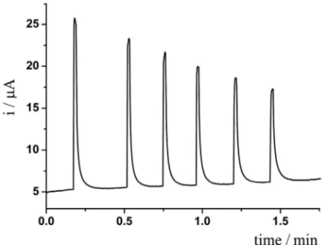
Next, constant-potential amperometry applying +1.3 V using the BIA system was investigated for the determination of naproxen. Figure 1 presents a series of six successive
injections of 50 µmol L-1 naproxen.
A constant decrease in the oxidation current was
verified for successive injections of 50 µmol L-1 naproxen,
clearly indicating adsorption of naproxen or its oxidized products on the electrode surface blocking active sites. Cyclic voltammetric recordings have also demonstrated the blocking of the electrode surface (the oxidation signal
of 1 mmol L-1 naproxen was completely depleted after the
third consecutive cyclic scan). Previous electroanalytical methods for naproxen determination did report electrode adsorption processes resulted from the electrochemical
oxidation of naproxen.12,13 However, those methods were
performed in non-aqueous media whilst the present method employs an aqueous electrolyte, which can be an explanation for the different behavior. Previous works have demonstrated that pulsed amperometry is able to solve such a drawback by the application of an additional potential pulse for cleaning the electrode surface from
adsorbed oxidation products.21,22 Thus, a sequence of two
pulses was studied taking into consideration the detection of naproxen and the efficient electrode cleaning. This strategy resulted in highly repetitive current responses for naproxen injections. Figure 2 presents a set of 10 successive
injections of 50 µmol L-1 naproxen applying +1.5 V for
200 ms and +1.0 V for 100 ms. The highest current response for naproxen and the most efficient cleaning were verified under this sequence of potential pulse. The relative standard deviation (RSD) was 3% (n = 10).
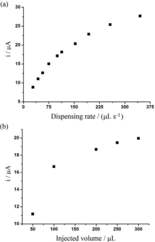
BIA parameters such as dispensing rate and injection volume controlled by the electronic micropipette were optimized. Figure 3 presents the variation of current response for naproxen in function of dispensing rate and injection volume.
The current peak increased, as long as the dispensing rate was increased (Figure 3a). A dispensing rate
of 160 µL s-1 was selected. Despite higher current
Figure 1. Amperometric recordings obtained from successive injections of 50 µmol L-1 naproxen. Working constant potential: +1.3 V; electrolyte:
0.05 mol L-1 phosphate buffer; injected volume: 200 µL; dispensing rate:
160 µL s-1.
Figure 2. Repeatability data obtained from successive injections of 50 µmol L-1 naproxen (n = 10). Working potentials: +1.5 V (200 ms) and
+1.0 V (100 ms); electrolyte: 0.05 mol L-1 phosphate buffer; injected
responses were verified at higher dispensing rates, low repeatability (high standard deviation) was verified at
higher dispensing rates than 160 µL s-1. The current peak
increased significantly with increasing injection volume, from 50 to 100 µL, and continued to increase slightly from 100 to 300 µL (Figure 3b). The injection volume of 200 µL was selected for further experiments.
The linear dynamic range under optimized conditions was
from 10 to 125 µmol L-1 naproxen. The current response did
not increase linearly for naproxen concentrations higher than
125 µmol L-1. The limits of detection (LOD) and quantification
(LOQ) under optimized conditions were estimated as 0.30 and
1.00 µmol L-1, respectively (LOD = 3s
B/S and LOQ = 10sB/S,
in which sB is the standard deviation of the intercept and S
is the slope of the calibration curve). The obtained LOD
values for naproxen determined by DPV at platinum12 and at
BDD electrodes13 were 0.9 and 0.03 µmol L-1, respectively.
However, both electroanalytical procedures employed
non-aqueous electrolyte.12,13 Figure 4 presents amperometric
responses for triplicate injections of solutions containing
increasing concentrations of naproxen (a-e: 10-100 µmol L-1).
The respective calibration curves (increasing and decreasing
order) are also presented (inset). The analytical frequency estimated in this amperometric recording is higher than
90 h-1 (much superior than the DPV methods previously
reported).12,13
A linear behavior, with a good correlation coefficient
(R > 0.99), was observed from 10 to 100 µmol L-1 naproxen
with similar slope values for both curves (0.357 and
0.336 µA L µmol-1). Electrode fouling was not verified
between injections of standard solutions, as evidenced by the fact that current responses were not diminished during amperometric measurements due to the application of a cleaning potential pulse (+1.0 V) using the pulsed amperometric detection technique.
The optimized BIA method with pulsed amperometric detection was applied for the determination of naproxen in pharmaceutical formulations. For comparison, the samples were also analyzed by spectrofluorimetry based on a
previous work.6 Results are presented in Table 1.
All results obtained by the proposed BIA method were in agreement with those obtained by spectrofluorimetry (Table 1). At the 95% confidence level, the calculated
t-values (paired student’s t-test) were smaller than the
critical value (2.78, n = 3), indicating that there are no
Figure 4. BIA amperometric responses for triplicate injections of (a) 10, (b) 25, (c) 50, (d) 75 and (e) 100 µmol L-1 naproxen standard solutions.
Inset: the corresponding calibration curves for increasing () and decreasing () injection order. Working potential: +1.5 V (200 ms) and +1.0 V (100 ms); electrolyte: 0.05 mol L-1 phosphate buffer; injected
volume: 200 µL; dispensing rate: 160 µL s-1.
Figure 3. Effect of (a) dispensing rate and (b) injected volume on the current response for 50 µmol L-1 naproxen. Working potential: +1.5 V
(200 ms) and +1.0 V (100 ms); electrolyte: 0.05 mol L-1 phosphate buffer;
injected volume: 200 µL in (a); dispensing rate: 154 µL s-1 in (b).
Table 1. Concentrations of naproxen obtained by the proposed BIA method and by spectrofluorimetry (mg per tablet) and the respective standard deviation values (n = 3).
Sample Label value / mg BIA / mg Spectrofluorimetry / mg
1 500 484 ± 33 507 ± 16
significant differences between the results. The presence of solid particles from sample matrix (excipients) in solution did not affect the amperometric measurement and then a filtration step was not necessary (advantage of electrochemical methods in comparison with optical ones).
Conclusions
It was demonstrated, for the first time, the application of BIA with amperometric detection for the determination of naproxen in pharmaceutical formulations. The proposed method is highly precise (RSD = 3%, n = 10), accurate (confirmed by comparison with the spectrofluorimetric
method), sensitive (LOD of 0.30 µmol L-1) and fast
(90 injections h-1). Therefore, this method can be applied
for routine analyses at a high analytical frequency.
Acknowledgements
The authors are grateful to CNPq (Conselho Nacional de Desenvolvimento Científico e Tecnológico, 478081/2010-3 and 305227/2010-6), FAPEMIG (Fundação de Amparo à Pesquisa do Estado de Minas Gerais, CEX-APQ-01856-10) and CAPES (Coordenação de Aperfeiçoamento de Pessoal de Nível Superior) for financial support. The authors also thank L. G. Silva for technical assistance in the spectrofluorimetric analyses.
References
1. Elsinghorst, P. W.; Kinzig, M.; Rodamer, M.; Holzgrabe, U.; Sorgel, F.; J. Chromatogr., B: Anal. Technol. Biomed. Life Sci.
2011, 879, 1686.
2. Sun, Y.; Zhang, Z.; Xi, Z.; Shi, Z.; Talanta2009, 79, 676. 3. Trelle, S.; Reichenbach, S.; Wandel, S.; Hildebrand, P.;
Tschannen, B.; Villiger, P. M.; Egger, M.; Jüni, P.; Rev. Port. Clin. Geral2011, 27, 118, DOI: 10.1136/bmj.d2218. 4. Holzbecher, M.; Ellenbeerger, H. A.; Marsh, J. M.; Boudreau, S.;
Clin. Biochem.1979, 12, 66.
5. Panderi, I.; Parissipoulou, M.; Analyst1994, 119, 697.
6. Damiani, P.; Bearzotti, M.; Cabezon, M. A.; J. Pharm. Biomed. Anal.2002, 29, 229.
7. Ibanez, G. A.; Escandar, G. M.; J. Pharm. Biomed. Anal.2005,
37, 149.
8. Junquera, E.; Aicart, E.; Int. J. Pharm.1999, 176, 169. 9. Sadecka, J.; Cakrt, M.; Hercegova, A.; Polonsky, J.; Skacani, I.;
J. Pharm. Biomed. Anal.2001, 25, 881.
10. Wainer, I. W.; Doyle, T. D.; J. Chromatogr.1984, 284, 117. 11. Kazemifard, A. G.; Moore, D. E.; J. Chromatogr., B: Anal.
Technol. Biomed. Life Sci.1990, 533, 125.
12. Adhoum, N.; Monser, L.; Toumi, M.; Boujlel, K.; Anal. Chim. Acta2003, 495, 69.
13. Suyanarayanan, V.; Zhang, Y.; Yoshihara, S.; Shirakashi, T.;
Electroanalysis2005, 17, 925.
14. Tormin, T. F.; Gimenes, D. T.; Richter, E. M.; Munoz, R. A. A.;
Talanta2011, 85, 1274.
15. Felix, F. S.; Angnes, L.; J. Pharm. Sci.2010, 99, 4784. 16. Gimenes, D. T.; Dos Santos, W. T. P.; Tormin, T. F.; Munoz,
R. A. A.; Richter, E. M.; Electroanalysis2010, 22, 74. 17. Quintino, M. S. M.; Angnes, L.; Electroanalysis2004, 16, 513. 18. Wang, J.; Taha, Z.; Anal. Chem.1991, 83, 1053.
19. Tormin, T. F.; Cunha, R. R.; Richter, E. M.; Munoz, R. A. A.;
Talanta2012, 99, 527.
20. Da Silva, R. A. B.; Gimenes, D. T.; Tormin, T. F.; Munoz, R. A. A.; Richter, E. M.; Anal. Methods2011, 3, 2804.
21. Tormin, T. F.; Gimenes, D. T.; Silva, L. G.; Ruggiero, R.; Richter, E. M.; Ferreira, V. S.; Munoz, R. A. A.; Talanta2010,
82, 1599.
22. Lacourse, W. R.; Johnson, D. C.; Rey, M. A.; Slingsby, R. W.;
Anal. Chem.1991, 63, 134.
23. Pedrotti, J. J.; Angnes, L.; Gutz, I. G. R.; Electroanalysis1996,
8, 673.
24. Silva, R. A. B.; Montes, R. H. O.; Richter, E. M.; Munoz, R. A. A.; Food Chem.2012, 133, 200.
25. Bosca, F.; Martinez-Manez, R.; Miranda, M. A.; Primo, J.; Soto, J.; Vano, L.; J. Pharmac. Sci.1992, 81, 479.