Investigation of phagotrophy in natural assemblages of the benthic
dinoflagellates
Ostreopsis
,
Prorocentrum
and
Coolia
Mixotrophy has been shown to be a common trait
among dinolagellates and its importance in the nu
-tritional ecology of harmful algae has been hypothe
-sized. Benthic harmful species have not been exten
-sively investigated as their planktonic counterparts
and there are major gaps in the knowledge of their
nutritional strategies. In this study the occurrence of
phagotrophy was investigated in natural assemblag
-es of benthic dinolagellat-es using epi-luor-escence
microscopy with DAPI and LysoSensor staining.
The study was conducted at ive sites along the coast
of Rio de Janeiro that were visited in January, Au
-gust and December 2010. In total, 1659 dinolagel
-late cells were observed. From these, only 0.4% of
1195
Ostreopsis
cf. ovata and 2.2% of 134
Coolia
spp. cells presented evidence of phagotrophy with
vacuoles stained by LysoSensor or a DAPI (4',6-di
-amidino-2-phenylindole) stained inclusion. Stained
vacuoles were not registered in the 330
Prorocen-trum
spp. cells observed. Few
O.
cf.
ovata
cells con
-tained round red inclusions ("red spots") that were
not stained either by DAPI or LysoSensor, suggest
-ing that these structures are not -ingested prey. The
results showed that phagotrophy was not a frequent
nutritional strategy in benthic dinolagellates during
the study period.
A
bstrAct
Eliliane Vasconcelos Corrêa Almada
1*, Wanderson Fernandes de Carvalho
2, Silvia Mattos
Nascimento
21Universidade Estadual do Norte Fluminense Darcy Ribeiro
(Av. Alberto Lamego, 2000 - Campos dos Goytacazes - 28035-200 - Rio de Janeiro - Brazil) 2Universidade Federal do Estado do Rio de Janeiro, UNIRIO
(Av. Pasteur, 436 - Urca - 22290-240 - Rio de Janeiro - Brazil)
*Corresponding author: [email protected]
Descriptors:
Dinolagellates,
LysoSensor,
Mixotrophy, Phagotrophy.
A mixotroia tem se mostrado uma característica comum
entre dinolagelados e a sua importância na ecologia nu
-tricional de algas nocivas vem sendo investigada. Espé
-cies bentônicas nocivas não foram tão estudadas quanto
as planctônicas, e existem grandes lacunas no conheci
-mento de suas estratégias nutricionais. A ocorrência de
fagotroia em comunidades naturais de dinolagelados
bentônicos foi investigada usando microscopia de epi
--luorescência e marcação por DAPI e LysoSensor. O
estudo foi realizado em cinco locais ao longo da costa
do Rio de Janeiro nos meses de janeiro, agosto e de
-zembro de 2010. No total, foram observadas 1659 cé
-lulas de dinolagelados. Destas, apenas 0,4% das 1195
células de
Ostreopsis
cf.
ovata
e 2,2% das 134 células
de Coolia
spp. observadas apresentaram evidências de
fagotroia com vacúolos marcados por LysoSensor ou
inclusão celular marcada por DAPI (4', 6-diamidino-2
--phenylindole). Não foram observados vacúolos marca
-dos nas 330 células de
Prorocentrum
spp. observadas.
Células de
O.
cf.
ovata
apresentaram inclusões esféricas
avermelhadas ("red spots") que não foram coradas por
DAPI e LysoSensor, sugerindo que estas estruturas não
correspondem a presas ou a vacúolos digestivos. Os re
-sultados mostraram que a fagotroia não foi uma estraté
-gia nutricional frequente nas espécies de dinolagelados
bentônicos observadas durante o período de estudo.
r
esumo
Descritores:
Dinoflagelados, LysoSensor, Mixotrofia,
Fagotrofia.
INTRODUCTION
Mixotrophs are organisms that are capable of combining phototrophic and heterotrophic nutrition to obtain energy (JONES, 1994). Algal mixotrophs vary from species with poor eiciency as phototrophs but high eiciency as phagotrophs to obligate phototrophs with minimal heterotrophy (STOECKER, 1998). Phagotrophy, consumption of particulate food or prey, refers to ingestion of discrete particles wherein digestion occurs in specialized phagocytic (food) vacuoles (GAINES; ELBRÄCHTER, 1987). Mixotrophy has been shown to be a common trait among dinolagellates and was observed in species of several orders (STOECKER, 1999). Dinolagellates represent 75 to 80% of all known harmful species (CEMBELLA, 2003) and form extensive blooms. It has been hypothesized that the association of mixotrophy with the allelopathic efects of toxins against predators and competitors may represent an ecological advantage in harmful algae bloom initiation and persistence (BURKHOLDER et al., 2008).
Studies regarding the nutritional strategies of benthic harmful dinolagellates such as species of the genera
Ostreopsis, Gambierdiscus, Prorocentrum and Coolia are scarce. Benthic harmful species have not been extensively investigated as their planktonic counterparts and there are major gaps in the knowledge of their physiology, nutritional requirements and life cycles (GEOHAB, 2012). Harmful events associated with outbreaks of benthic dinolagellates have been reported more frequently over the last decade, including in areas where benthic harmful genera were hardly known (GEOHAB, 2012).
Several species of Ostreopsis produce potent
neurotoxins of the palytoxin group (CIMINIELLO et al., 2008; 2010) that have been associated with human seafood poisoning through the consumption of fish (TANIYAMA et al., 2003; AMZIL et al., 2012). Recurrent massive blooms of Ostreopsis cf. ovata have been reported in the Mediterranean Sea
where they have been associated with impacts on marine invertebrate (FAIMALI et al., 2012) as well as humans health through inhalation (CIMINIELLO et al. 2006; 2010) and severe skin irritations (DEEDS; SCHWARTZ, 2010). In Brazil, O. cf. ovata blooms
are frequent in Rio de Janeiro (NASCIMENTO et al., 2012a) and have also been reported at Saint Paul’s
Rocks (NASCIMENTO et al., 2012b). Among the benthic species of Prorocentrum, at least nine produce
harmful metabolites or toxins including okadaic acid (OA) and its analogues, dinophysistoxins (DTXs) (HOPPENRATH et al., 2013) that are responsible for shellfish poisoning (DSP). DSP is a human illness caused by the consumption of shellfish contaminated with OA and DTXs. In the genus Coolia, C. monotis
from Australia, was reported to produce cooliatoxin (HOLMES et al., 1995) and WAKEMAN et al. (2015) reported that Coolia malayensis produces unique
metabolites related to cooliatoxin and yessotoxin analogs.
Mixotrophy has been previously reported in benthic dinolagellates such as Ostreopsis cf. ovata, Ostreopsis lenticulares, Ostreopsis siamensis, Ostreopsis labens,
Prorocentrum belizeanum, Prorocentrum hofmannianum,
Prorocentrum arenarium and Gambierdiscus toxicus
through the identiication of prey inside cells (FAUST; MORTON, 1995; FAUST et al., 1996; FAUST, 1998). Using light microscopy, organisms such as cyanobacteria, centric diatoms, ciliates and small microalgae have been identiied as prey for benthic dinolagellates in Belize (FAUST, 1998; FAUST; MORTON, 1995; FAUST et al., 1996), although the feeding behavior has not been observed. These authors suggested that mixotrophy may be common among Ostreopsis species.
Mixotrophy is often difficult to detect in dinoflagellates for several reasons. The use of markers for digestive vacuoles can be an alternative to facilitate the investigation of phagotrophy. LysoSensor probes are selectively concentrated and become more fluorescent in acidic organelles such as food vacuoles. This probe has been successfully used to study phagotrophy in the dinoflagellates Oxyrrhis marina and Dinophysis norvegica and in the haptophyte Prymnesium parvum
and food vacuoles were detected by both fluorescence microscopy and flow cytometry (BOWERS et al., 2010; CARVALHO; GRANÉLI, 2006; CARVALHO; GRANÉLI, 2010).
MATERIAL AND METHODS
Two luorescence methods (staining the nucleus of potentially ingested prey and staining food vacuoles) were used to investigate phagotrophy in cells of benthic dinolagellates associated to macroalgae at the Rio de Janeiro coastline. Sampling sites comprised rocky shores on shallow embayment along Armação dos Búzios (22°45’18’’ S, 41°54’07’’ W) and Arraial do Cabo (22°57’58’’S, 42°01’40’’W) (Fig. 1, Table 1).
DAPI staining of ingested prey
In January 2010 macroalgal samples were collected from a depth of 1-3 m by snorkel diving from sites 1 and 2 (Table 1). Specimens of macroalgae were placed in sealable plastic bags with 50 mL of filtered seawater and were vigorously shaken for 2 minutes to detach the associated epiphytic cells. The epiphytic suspension was filtered through a 125 µm mesh to remove larger particles and debris. Pigments from microalgae cells present in the epiphytic suspension were removed following the procedure described by SALOMON et al. (2003) with slight modifications (fixation with phosphate buffered saline solution containing paraformaldehyde) previous to dinoflagellate cells observation under fluorescence microscopy with UV (365 nm) filter (Olympus BX51, USA). This procedure was applied to avoid the interference of chlorophyll auto fluorescence on the visualization of DAPI staining. The fluorochrome DAPI was used to stain the nucleus of ingested prey inside benthic dinoflagellates, although it is possible that the nucleus of parasites would be stained as well. Two µL of DAPI (50 µg mL-1) were added to 100 µl of the solution containing
benthic dinoflagellates.
The solution was kept in the dark until observation. Benthic dinolagellates were observed using an Olympus microscope (Olympus BX51, USA) with phase contrast, diferential interference contrast under light and epiluorescence microscopy with UV (365 nm) ilter (Olympus BX51, USA). The presence of blue stained inclusions or other structures like vesicles or red inclusions was registered when present.
LysoSensor staining of food vacuoles
Macroalgal samples were collected in sites 2, 3 and 4 in August 2010 while sites 3, 4 and 5 were visited in
December 2010 (Table 1, Fig. 1). Benthic dinolagellates were detached from their substrates as described above. Cells of Ostreopsis, Prorocentrum and Coolia present in the epiphytic suspension were concentrated in a 20 µm mesh and were then resuspended in 30 mL of iltered (glass-iber ilter, Millipore AP-40, Millipore Brazil) seawater. Two µL of the LysoSensor™ Yellow/Blue DND-160 (PDMPO) solution (1mM) (Molecular Probes, Oregon, USA) were added to 1 mL of the concentrated epiphytic suspension (probe inal concentration: 1 µM) that was left in the dark for 5 min before observation under light and epi-luorescence microscopy with UV (365 nm) ilter (Olympus BX51, USA). According to the manufacturers, LysoSensor Probes are weak bases that are selectively concentrated in acidic organelles after protonation, turning bright luorescent in acidic environments. The LysoSensor™ Yellow/Blue probe is unique in that it exhibits both excitation and dual-emission spectral peaks that are pH-dependent and have predominantly yellow luorescence in acidic organelles when excited by blue light (488nm), and blue luorescence in less acidic organelles when excited by UV light (360 nm).
DAPI and LysoSensor were not applied in combination because epi-luorescence microscopy was used to observe the cells and LysoSesor should be used in live cells during short incubations (one to two minutes) and as soon as samples are collected. This prevents the use of procedures necessary to remove pigments from the cells to avoid their auto luorescence from masking DAPI staining.
RESULTS
In total, 1659 dinolagellate cells were counted during this study; most of them O. cf. ovata (1195 cells). In January
2010, the DAPI luorochrome worked successfully as the nucleus of O. cf. ovata cells was stained (Fig. 2). Among
the 255 O. cf. ovata cells examined in January 2010,
only one presented a DAPI-stained inclusion close to the ventral area of the cell (Fig. 2). Colorless and irregularly shape vesicles were observed in 42 O. cf. ovata cells, but
were not stained by DAPI (Fig. 3).
Table 1. Sampling stations, list of macroalgae species collected in January (Summer), August (Winter) and December
(Summer) 2010 along the Rio de Janeiro coastline and staining method used to identify phagotrophy. Numbers in brackets represent the number of specimens collected of each species.
Month/
2010 Sampling station Macroalgae species Staining method
January
Tartaruga site 1 Sargassum vulgare (8)
DAPI Pedra Vermelha,
Cabo Frio Island site 2 Sargassum vulgare (4), Spyridia sp. (1), Amphiroa spp. (4)
August
Prainha site 3 Jania capillacea + Amphiroa fragilissima (1), Amphiroa fragilissima (3)
LysoSensor
Sargassum bank site 4 Amphiroa fragilissima(1), (2), Jania capillacea + Amphiroa fragilissima Spyridia sp. (1)
Pedra Vermelha,
Cabo Frio Island site 2 Amphiroa spp. (1), Amphiroa spp. + Hypnea spinella (1)
December
Prainha site 3 Amphiroa spp. (2), Spyridia sp. (1)
LysoSensor
Sargassum bank site 4 Sargassum vulgare (1), Padina gymnospora (1), Amphiroa spp. (1) Anjos site 5 Sargassum vulgare (1), Spyridia sp. (1)
Figure 1. Map of South America showing the location of Brazil and Rio de Janeiro state. The “inlet” shows a detailed map of the sampling stations in Armação dos Búzios and Arraial do Cabo cities.
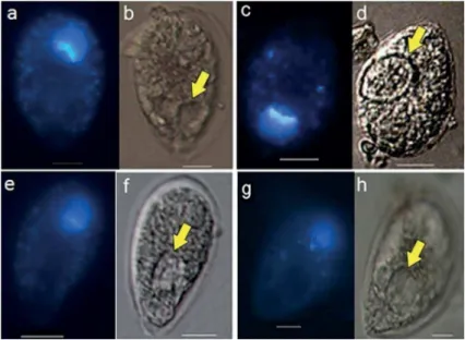
Figure 3. a,c, e, g - Epi-luorescence microscopy with UV emission. b, d, f, h - Light microscopy. Micrographs of O. cf. ovata cells with round vesicles (yellow arrows), January 2010. Note posterior nucleus stained by DAPI. Scale bar = 10 µm.
Figure 4. a, b - Epi-luorescence microscopy with UV emission. c - Light microscopy. Micrographs of O. cf. ovata cell with vesicle stained by LysoSensor, August 2010. a- LysoSensor (blue) stained vesicle (yellow arrow). b- Fading luorescence approximately 3 min later c. vesicle under light microscopy. Scale bar = 10 µm.
cell presented a blue-stained vesicle resembling a food vacuole (Fig. 4).
In December 2010, 713 O. cf. ovata cells and 134 Coolia spp. cells were observed and only three cells of
each species showed stained structures similar to food vacuoles (Fig. 5). Food items could not be identiied inside the stained digestive vacuoles. Stained structures were not registered in the 330 cells of Prorocentrum spp. (mainly Prorocentrum lima, but also Prorocentrum mexicanum).
Ostreopsis cf. ovata cells containing a round red
inclusion under light microscopy and natural orange luorescence under epi-luorescence (Fig. 6) were observed in January and December 2010. The structure resembles what some authors call a “red spot” or “red body”. In total, eight cells containing red bodies were recorded, three in
site 5). None of these red spots were stained either by DAPI or LysoSensor (Fig. 6). The red spots were not seen in cells of Prorocentrum spp. and Coolia spp.
DISCUSSION
In the current study, only a small percentage (0.4% of
O. cf. ovata and 2.2% of Coolia) of the dinolagellate cells
presented evidence of phagotrophy (vesicles stained by LysoSensor or a DAPI stained inclusion) and no prey was discernible inside the cells of Ostreopsis, Prorocentrum
and Coolia. FAUST (1998) reported that between 7 and
55% of the Ostreopsis species from Belize contained a
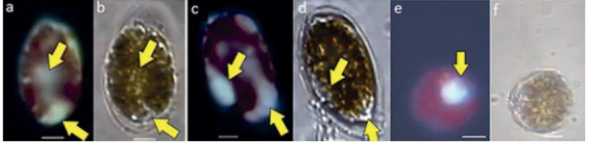
Figure 5. a, c, e - Epi-luorescence microscopy with UV emission. b, d & f- Light microscopy. Micrographs of O. cf. ovata and Coolia sp. cells with vesicles stained by LysoSensor (yellow arrows), December 2010. a, b, c, d- O. cf. ovata cells; e, f Coolia sp. Scale bar = 10 µm.
Figure 6. a, c, e - Epi-luorescence microscopy with UV emission. b, d, f - Light microscopy. Micrographs of O. cf. ovata cells with round reddish inclusions with orange luorescence. a, b- DAPI staining assay. c, d, e, f- LysoSensor assay, note that the red spots are not stained by LysoSensor. Scale bar = 10 µm.
2006). Chloroplasts may mask food vacuoles and it can be problematic to distinguish food vacuoles from other types of inclusions such as gametes or parasites (GRANÉLI; CARLSSON, 1998; LEGRAND et al., 1998; CARVALHO and GRANÉLI, 2006). In an early stage of the infection, parasites can be easily misidentiied as prey. Moreover, the cytoplasm of dinolagellates is highly vesiculate and many species contain “accumulation bodies” or “polyvesicular bodies” whose function is probably the breakdown of superluous organelles and membranes (VAN DEN HOEK et al., 1995). The cytoplasm of the most conspicuous benthic dinolagellate, O. cf. ovata was shown to contain accumulation bodies, cytosolic lipid droplets, rounded translucent bodies, membranous material often surrounded by smooth endoplasmic reticulum and a large number of ibrous material containing vesicles delimited by two membranes, and likely to represent the pusule (HONSELL et al., 2013). This complex cytoplasmic structure adds some diiculty to the observation of food vacuoles or ingested prey through the observation solely by light microscopy.
Therefore, it is likely that early investigations of mixotrophy in benthic dinolagellates (FAUST; MORTON, 1995) based on light microscopy observations may have overestimated the occurrence of phagotrophy in these species. In the current study, vesicles not stained by LysoSensor were observed.
FAUST and MORTON (1995) reported the presence of large ingested particles or food vacuolated cells containing engulfed prey with an intense red color in O.
labens. Those authors speculated that the ventral plate
with associated opening is a peduncle-like structure that is the feeding apparatus of O. labens, although prey
ingestion was not described or illustrated by them. More recently, ESCALERA et al. (2014) interpreted a strand of tightly adpressed microtubules within the sulcal area of the closely related species O. cf. ovata as the micro-tubular strand of the peduncle (MSP). However, these authors stated that a peduncle was never observed in culture or ield samples of O. cf. ovata, nor was it detected
in any TEM sections and that a peduncle-homologous microtubular strand was reported in Palatinus apiculatus
(CRAVEIRO et al., 2009), a species that lacks a peduncle (ESCALERA et al., 2014). ESCALERA et al. (2014) provided evidence that the ventral opening of O. cf. ovata
plays a role in mucus excretion.
The red spots that were observed in O. cf. ovata cells
in the current study were not stained by LysoSensor, suggesting that they are not digestive vacuoles.
FAUST and MORTON (1995) and FAUST (1998) described these reddish round inclusions and considered them as prey in O. lenticulares and O. labens observed
under light microscopy. The presence of a red-pigmented structure is widely described in the literature on dinolagellate cysts (GRAHAM; WILCOX, 2000). It has been called as red-spots, red-brown spots or dark-brown material by a number of authors. The cells with red spots observed in the current study are similar to the O. cf. ovata
ACCORONI et al. (2014). Red bodies in Ostreopsis cysts enclosed in hyaline membranes were observed mainly at the end of the bloom by ALIGIZAKI and NIKOLAIDIS (2006). MEKSUMPUN and MONTANI (1995) reported that red-brown inclusions in the marine dinolagellate
Scrippsiella trochoidea are composed of fucoxanthin, 19’ -
hexanoyl - fucoxanthin, diadinoxanthin and diatoxanthin. The authors suggested that these pigments are useful for growth of vegetative cells after germination from cysts.
Diferences in environmental conditions such as irradiance; nutrient concentrations (limited/suicient) and prey availability have been reported to inluence the occurrence of phagotrophy in dinolagellate species (LEGRAND et al., 1998; STOECKER et al., 2006). The planktonic dinolagellate Prorocentrum minimum
was induced to prey in limiting nitrogen and phosphorus experimental conditions and feeding was never observed under nutrient replete conditions (JOHNSON, 2015). The study area is among the most productive regions in the Brazilian littoral due to the occurrence of coastal upwelling, particularly during austral summer that sustains high primary productivity and marine biodiversity (VALENTIN, 1984; GONZALEZ-RODRIGUEZ et al., 1992). Therefore, it is likely that benthic dinolagellates from the study area were nutrient suicient, a condition that would not stimulate the occurrence of phagotrophy. Data presented herein did not support phagotrophy as a common nutritional strategy among benthic dinolagellates. Further studies including laboratory experiments under diferent environmental conditions are necessary to assess the capability of phagotrophy in benthic dinolagellates.
ACKNOWLEDGEMENTS
The authors are indebted to the Instituto de Estudos do Mar Almirante Paulo Moreira (IEAPM) and to Dra. Eliane González-Rodríguez for all the assistance provided during ield trips. This research was funded by Fundação Carlos Chagas Filho de Amparo à Pesquisa do Estado do Rio de Janeiro (FAPERJ- Brazil to SMN, grant number E26/111.925/2012) and Conselho Nacional de Desenvolvimento Cientíico e Tecnológico (CNPq Brazil to SMN, grant number 481150/07-2).
REFERENCES
ALIGIZAKI, K.; NIKOLAIDIS, G. The presence of the poten -tially toxic genera Ostreopsis and Coolia (Dinophyceae) in
the North Aegean Sea, Greece , v. 5, n. 3, p.
ACCORONI, E.; ROMAGNOLI, T.; PICHIERRI, S.; TOTTI, C. New insights on the life cycle stages of the toxic benthic dinolagellate Ostreopsis cf. ovata. Harmful Algae, v. 34, p.
7-16, 2014.
AMZIL, Z.; SIBAT, M.; CHOMERAT, N.; GROSSEL, H.; MAR
-CO-MIRALLES, F.; LEMEE, R.; NEZAN, E.; SECHET, V. Ovatoxin-a and palytoxin accumulation in seafood in relation
to Ostreopsis cf. ovata blooms on the French Mediterranean coast. Mar. Drugs, v. 10, n. 2, p. 477-496, 2012.
BOWERS, H. A.; BRUTEMARK, A.; CARVALHO, W. F.; GRANÉLI, E. Combining Flow Cytometry and Real-Time PCR Methodology to Demonstrate Consumption by Prym-nesium parvum. J. Am. Water Works Assoc., v. 46, n. 1, p.
133-143, 2010.
BRAVO, I.; VILA, M.; CASABIANCA, S.; RODRIGUEZ, F.; RIAL, P.; RIOBÓ, P.; PENNA, A. Life cycle stages of the benthic palytoxin-producing dinolagellate Ostreopsis cf. ovata (Dinophyceae). Harmful Algae, v. 18, p. 24-34, 2012.
BURKHOLDER, J. M.; GLIBERT, P. M.; SKELTON, H. M. Mi
-xotrophy, a major mode of nutrition for harmful algal species in eutrophic waters. Harmful Algae, v. 8, n. 1, p. 77-93, 2008.
CARVALHO, W. F.; GRANÉLI, E. Acidotropic probes and low cytometry: a powerful combination for detecting phagotro -phy in mixotrophic and heterotrophic protists. Aquat. Microb. Ecol., v. 44, n. 1, p. 85-96, 2006.
CARVALHO, W. F.; GRANÉLI, E. Contribution of phagotrophy versus autotrophy to Prymnesium parvum growth under ni
-trogen and phosphorus suiciency and deiciency. Harmful Algae, v. 9, n. 1, p. 105-115, 2010.
CEMBELLA, A. D. Chemical ecology of eukaryotic microalgae in
marine ecosystems. Phycologia, v. 42, n. 4, p. 420-447, 2003.
CIMINIELLO, P.; DELL’AVERSANO, C.; FATTORUSSO, E.; FORINO, M.; TARTAGLIONE, S.; GRILLO, C.; ME
-LCHIORRE, N. Putative palytoxin and its new analogue, ovatoxin-a, in Ostreopsis ovata collected along the Ligurian
coasts during the 2006 toxic outbreak. J. Am. Soc. Mass Spec-trom., v. 19, n. 1, p. 111-120, 2008.
CIMINELLO, P.; DELL’AVERSANO, C.; FATTORUSSO, E.; FORINO, M.; MAGNO, G. S.; TARTAGLIONE, L.; GRI
-LLO, C.; MELCHIORRE, N. The Genoa 2005 outbreak. De
-termination of putative palytoxin in Mediterranean Ostreopsis ovata by a new liquid chromatography tandem mass spectro -metry method. Anal. Chem., v. 78, n. 17, p. 6153-6159, 2006.
CIMINELLO, P.; DELL’AVERSANO, C.; DELLO IACOVO E.; FATTORUSSO, E.; FORINO, M.; GRAUSO, L.; TAR
-TAGLIONE, L.; GUERRINI, F.; PISTOCCHI, R. Complex palytoxin-like proile of Ostreopsis ovata. Identiication of
four new ovatoxins by high-resolution liquid chromatogra
-phy/mass spectrometry. Rapid Commun. Mass Spectrom., v.
24, n. 18, p. 2735-2744, 2010.
CRAVEIRO, S. C.; CALADO, A. J.; DAUGBJERG, N.; MOES
-TRUP, O. Ultrastructure and LSU rDNA-based revision of
Peridinium group Palatinum (Dinophyceae) with the descrip
-tion of Palatinus gen. nov. J. Phycol., v. 45, n. 5, p.
1175-1194, 2009.
DEEDS, J. R.; SCHWARTZ, M. D. Human risk associated with palytoxin exposure. Toxicon, v. 56, n. 2, p. 150-162, 2010.
ESCALERA, L.; BENVENUTO, G.; SCALCO, E.; ZINGONE, A.; MONTRESOR, M. Ultrastructural Features of the Ben
Pro-FAIMALI, M.; GIUSSANI, V.; PIAZZA, V.; GARAVENTA, F.; CORRÀ, C.; ASNAGHI, V.; PRIVITERA, D.; GALLUS, L.; CATTANEO-VIETTI, R.; MANGIALAJO, L.; CHIANTORE, M. Toxic efects of harmful benthic dinolagellate Ostreopsis ovata on invertebrate and vertebrate marine organisms. Mar. Environ. Res., v. 76, p. 97-107, 2012.
FAUST, M. A. Mixotrophy in tropical benthic dinolagellates. In: REGUERA, B.; BLANCO, J.; FERNÁNDEZ, M. L.; WYATT, T. (Ed.). VIII International Conference on Harmful Algae.
Vigo: International Oceanographic Comission of UNESCO, 1998. p. 390-393.
FAUST, M. A.; MORTON, S. L. Morphology and ecology of the marine dinolagellate Ostreopsis labens sp. nov. (Dinophy
-ceae). J. Phycol., v. 31, n. 3, p. 456-463, 1995.
FAUST, M. A.; MORTON, S. L.; QUOD, J. P. Further SEM stu
-dy of marine Dinolagellates: the genus Ostreopsis (Dinophy
-ceae). J. Phycol., v. 32, n. 6, p.1053-1065, 1996.
GAINES, G.; ELBRÄCHTER, M. Heterotrophic nutrition. In: TAYLOR, F. J. R. (Ed.). The Biology of Dinolagellates. Palo Alto: Blackwell Scientiic Publications, 1987. p. 224-268. GEOHAB 2012. Global Ecology and Oceanography of Harmful
Algal Blooms, GEOHAB Core Research Project: HABs in Benthic Systems. E. Berdalet, P. Tester, A. Zingone (Eds.) IOC of UNESCO and SCOR, Paris and Newark, 64 pp.
GONZALEZ-RODRIGUEZ, E., VALENTIN, J. L.; ANDRÉ, D. L.; JACOB, S. A. Upwelling and downwelling at Cabo Frio (Brazil): comparison of biomass and primary production res -ponses. J. Plankton Res., v. 14, n. 2, p. 289-306, 1992.
GRAHAM, L. E.; WILCOX, L. W. Dinolagellates. In: GRA
-HAM, J. E.; WILCOX, L. W.; GRA-HAM, L. E. Algae. Upper
Saddle River: Prentice Hall, 2000.
GRANÉLI, E.; CARLSSON, P. The ecological signiicance of phagotrophy in photosynthetic lagellates. In: ANDERSON, D. M.; CEMBELLA, A. D.; HALLEGRAEFF, G. M. (Eds.).
Physiological ecology of harmful algal blooms, NATO ASI Se-ries G41. Berlin: Springer-Verlag, 1998, p. 539-557.
HOLMES, M. J.; LEWIS, R. J.; JONES, A.; HOY, A. W. Coolia
-toxin, the irst toxin from Coolia monotis (Dinophyceae). Nat. Toxins, v. 3, n. 5, p. 355-362, 1995.
HONSELL, G.; BONIFACIO, A.; DE BORTOLI, M.; PENNA, A.; BATTOCCHI, C.; CIMINIELLO, P.; DELL’AVERSANO, C.; FATTORUSSO, E.; SOSA, S.; YASUMOTO, T.; TUBARO, A. New Insights on Cytological and Metabolic Features of Os-treopsis cf. ovata Fukuyo (Dinophyceae): A Multidisciplinary Approach. PLoS ONE, v. 8, n. 2, p. e57291, 2013.
HOPPENRATH, M.; CHOMÉRAT, N.; HORIGUCHI, T.; SCHWEI
-KERT, M.; NAGAHAMA, Y.; MURRAY, S. Taxonomy and phylogeny of the benthic Prorocentrum species (Dinophyceae) - A proposal and review. Harmful Algae, v. 27, p. 1-28, 2013.
JOHNSON, M. D. Inducible mixotrophy in the dinolagellate Prorocen-trum minimum. J. Eukaryot Microbiol., v. 62, n. 4, p. 431-443, 2015.
JONES, R. I. Mixotrophy in planktonic protists as a spectrum of nutri
-tional strategies. Mar. Microb Food Webs, v. 8, n. 1, p. 87-96, 1994.
LEGRAND, C.; GRANÉLI, E.; CARLSSON, P. Induced phago
-trophy in the photosynthetic dinolagellate Heterocapsa trique-tra. Aquat. Microb. Ecol., v. 15, p. 65-75, 1998.
MEKSUMPUN, S.; MONTANI, S. Chemical components of red
--brown material in cyst of Scrippsiella trochoidea (Dinophy
-ceae). Kasetsart Univ. Fish Res. Bull., v. 21, p. 1-16, 1995.
NASCIMENTO, S. M.; CORRÊA, E. V.; MENEZES, M; VARE
-LA, D.; PAREDES, J.; MORRIS, S. Growth and toxin proile of Ostreopsis cf. ovata (Dinophyta) from Rio de Janeiro, Bra
-zil. Harmful Algae, v. 13, p. 1-9, 2012a.
NASCIMENTO, S. M.; FRANÇA, J. V.; GONÇALVES, J. E. A.; FERREIRA, C. E. L. Ostreopsis cf. ovata (Dinophyta) bloom
in an equatorial island of the Atlantic Ocean. Mar. Pollut. Bull.,
v. 64, n. 5, p. 1074-1078, 2012b.
SALOMON, P. S.; JANSON, S.; GRANÉLI, E. Parasitism of
Dinophysis norvegica by Amoebophrya sp. in the Baltic Sea. Aquat. Microb. Ecol., v. 33, n. 2, p. 163-172, 2003.
STOECKER, D. K. Conceptual models of mixotrophy in plankto
-nic protists and same ecological and evolutionary implications.
Eur. J. Protistol., v. 34, n. 3, p. 281-290, 1998.
STOECKER, D. K. Mixotrophy among dinolagellates. J. Euka-ryot. Microbiol., v. 46, n. 4, p. 397-401, 1999.
STOECKER, D. K.; TILLMANN, U.; GRANÉLI, E. Phagotrophy in harmful algae. In: GRANÉLI, E.; TURNER, J. (Eds.). Ecology of Harmful Algae. Berlin: Springer-Verlag, 2006. p. 177-187.
TANIYAMA, S.; ARAKAWA, O.; TERADA, M.; NISHIO, S.; TAKATANI, T.; MAHMUDA, Y.; NOGUCHI, T. Ostreopsis
sp., a possible origin of palytoxin (PTX) in parrotish Scarus ovifrons. Toxicon, v. 42, n. 1, p. 29-33, 2003.
VALENTIN, J. L. Analyse des paramètres hydrobiologiques dans la remontée de Cabo Frio (Brésil). Mar. Biol., v. 82, n. 3, p.
259-276, 1984.
VAN DEN HOEK, C.; MANN, D. G.; JAHNS, H. M. Algae an introduction to phycology. Cambridge: Cambridge University
Press, 1995. 623 p.
WAKEMAN, K. C.; YAMAGUCHI, A.; ROY, M. C.; JENKE
--KODAMA, H. Morphology, phylogeny and novel chemical compounds from Coolia malayensis (Dinophyceae) from Oki