Article
J. Braz. Chem. Soc., Vol. 28, No. 5, 707-723, 2017. Printed in Brazil - ©2017 Sociedade Brasileira de Química 0103 - 5053 $6.00+0.00
*e-mail: [email protected] †In memoriam
Chemical Constituents of
Psychotria nemorosa
Gardner and Antinociceptive Activity
Nivea O. Calixto,a Millena S. Cordeiro,b Thais B. S. Giorno,b Gibson G. Oliveira,c
Norberto P. Lopes,c Patricia D. Fernandes,b Angelo C. Pinto†,a and Claudia M. Rezende*,a
aInstituto de Química and bInstituto de Ciências Biomédicas, Universidade Federal do Rio de Janeiro,
21949-900 Rio de Janeiro-RJ, Brazil
cNPPS - Faculdade de Ciências Farmacêuticas de Ribeirão Preto, Universidade de São Paulo,
14040-903 Ribeirão Preto-SP, Brazil
Through dereplication strategies using gas chromatography-mass spectrometry (GC-MS) and ultra fast liquid chromatography-tandem mass spectrometry (UFLC-MS/MS), the ethanol extract from Psycotria nemorosa leaves (Rubiaceae) showed to be composed of: cinnamic acid,
dihydroactinidiolide, 4-hydroxy-β-ionone, phytol, isophytol, 4,8,12,16-tetramethylheptadecan-4-olide, lupeol, a mixture of α/β-amyrin, the keto and acetylated derivatives, besides stigmast-4-en-3-one, campesterol, stigmasterol and γ-sitosterol by GC-MS. Likewise, by UFLC-MS/MS, the main compounds identiied were: butin, resveratrol, rutin, kaempferol 7-O-β-D-glucopyranoside,
deacetylasperuloside, epiloganin, hordenine, strictosidine, N-methyl-1,2,3,4-tetrahydro-β-carboline
and N-formyl-tryptamine. The antinociceptive activity of the crude extract ant its fractions was
reported.
Keywords: Psychotria nemorosa, hyphenated chromatography-mass spectrometry,
antinociceptive activity
Introduction
Herbal drugs have been used since ancient times as medicines, for the treatment of a range of diseases. Many modern drugs have been evolved from ethnopharmacological approaches.1,2 Discovery of novel
bioactive compounds from plants is a challenging, laborious, expensive process and requires long time to isolate and characterize an active compound. If the isolated bioactive is a known compound, then the time and resources spent on it will be quite a wasted
effort.3 To overcome these issues, new techniques for
direct identiication of bioactive natural products from extracts were developed. Recent high throughput assays technologies to study plant extracts for biological activity has intensiied the need for appropriate dereplication strategies.4 Dereplication is the process that allows a rapid
identiication of secondary metabolites in crude extracts by distinguishing previously identiied compounds from novel ones. The dereplication process involves separation of single metabolites by chromatographic methods,
identiication by spectroscopic methods, bioassay for evaluation of the biological activity, and search in databases to verify the novelty of these compounds.5
Hyphenated chromatographic techniques possess characteristics that include efficient separation and rapid identiication, among several other advantages in comparison with other analytical methods. These techniques bring signiicant improvements in the identiication of novel natural compounds in complex matrices.6,7
Psychotria comprises an important genus of Rubiaceae
family and contains approximately 1700 species of lowering plants with tropical and subtropical distribution.8
The polyphyly of the genus has been demonstrated through recent worldwide molecular phylogenetic
studies. Razafimandimbison et al.9 established that
Psychotria includes all its allied genera, rendering the tribe
Psychotrieae monogeneric. It is commonly used in folk herbal medicine for regulating menstrual disturbance, fever, against microbial infections, anti-inlammatory, analgesic, anticonvulsant, anti-rheumatic and even as cardiovascular, mental and eating disorders.10-13 Psychotria genus is a
Psychotria nemorosa Gardner (Rubiaceae), popularly
known as “casca d’anta”, is a native terrestrial substrate shrub measuring 1-2 m in height with endemic growth in Brazil, found in Rain Forests in the states of Bahia, Paraíba (Northeast), Goiás (West-Central), Espírito Santo, Minas Gerais, Rio de Janeiro, São Paulo (Southeast), Paraná, Santa Catarina and Rio Grande do Sul (South).18 It is characterized
by persistent bilobed stipules containing 8 to 10 arched secondary ribs on each side and visibles cross linked tertiary ribs. Flowering and fruiting occurs in January and March, respectively.19
Literature review showed that the extracts of many
Psychotria species have anti-inlammatory and analgesic
activity, and preliminary tests pointed to alkaloids as the major responsible for the effect, besides other compounds.20,21 Pain is a protective mechanism, triggered
by potentially injurious stimuli, that inluences the quality of life and is directly related to a high amount of medical licenses all over the world.22,23 Usually, the therapeutic
proile is based on analgesic and anti-inlammatory drugs that present several side effects as ulcers, vomiting, tolerance or dependence.24 Due to these problems, there
is continuous interest in the search of new substances that could act with lesser side effects.
As far as we know, no record on phytochemical studies nor biological approach of P. nemorosa is described in
scientiic literature. So, in this work, our objective was to investigate the chemical composition of the P. nemorosa
leaves on a metabolomic approach and its antinociceptive property in mice.
Experimental
General experimental procedures
All solvents were high-grade purity purchased from Tedia (Brazil). Infrared (IR) spectra were recorded on a Nicolet Magna IR 760 spectrometer using KBr pellets. Column chromatography was performed over silica gel (25 g, 200-300 mesh, Silicycle, Canada) and thin layer
chromatography (TLC) on precoated silica gel GF254
aluminum plates (Merck, Germany). Spots were visualized by spraying with acidic cerium (IV) sulfate reagent followed by heating at 110 °C and usual speciic reagents for different classes of natural product.
Plant material
Leaves of P. nemorosa Gardner were collected in
November (2012) at Serra dos Órgãos (Rio de Janeiro State, Southeast of Brazil, −22.464042, −43.011746). The access
authorization number is 27035-4 emitted by Ministério do Meio Ambiente. A voucher specimen (Trovó 389 RB) was deposited at the herbarium of the Jardim Botânico do Rio de Janeiro and identiied by Marcelo Trovó Lopes de Oliveira.
Extraction and isolation
Dried leaves (60 g) were exhaustively extracted with a 90% aqueous ethanol (EtOH) solution at room temperature for 7 days. The resulting extracts were combined and concentrated under low pressure at 40 °C in a rotaevaporator to yield a dark green liquid (crude ethanolic extract (EE), 7 g). EE (1.0 g) was dissolved in MeOH and subjected to silica gel column chromatography (CC) (21 × 2.5 cm) eluted with hexane, a gradient of hexane/ethyl acetate (EtOAc) (5, 10, 20, 50 and 100%) and methanol (MeOH) to afford thirty-ive fractions, which were combined in twelve resulting fractions according to TLC analysis (Table S1, Supplementary Information).
Gas chromatography-mass spectrometry (GC-MS)
GC-MS analyzes were performed on a gas chromatograph coupled to a mass spectrometry (GCMS-QP 2010 Ultra, Shimadzu, Kyoto, Japan) equipment with a quadrupole mass analyzer, electron impact ion source (at 70 eV), auto sampler AOC-20s and auto injector AOC-20i. Analyzes were carried out using a DB-1 capillary column of 30 m × 0.25 mm × 0.25 µm, helium as carrier gas at 1.0 mL min-1. The GC temperature was programmed as: 60 °C
(2 min), 60-120 °C (6 °C min-1), 120-290 °C (15 °C min-1)
then 290 °C for 17 min, according to Patitucci et al.25 Injector,
ion source and interface temperatures were 280, 230 and 290 °C, respectively. Data acquisition was performed by Lab Solutions Software and data analysis by NIST MS search database. The sample injection volume was 1.0 µL, all diluted with EtOAc. The triterpenes were identiied by comparison of the retention time with authentic standards. N-Octadecane
was used as internal standard in gas chromatography coupled to a lame ionization detector (GC-FID) with the same conditions described above.
Oxidation of triterpenes
β/α-Amyrin acetate (20a, 20b)
β/α-Amyrin (31a, 31b) (0.05 g), acetic anhydride
(1.5 mL) and DMAP (0.001 g) were mixed, stirred and heated under relux. The reaction was monitored by TLC (2 h), then diluted with H2O (4 mL) and extracted with
dichloromethane (CH2Cl2) (3 × 10 mL). The combined
a white solid of β/α-amyrin acetate (20a, 20b) was obtained
(49.0 mg, 98%). IR (KBr) νmax / cm-1: 2949 (=C−H), 1734
(C=O), 1379, 1367 (C−H), 1246 (C−O); β-amyrin acetate (20a) MS m/z (%): 468 (M+, 3), 218 (100), 203 (52),
189 (24); α-amyrin acetate (20b) MS m/z (%): 468 (M+, 4),
218 (100), 203 (23), 189 (27). Lupeol acetate (48 mg, 96%). IR (KBr) νmax / cm-1: 2943 (=C−H), 1732 (C=O), 1639
(C=C), 1454, 1367 (C−H), 1249 (C−O); MS m/z (%): 468
(M+, 20), 408(20), 218(36), 204(44), 189(100).
β/α-Amyrone (18a, 18b)
Jones’ reagent was added dropwise to the β/α-amyrin (31a, 31b) (0.05 g) in acetone (5.0 mL) at room temperature
until the red-orange color of the oxidant persisted. Drops of isopropanol were added until the appearance of a blue-green color, followed by water addition (3.0 mL), a Na2CO3
saturated solution and extraction with CH2Cl2 (3 × 10 mL).27
The combined organic layers were dried over Na2SO4 and
evaporated to give off-white crystals of amyrones (48.5 mg, 97%). IR (KBr) νmax / cm-1: 2948 (=C−H), 2853 (C−H), 1706
(C=O), 1456, 1380 (−C−H); β-amyrone (18a) MS m/z (%):
424 (M+, 11), 218 (100), 203 (57), 189 (19); α-amyrone (18b)
MS m/z (%): 424 (M+, 11), 218 (100), 203 (23), 189 (19).
3,11-Dioxo-β/α-amyrene (21a, 21b)
tert-Butyl chromate (3.0 mL) was added to a solution of β/α-amyrone (18a, 18b) (20 mg) in acetone (2.0 mL), acetic
anhydride (3.0 mL) and acetic acid (6.0 mL), reluxed for 6 h and at the end diluted with H2O (2.0 mL) and extracted
with CH2Cl2 (3 × 10 mL). The organic phase was washed
with aqueous oxalic acid solution 5% (2 × 10 mL), Na2CO3
saturated solution (10.0 mL) and H2O (10.0 mL), then dried
over Na2SO4 and evaporated.28 The product was a yellowish
oil (17.6 mg, 88%). IR (KBr) νmax / cm-1: 2976 (=C−H), 2872
(−C−H), 1709, 1663 (C=O), 1457 (C=C); 3,11-dioxo-β -amyrene (21a) MS m/z (%): 438 (M+, 31), 273 (75), 232 (93),
217 (28), 135 (100); 3,11-dioxo-α-amyrene (21b) MS m/z
(%): 438 (M+, 19), 273 (100), 232 (54), 217 (9), 135 (76).
Ultra fast liquid chromatography-mass spectrometry (UFLC-MS)
UFLC-MS analyzes were performed on a Shimadzu UFLC (UFLC Shimadzu System Proeminene, Kyoto, Japan), consisting of a binary pump solvent management system, an online degasser, an auto-sampler, a column oven and a photo diode array detector (DAD). Separation was performed on a Phenomenex Luna C18 (2) 100 A (250 × 4.6 mm i.d., 5 µm) column equipped with a Phenomenex Luna C18 guard column (4.3 × 5 mm; Phenomenex, California, USA). The mobile phase consisted of water (A) and acetonitrile (B) both
containing 0.1% formic acid (v/v), with a gradient elution: 0-50 min: 5-100% B (linear gradient); 50-55 min: 100% B (isocratic gradient); 55-60 min: 100-5% B (column washing); 60-65 min: 5% B (isocratic, column equilibration). The low rate was 1.0 mL min-1. The column temperature was set at
35 °C and the injection volume was 20 µL. The UV spectra were recorded from 200 to 800 nm. The UFLC system was coupled to a micrOTOF-II mass spectrometer equipped with an electrospray interface (ESI) (Bruker Daltonics Inc., Billerica, MA, USA) operating in both positive and negative ion mode using a capillary voltage of 3.5 kV, end plate 500 V to obtain the accurate mass data. The parameters for ESI coupled to quadrupole-time-of-light mass spectra (ESI-QTOF-MS) were: drying gas temperature, 320 °C; drying gas low, 10 L min-1 and nebulizing gas pressure, 60 psi.
Detection was carried out within a mass range of 50-1300 Da. Nitrogen was used as drying, nebulizing and collision gas.
Similar UFLC equipment and chromatographic conditions were used, as described above, coupled to a mass spectrometer ESI-IT (Bruker Daltonicx, Billerica, MA, USA), fitted with an electrospray ionization source operating in positive and negative mode and
ion-trap analyzer. Tandem mass spectrometry (MS2)
analyzes were developed in this equipment. The mass spectrometer parameters used were: capillary voltage, 3.5 kV; dessolvation temperature, 330 °C; gas flow, 10 L min-1 and pressure at 60 psi. Nitrogen was used as
both drying and nebulizing gas. Amplitude fragmentation energy for MS/MS experiments was 0.70 V. Data analysis was carried out with Bruker Daltonics software version 4.1.
Animals
All experiments were performed with male Swiss Webster mice (20-25 g) obtained from our own animal facilities. Animals were kept in a room with controlled temperature 22 ± 2 °C for 12 h light/dark cycle with free access to food and water. Twelve hours before each experiment, the animals received only water, in order to avoid food interference with substances absorption. Animal care and research protocols were in accordance with the principles and guidelines adopted by the Brazilian College of Animal Experimentation (COBEA), approved by the Biomedical Science Institute/UFRJ, Ethical Committee for Animal Research, and received the number DFBCICB015-04/16.
Psychotria nemorosa fractions administration
P. nemorosa (leaves) fractions were dissolved in dimethyl
concentration of 100 mg mL-1. In all experiments, the inal
concentration of DMSO did not exceed 0.5% at which it has no effect per se. The crude extract (EE), hexane, hexane/
EtOAc 5%, hexane/EtOAc 10%, EtOAc and MeOH fractions were administered by oral gavage at 10, 30 or 100 mg kg-1
in a inal volume 0.1 mL. The control group was composed by vehicle (ultrapure water), which was the same used to solubilize the EE and fractions that were administered on the day of the experiment.
Formalin-induced licking response
This procedure was similar to the method described by Gomes et al.29 Mice received an injection of 20 µL
of formalin (2.5% v/v) into the dorsal surface of the left hind paw. The time that the animal spent licking the injected paw was immediately recorded. The nociceptive and inlammatory response consists of the following two phases: the irst phase lasts until 5 min after the formalin injection (irst phase, neurogenic pain response), and the second phase occurs 15-30 min after the formalin injection (second phase, inlammatory pain response). The animals were pre-treated with oral doses of EE, hexane, hexane/ EtOAc 5% and hexane/EtOAc 10% fractions, EtOAc and MeOH 60 min before the administration of formalin.
Statistical analysis
Each experimental group consisted of 6 mice. The results are presented as mean ± standard deviation (S.D.). The area under the curve (AUC) was calculated using Prism Software 5.0 (GraphPad Software, La Jolla, CA, USA). Statistical signiicance between groups was determined using the application of analysis of variance (ANOVA) followed by Bonferroni’s test. p-Values less than 0.05 were
considered to be signiicant.
Results and Discussion
The crude ethanolic extract (EE) from leaves of
P. nemorosa was fractionated in a silica gel open column
chromatography affording twelve fractions, all analyzed by TLC. Each fraction was evaluated by GC-MS or liquid chromatography coupled to mass spectrometry (LC-MS) on a metabolomic perspective based on their polarities. Mass spectrometry (MS) allows a direct screening of the compounds, providing structural information without the need of laborious isolation since each family of metabolites may have a characteristic mass fragmentation pattern and retention times in GC-qMS and typical UV absorption and
MS/MS proile in LC-DAD-MS/MS systems.30
In addition, fractions and EE were evaluated for antinociceptive properties on formalin-induced licking response in mice due the occurrence of two distinct phases of nociceptive behavior: one phase immediately after the injection, lasting for about 5 min, and the other phase starting approximately 20 min after the injection.31
The fractions resulting from the CC silica gel can be seen in Table S1 (Supplementary Information). Only those with suficient amount to access biological activity were investigated in the phytochemical approach. To identify the less polar compounds, GC-MS was used and their proiles are shown in Figures 1A-1D.
GC-MS chemical compounds present in fractions 1, 5, 8 and 9 can be seen in Tables S2 and S3 (Supplementary Information). The MS of the compounds showed more than 90% of similarity with NIST database. These results attested to the major occurrence of cinnamic acid, sterols (campesterol, stigmasterol, γ-sitosterol and stigmast-4-en-3-one) and pentacyclic triterpenes such α/β-amyrone, α/β-amyrin acetate,
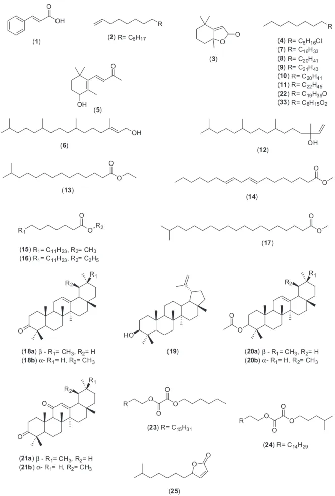
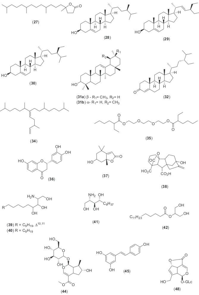
3,11-dioxo-α/β-amyrene, lupeol and α/β-amyrin, besides some fatty acids, diterpene alcohols and hydrocarbons. Co-injection with authentic materials additionally confirmed the identiication. It is important to report that fractions 15-22 were not investigated by GC-MS due to their increased polarity and also due to the small amounts for biological assays. Figure 2 shows the chemical structures of the compounds identiied in this work.
Although Psychotria genus is characterized mainly
as a source of alkaloids, the occurrence of terpenoids is
well known. In P. yunnamensis, norisoprenoids and the
monoterpenoid (6S)-menthiafolic acid were isolated, in
addition to the ancorane sesquiterpene psycacoraone A.32,33
Volatile compounds were also detected in P. leiocarpa.34
β-Sitosterol was found in P. mariniana and P. hainanensis,
together with stigmasterol in P. vellosiana.17,35,36 These
steroids, in the glycosylated form, were also reported.36-38
Lupeol was found in P. vellosiana and P. mariniana.17,35
Phytochemical studies of P. adenophylla allowed the
caracterization of other triterpenes such as bauerenol, bauerenol acetate, friedelin, betulin, ursolic acid, traces of
α- amyrin and betulinic acid.39 Ursolic acid was isolated
from P. serpens.40 Besides P. adenophylla, α- amyrin was also found in P. stachyoides.37 It should be emphasized
that the occurrence of α/β-amyrone, α/β-amyrin acetate and 3,11-dioxo-α/β-amyrene is being reported for the irst time in Psychotria genus. α/β-Amyrone (m/z 424,
[M]+•) and α/β-amyrin acetate (m/z 468, [M]+•) showed
ions at m/z 218 due to a typical retro Diels-Alder reaction
([M]+• – C
14H22O and [M]+• – C16H26O2, respectively).
([M]+• – CH
3), derived from the ions m/z 218, were also
observed. A McLafferty rearrangement in α/β-amyrin
acetates ([M]+• – C
2H4O2) at m/z 408 was also seen.
The same takes place with 3,11-dioxo-α/β-amyrene
(m/z 438, [M]+•), generating the characteristic fragments
at m/z 273 ([M]+• – C11H17O), 232 ([M]+• – C14H22O), 217
([M]+• – C
14H22O – CH3), 189 ([M]+• – C14H22O – CH3 – CO)
and 135 ([M]+• – C
11H17O – C10H18).41
Figure 1. Total ion count mass spectrometry analysis of P. nemorosa leaves: (A) hexane; (B) hexane/EtOAc 5% and (C and D) hexane/EtOAc 10% fractions
The mixture of α/β-amyrin (31a, 31b), previously
puriied from Protium sp. resin,42 and lupeol (19), derived
from Vellozia sp.,43 were subjected to classic esteriication
and oxidation reactions to give derivatives in good yields (Schemes 1 and 2). These reference compounds were used in co-injection assays or for comparison of retention time as an additional tool to confirm GC-MS results (Supplementary Information).
As α-amyrin acetate (20b) and lupeol acetate exhibit
the same retention times in this condition (34.8 min), the mass fragmentation proile was determined to conirm the presence of α-amyrin acetate in fraction 5 (Supplementary Information).
Besides the triterpenes conirmation, GC-MS allowed us to detect a further series of hydrocarbons, long chain fatty acids and their esters.
Nowadays, ultra fast liquid chromatography/tandem mass spectrometry (UFLC-MS/MS) is one of the most
convenient techniques for online characterization, due to its superior sensitivity, high selectivity, short run time and resolution power which allow direct screening of natural
products.44 UFLC coupled to quadrupole-time-of-light
(Q-TOF) is widely applied in herb research and has brought a big convenience for qualitative analysis, allowing quick and effective data acquisition. In terms of the accurate mass measurement, it gives characteristic ions for the identiication of molecular formula and the gas phase decomposition reactions activated by collision induction dissociation (CID) may furnish key information for a safe structural elucidation.45,46
The most polar fractions eluted with EtOAc and MeOH from the silica gel column chromatography of EE were analyzed by UFLC coupled with a Q-TOF and a quadrupole ion trap MS apparatus, to obtain high mass accuracy measurements of both parent and fragment ions, as also to deliver suficient information to detect and reliably identify
Scheme 2. (a) Ac2O, DMAP, relux, 2 h (96%).26
compounds in this complex mixture. Tables 1 to 4 show retention times, formulae, UV features, compound names, mass spectrum features in both positive and negative ions modes, and fragment ions results produced by MS2.
In the EtOAc fraction, it was possible to identify some
compounds previously described in other Psychotria
species, such as: loliolide, in P. cadigensis47 and
P. yunnanensis;33 butin, also in P. yunnanensis;33 epiloganin,
as part of the alkaloid brachycerin in P. brachyceras48
and deacetylasperuloside, in P. leiocarpa.15 The fraction
eluted with MeOH yielded rutin and kaempferol-7-O
-glucopyranoside, as in P. haianensis,36 and epiloganin.
Phenolic compounds closely related with syringol/vannilic alchool/hydroxytyrosol were described in P. yunnanensis.33
Characteristic UV spectra existed in different types of compounds, and can be an important tool to precisely identify isomers with distinguishable chromophores.
Data from mass spectra, such as [M – H]−, [M + H]+,
[M + Na]+, [M + K]+ ions with their characteristic fragment
ions reinforce identiication. In our analyzes, butin (m/z
273.1108, [M + H]+) was detected in positive mode
showing that loss of H2O (m/z 255.09 [M + H – H2O]+)
is characteristic of two OH groups in ortho. Ions at m/z
245.05 [M + H – CO]+ and m/z 229.08 [M + H – CO
2]+
were observed, consistent with the literature.49 MS
and UV data are according to literature50,51 (Table 1).
Loliolide (m/z 197.1163, [M + H]+) was identiied by
comparison of its mass spectra with previously reported values. Strong UV-Vis absorption at 218 nm suggested an α-β unsaturated ester/lactone.52 Ions at m/z 179.06 and
m/z 111.92 conirmed the loss of H2O [M + H – H2O]+,
and also the loss of the ring adjacent to the lactone [M + H – H2O – C5H8]+ (Table 1).
The ion at m/z 385.1258 [M + Na]+ was related to a
gibberellin nucleus, with a fragmentation similar to that reported by Takahashi et al.53 The ions at m/z 316.2820
[M + H]+ and m/z 318.2974 [M + H]+ led to the identication
of dehydrophytosphingosine and phytosphingosine, respectively, according to the literature.54,55 The ceramide
2-amino-1,3,4-docosanetriol (m/z 412.3161, [M + K]+)
and 2-palmitoylglycerol (m/z 353.2645, [M + Na]+), a
glycerolipid, were observed. The mass spectra data are in agreement with previous reports56,57 (Table 1).
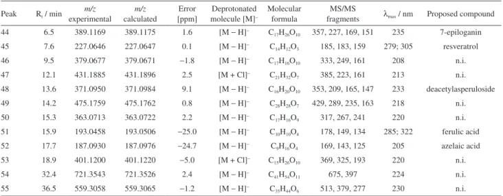
The ESI-MS spectra (negative ion mode) of fraction EtOAc (Table 2) revealed 7-epiloganin, resveratrol, deacetylasperuloside, ferulic and azelaic acids. Based
on the fragmentation characteristics of iridoid-O
-glycosides, the assignment of 7-epiloganin (m/z 389.1169,
[M – H]–) was consistent with fragments at m/z 357.20
and m/z 226.95 in the MS/MS spectrum, due to the
loss of methoxy group and glucose moiety from the
precursor ion, respectively. Elimination of methyl ester from m/z 226.95 [M – H – C2H4O2]– with subsequent
dehydration showed the major product ion at m/z 151.11
[M – H – C2H4O2 – H2O]–.58,59 The UV spectrum and
fragmentation patterns of [M – H]– of resveratrol
(m/z 227.0646) is consistent with previous reports.60,61 The
fragment ion at m/z 185.05 [M – H – CHCOH]– corresponds
to a loss of 42 Da with H rearrangement from the precursor ion (m/z 227.0646). The ions at m/z 159.08 [M – H – C3O2]–
and m/z 183.18 [M – H – CO2]– were formed by a cyclization
reaction of the precursor ion with loss of C3O2 and OH
rearrangement with loss of CO2, respectively.
Based on literature published data, the presence of deacetylasperuloside (m/z 371.0950 [M – H]–) was suggested
by the following ions: m/z 353.04 [M – H – H2O]–, m/z
209.06 [M – H – Glc]–, m/z 165.06 [M – H – Glc – CO 2]–
and m/z 147.05 [M – H – Glc – CO2 – H2O]ˉ.62,63
Ferulic acid was identiied through the deprotonated molecule [M – H]– (m/z 193.0458) that gave the major
fragment ion at m/z 148.92 [M – H – CO2]–. Other fragments
were at m/z 178.08 [M – H – CH3]– and m/z 134.12
[M – H – CH3 – CO2]–.64,65 The UV values correspond to
this phenolic compound.66
The deprotonated molecule at m/z 187.0930 [M – H]–
for azelaic acid produced the fragment ions at m/z: 169.03
[M – H – H2O]ˉ, 143.10 [M – H – CO2]ˉ and 124.98
[M – H – CO2 – H2O]ˉ according to mass bank record and
UV spectrum.67,68
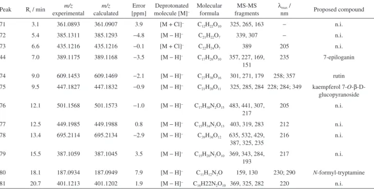
In the polar fraction eluted with MeOH it was detected hordenine, eusterol, 8-amino-7-oxononanoic acid, syringol/vanillic alchool/hydroxytyrosol, stryctosidine,
N-methyl-1,2,3,4-tetrahydro-β-carboline and a pyrogallol related compound, in positive mode. 7-Epiloganin, rutin,
kaempferol 7-O-β-D-glucopyranoside and N
-formyl-tryptamine were identiied in the negative mode (Tables 3 and 4). The alkaloids stryctosidine, N
-methyl-1,2,3,4-tetrahydro-β-carboline and the flavonoids rutin and
kaempferol 7-O-β-D-glucopyranoside have been reported in other Psychotria species.36,69,70
Hordenine yielded [M + H]+ at m/z 166.1215 and a
major fragment at m/z 121.01 [M + H – HN(CH3)2]+,
which then loses H2O yielding an ion at m/z 103.07
[M + H – HN(CH3)2 – H2O]+. Stryctosidine, already
reported in genus Psychotria, was detected as [M + H]+
at m/z 531.2283, that loses OH [M + H – OH]+ yielding a
fragment at m/z 514.19 and at m/z 369.17, due to loss of
the sugar moiety [M + H – Glc]+.71 The alkaloid N
-methyl-1,2,3,4-tetrahydro-β-carboline [M + H]+ at m/z 187.1226
yielded ions at m/z 158.09 and m/z 144.16 due to loss of
CH3N [M + H – CH3N]+ and C2H5N [M + H – C2H5N]+
The lavonoid rutin was detected as the deprotonated molecule [M – H]– at m/z 609.1453. The fragment ions,
resulting from the cleavage of the glycosidic bond with the loss of 308 Da corresponds to a rhamnose (146 Da) plus a glucose (162 Da) moiety followed by loss of CH2O
fragment, at m/z 301.06 [M – H – Rham – Glc]– and 271.10
[M – H – Rham – Glc – CH2O]–, respectively. All spectral
data, mass and UV, are in according to Tiberti et al.73
and Lopes-Lutz et al.74 The molecular formula C21H20O11
([M – H]– at m/z 447.1827) was associated to kaempferol
7-O-β-D-glucopyranoside, that gave an ion at m/z 285.08
[M – H – Glc]– due to known O-glucosides fragmentation
pattern.75 Consistent with the UV spectra and fragmentation
behavior, a homolytic cleavage is observed, generating an aglycone ion at m/z 284.13.76
The alkaloid N-formyl tryptamine was detected in
negative mode ([M – H]ˉ at m/z 187.0934) and exhibited
two fragments: one major at m/z 159.05 due the loss of
CO moiety [M – H – CO]ˉ, and other at m/z 130.11 after a
supposing loss of CHNH2 [M – H – CHNH2]ˉ.
The occurrence of hordenine, resveratrol and ferulic acid are being reported for the irst time in Psychotria
genus.
This preliminary phytochemical screening by GC-MS and LC-MS proved to be an useful tool to identify secondary metabolites from P. nemorosa leaves, both polar
and nonpolar bioactive compounds successfully mapped (Figure 2).
Antinociceptive activity
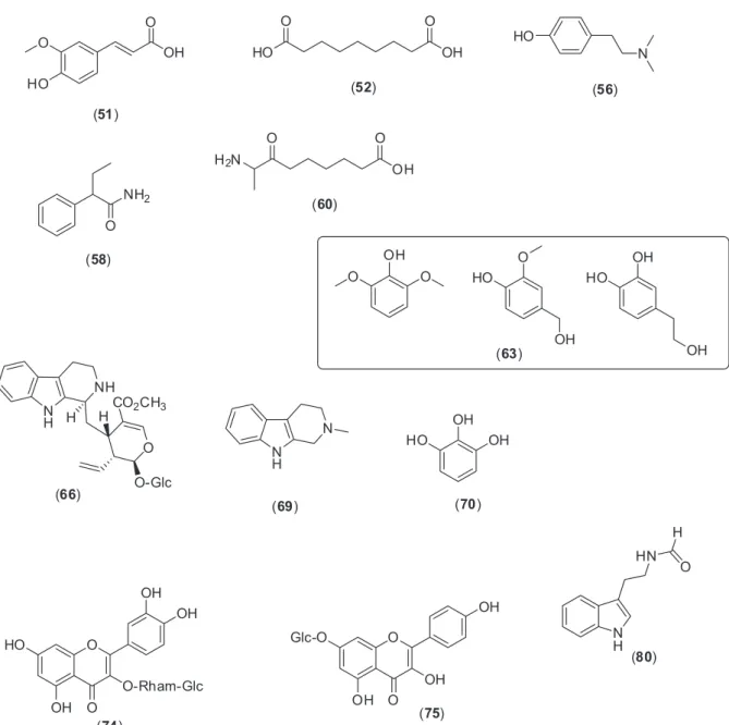
As shown in Figure 3, the EE of P. nemorosa leaves
exhibited a signicant antinociceptive activity on formalin-induced licking response test in mice, but the EE from branches did not (data not shown). The injection of formalin (2.5%) leads to a biphasic licking response of the injected
Table 1. Characterization of the chemical composition of EtOAc fraction of P. nemorosa leaves by UFLC-DAD-ESI-MS2 in positive mode
Peak Rt / min experimentalm/z calculatedm/z
Error [ppm]
Protonated molecule [M]+
Molecular formula
MS/MS
fragments λmax / nm Proposed compound 36 14.8 273.1108 273.1112 −1.5 [M + H]+ C
15H12O5 255, 245, 229 274; 318 butin 37 16.8 197.1163 197.1172 4.6 [M + H]+ C
11H16O3 179, 164, 112 217 loliolide 38 19.9 385.1259 385.1258 0.4 [M + Na]+ C
19H22O7 353, 327, 309 210 gibberellin related compound 39 25.4 316.2820 316.2842 −8.2 [M + H]+ C
18H37NO3 298, 280, 250 223 dehydrophytosphingosine 40 26.5 318.2974 318.3003 9.1 [M + H]+ C
18H39NO3 300, 282, 265 223 phytosphingosine 41 28.9 412.3161 412.3188 6.4 [M + K]+ C
22H47NO3 394, 376, 346 223 2-amino-1,3,4-docosanetriol 42 31.8 353.2645 353.2662 −5.0 [M + Na]+ C
19H38O4 335, 262, 248 224 2-palmitoylglycerol
43 34.4 629.3601 629.3601 0 [M + Na]+ C
41H50O4 611, 447, 429 223 n.i. n.i.: not identiied.
Table 2. Characterization of the chemical composition of EtOAc fraction of P. nemorosa leaves by UFLC-DAD-ESI-MS2 in negative mode
Peak Rt / min m/z experimental m/z calculated Error [ppm] Deprotonated molecule [M]−
Molecular formula
MS/MS
fragments λmax / nm Proposed compound 44 6.5 389.1169 389.1175 1.6 [M − H]− C
17H26O10 357, 227, 169, 151 235 7-epiloganin 45 7.6 227.0646 227.0647 0.1 [M − H]− C
14H12O3 185, 183, 159 279; 305 resveratrol 46 9.5 379.0677 379.0671 −1.8 [M − H]− C
17H16O10 333, 249, 161 208 n.i. 47 12.1 431.1885 431.1896 2.5 [M + Cl]− C
21H32O7 385, 223, 161 213 n.i. 48 13.6 371.0950 371.0984 9.1 [M − H]− C
16H20O10 353, 209, 165, 147 233 deacetylasperuloside 49 14.2 475.1759 475.1762 0.8 [M − H]− C
28H28O7 429, 289, 235, 163 218 n.i. 50 15.3 363.0713 363.0722 2.2 [M − H]− C
17H16O9 317, 267, 241 220 n.i. 51 15.9 193.0458 193.0506 −25.0 [M − H]− C
10H10O4 178, 149, 134 285; 322 ferulic acid 52 17.7 187.0930 187.0976 −24.7 [M − H]− C
9H16O4 169, 143, 125 205 azelaic acid 53 18.9 401.1200 401.1220 −5.0 [M + Cl]− C
15H26O10 369, 325, 193 220 n.i. 54 32.4 721.3543 721.3526 2.4 [M − H]− C
41H54O11 675, 397 224 n.i. 55 36.5 559.3058 559.3065 −1.2 [M − H]− C
paw. The irst phase lasts until 5 min after injection due to a direct stimulation of nociceptors, and the second phase occurs between 15 and 30 min after formalin injection due to a combination of an inlammatory reaction in the peripheral tissue and changes in central processing.31
The anti-inlammatory non-steroidal drug acetylsalicylic acid (ASA) reduced the time by 13.0% and the opioid analgesic morphine by 44.8%. Ethanol extract (EE) inhibited in 64.4, 62.5 and 67.2% the licking time at the doses of 10, 30, and 100 mg kg-1, respectively (Figure 3), Table 3. Characterization of the chemical composition of MeOH fraction of P. nemorosa leaves by UFLC-DAD-ESI-MS2 in positive mode
Peak Rt / min
m/z experimental m/z calculated Error [ppm] Protonated molecule [M]+
Molecular formula
MS/MS fragments
λmax /
nm Proposed compound 56 3.2 166.1215 166.1226 −7.2 [M + H]+ C
10H15NO 121, 103, 91 278 hordenine 57 4.4 328.1719 328.1731 −3.6 [M + Na]+ C
14H27NO6 166, 121 − n.i. 58 6.0 164.1054 164.1046 4.7 [M + H]+ C
10H13NO 147, 119, 91 260 eusterol 59 6.4 310.1254 310.1261 2.3 [M + Na]+ C
13H21NO6 292, 264, 166 206 n.i. 60 6.8 210.1111 210.1101 −4.9 [M + Na]+ C
9H17NO3 192, 166 215 8-amino-7-oxononanoic acid 61 8.0 244.0309 244.0330 8.5 [M + K]+ C
6H11N3O5 205, 202 202 sugar containing azide 62 8.5 360.1757 360.1765 2.3 [M + H]+ C
15H25N3O7 315, 297, 191 202 n.i. 63 11.2 177.0533 177.0546 7.2 [M + Na]+ C
8H10O3 145, 159 229; 282 syringol/vanillic alcohol/ hydroxytyrosol 64 11.5 207.1367 207.1356 −5.4 [M + Na]+ C
11H20O2 190, 152, 139 209 n.i. 65 11.8 374.1917 374.1922 −1.1 [M + H]+ C
16H27N3O7 329, 297, 209, 173 214 n.i. 66 14.1 531.2283 531.2313 5.7 [M + H]+ C
27H34N2O9 514, 369 217 stryctosidine 67 16.6 261.1085 261.1081 1.7 [M + H]+ C
10H16N2O6 243, 201, 177 219 n.i. 68 17.5 461.1913 461.1918 −1.1 [M + H]+ C
23H28N2O8 371, 299, 158 219 n.i. 69 18.6 187.1226 187.1233 2.2 [M + H]+ C
12H14N2 158, 144 219 N
-methyl-1,2,3,4-tetrahydro-β-carboline 70 20.4 149.0207 149.0209 1.8 [M + Na]+ C
6H6O3 132, 131 275 pyrogallol related compound n.i.: not identiied.
Table 4. Characterization of the chemical composition of MeOH fraction of P. nemorosa leaves by UFLC-DAD-ESI-MS2 in negative mode
Peak Rt / min
m/z experimental m/z calculated Error [ppm] Deprotonated molecule [M]−
Molecular formula
MS-MS fragments
λmax /
nm Proposed compound
71 3.1 361.0893 361.0907 3.9 [M + Cl]− C
12H22O10 325, 265, 163 − n.i. 72 5.4 385.1311 385.1293 −4.8 [M − H]− C
21H22O7 339, 307 − n.i.
73 6.6 435.1216 435.1216 −0.1 [M + Cl]− C
22H24O7 389 205 n.i.
44 7.0 389.1175 389.1168 −3.5 [M − H]− C
17H26O10 357, 227, 169, 151
235 7-epiloganin
74 9.0 609.1453 609.1469 −2.1 [M − H]− C
27H30O16 301, 271, 179 258; 357 rutin 75 9.5 447.1827 447.1832 −0.9 [M − H]− C
21H20O11 325, 285, 284 228; 284; 349 kaempferol 7-O-β -D-glucopyranoside 76 12.1 501.1568 501.1573 −1.0 [M − H]− C
17H30N2O15 483, 441, 307, 217
205 n.i.
77 12.5 449.1985 449.1988 0.8 [M − H]− C
15H34N2O13 403, 319, 283 212 n.i. 78 13.4 695.2114 695.2134 −2.9 [M − H]− C
39H36O12 635, 532, 429, 387, 325, 235
216 n.i.
79 15.5 387.1059 387.1045 3.5 [M − H]− C
15H20N2O10 369, 343, 284, 193
217 n.i.
80 18.1 187.0934 187.0949 7.9 [M − H]− C
11H12N2O 159, 130 230; 290 N-formyl-tryptamine 81 20.7 401.1213 401.1202 1.9 [M − H]− C
when compared with the vehicle group (42.4 ± 7.7) in the irst phase. Analysis of the 2nd phase of the response
to formalin showed a signiicant inhibition at doses of 30 and 100 mg kg-1, with the following inhibition values: 44.2
and 62.4%, respectively. The positive control groups ASA and morphine showed 46.7 and 33.3% of reduction in the licking response, respectively.
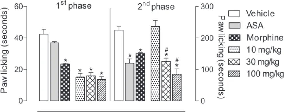
In the search for compounds exhibiting antinociceptive activity, the fractions obtained from silica gel column chromatography were tested on formalin-induced licking response in mice, and all of them were able to inhibit the phases of response to formalin (Figures 4 and 5). Only hexane/EtOAc 10% (fraction 9 that contains the sterols) and hexane/EtOAc 20% were not tested due to the small amount.
Figure 4A shows that the hexane fraction (fraction 1) was able to signiicantly inhibit the irst phase at the dose of 100 mg kg-1 (41.3% of inhibition) and in the second phase
at the dose of 30 mg kg-1 (41.7% of inhibition). The hexane/
EtOAc 5% fraction (fraction 5) was able to signiicantly inhibit only the irst phase at the dose of 30 and 100 mg kg-1
(45.7 and 40.1% of inhibition, respectively) (Figure 4B). The hexane/EtOAc 10% fraction (Fraction 8) was able to signiicantly inhibit the irst phase at the doses of 30 and 100 mg kg-1 (47.2 and 55.6% of inhibition, respectively)
and in the second phase at the doses of 10 and 30 mg kg-1
(50.0 and 55.3% of inhibition, respectively) (Figure 4C). As previously mentioned, fraction 1 contains cinnamic
acid, dihydroactinidiolide, 4-hydroxy-β-ionone and
phytol, compounds that previously showed antinocicepive
activity.77-80 A mixture of oxidized and esteriied triterpenes
were observed in fraction 5, as also α/β-amyrin triterpenes in fraction 8, well known for their analgesic and anti-inlammatory roles.81-84
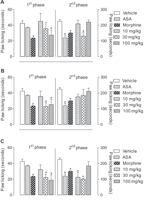
Figure 5A shows that EtOAc fraction was able to signiicantly inhibit the irst phase at the doses of 10, 30 and 100 mg kg-1 (61.7, 46.7 and 63.7% of inhibition,
respectively) and in the second phase at the dose of
100 mg kg-1 (36.7% of inhibition). On the other hand,
MeOH fraction was able to signiicantly inhibit the irst phase and the second phase at the doses of 10, 30 and 100 mg kg-1 (71.2, 59.7 and 52.4%; 41.7, 63.5 and 59.5%
of inhibition, respectively) (Figure 5B). This effect may be attributed, at least partially, to the phenolic compounds butin,85 resveratrol,86 ferulic acid87 and the glycosilated
iridoid loganin,88 present in EtOAc fraction. Next, in the
MeOH fraction, the great inhibition may be due to the presence of alkaloids,21,89,90 lavonoids91,92 and phenolic
compounds.79,93 We also tested whether the extracts could
cause some effect on motor activity. The results obtained after treatment of the mice with doses of 100 mg kg-1 did
not show any effect on motor performance of mice (data not shown).
The model used to start the studies was the formalin-induced licking response, as it can discriminate pain in its central and/or peripheral components; this test studies moderate and tonic pain.94 It has two distinct phases,
which relect two different types of pain. The irst phase (neurogenic pain) is characterized by direct chemical stimulation of nociceptors, afferent ibers predominantly
type C and, in part, type Aδ, while the second phase
(inlammatory pain) is characterized by the appearance of a local inlammatory process, where various proinlammatory chemical mediators are released and that can be inhibited by nonsteroidal anti-inlammatory drugs.95,96
The results showed that the EE and MeOH fraction (10,
Figure 3. Effects of EE extract from P. nemorosa on formalin-induced licking response in mice. Animals were pre-treated with different doses (10, 30 or 100 mg kg-1, p.o.) of EE, ASA (200 mg kg-1), morphine (2.5 mg kg-1) or vehicle. The results are presented as mean ± S.D. (n = 6) of the time that the animal spent licking the formalin-injected paw. Statistical signiicance was calculated by ANOVA followed by Bonferroni’s test. *p < 0.05 when compared
to vehicle-treated mice; #p < 0.05 when compared to morphine-treated mice.
30 and 100 mg kg-1) signiicantly inhibit both phases of the
formalin-induced licking response. The fractions hexane
(100 mg kg-1), hexane/EtOAc 5%, 10% and EtOAc (10,
30 and 100 mg kg-1) signiicantly inhibit the irst phase.
The results of the irst phase suggest an antinociceptive activity that can result in both direct action on opioid receptors (predominantly type µ), present in primary afferent ibers (type C) or even an inhibition in the release of mediators such as serotonin, substance P, kinins, histamine and calcitonin gene-related peptide (CGRP). An inhibitory effect shown by EE (30 and 100 mg kg-1),
hexane (30 mg kg-1) and hexane/EtOAc 10% (10 and
30 mg kg-1), EtOAc (100 mg kg-1) and MeOH (10, 30 and
100 mg kg-1) in the second phase of formalin may suggest
an inhibition in the formation and/or release of arachidonic acid metabolites such as prostaglandins and leukotrienes and other inlammatory mediators such as bradykinin, histamine and serotonin, as well as cytokine, eicosanoids, kinins, glutamate and nitric oxide (NO).94,95
Conclusion
In summary, it was disclosed a concise and useful way to analyze both nonpolar and polar fractions from P. nemorosa
leaves through dereplication strategy. Compounds as cinnamic acid, dihydroactinidiolide, 4-hydroxy-β-ionone, phytol, isophytol, and 4,8,12,16-tetramethylheptadecan-4-olide and mainly the triterpenoids α/β-amyrin, α/β
-amyrone, lupeol, α/β-amyrin acetate,
3,11-dioxo-α/β-amyrene and the steroids stigmast-4-en-3-one,
Figure 4. Effects of P. nemorosa (A) hexane fraction (fraction 1); (B) hexane/EtOAc 5% fraction (fraction 5) and (C) hexane/EtOAc 10% fraction (fraction 8), on formalin-induced licking response in mice. Animals were pre-treated with different doses (10, 30 or 100 mg kg-1, p.o.) of fractions, ASA (200 mg kg-1), morphine (2.5 mg kg-1) or vehicle. The results are presented as mean ± S.D. (n = 6) of the time that the animal spent licking the formalin-injected paw. Statistical signiicance was calculated by ANOVA followed by Bonferroni’s test. *p < 0.05 when compared to vehicle-treated mice.
kaempferol 7-O-β-D-glucopyranoside; the iridoid
glycosides deacetylasperuloside, epiloganin, and alkaloids as hordenine, stryctosidine, N -methyl-1,2,3,4-tetrahydro-β-carboline and N-formyl-tryptamine.
Furthermore, to the best of our knowledge, this is the irst work describing the phytochemical proile and antinociceptive properties of P. nemorosa leaves. The extract
and fractions were able to produce an oral antinociceptive effect in acute pain model in mice (formalin-induced licking response).
Supplementary Information
Supplementary data are available free of charge at http://jbcs.sbq.org.br as a PDF ile.
Acknowledgments
Figure 5. Effects of P. nemorosa (A) EtOAc and (B) MeOH fractions on formalin-induced licking response in mice. Animals were pre-treated with different doses (10, 30 or 100 mg kg-1, p.o.) offractions, ASA (200 mg kg-1), morphine (2.5 mg kg-1) or vehicle. The results are presented as mean ± S.D. (n = 6) of the time that the animal spent licking the formalin-injected paw. Statistical signiicance was calculated by ANOVA followed by Bonferroni’s test. *p < 0.05
when compared to vehicle-treated mice; #p < 0.05 when compared to morphine-treated mice.
Sintéticos (NPPS), Department of Physics and Chemistry, Faculty of Pharmaceutical Sciences, University of São Paulo) for mass spectrometer assistance.
References
1. Calixto, J. B.; Braz. J. Med. Biol. Res. 2000, 33, 179.
2. Patwardhan, B.; J. Ethnopharmacol. 2005, 100, 50.
3. Cordell, G. A.; Shin, Y. G.; Pure Appl. Chem. 1999, 71,
1089.
4. Ernst, M.; Silva, D. B.; Silva, R. R.; Vêncio, R. Z.; Lopes, N. P.; Nat. Prod. Rep. 2014, 31, 784.
5. Lang, G.; Mayhudin, N. A.; Mitova, M. I.; Sun, L.; van der Sar, S.; Blunt, J. W.; Cole, A. L. J.; Ellis, G.; Laatsch, H.; Munro, M. H. G.; J. Nat. Prod.2008, 71, 1595.
6. He, M.; Lv, H.-Y.; Li, Y.-P.; Gonçalves, C. M. V.; Dong, N.-P.; Pan, L.-S.; Liu, P.-L.; Liang, Y.-Z.; Anal. Methods 2014, 6, 2239.
7. Pinto, A. C.; Vessecchi, R.; Silva, C. G.; Amorim, A. C. L.; Santos Júnior, H. M.; Rezende, M. J. C.; Gates, P. J.; Rezende, C. M.; Lopes, N. P.; Rapid Commun. Mass Spectrom.2015, 29, 1. 8. Davis, A. P.; Bridson, D.; Jarvis, C.; Govaerts, R. L.; Bot. J.
Linn. Soc. 2001, 135, 35.
9. Razaimandimbison, S. G.; Taylor, C. M.; Wikström, N.; Pailler, T.; Khodabandeh, A.; Bremer, B.; Am. J. Bot. 2014, 101, 1102. 10. Watt, J. M.; Breyer-Brandwijk, M. G.; The Medicinal and
Poisonous Plants of Southern and Eastern Africa, 2nd ed.; E.
& S. Livingstone: London, United Kingdom, 1962.
11. Locher, C. P.; Burch, M. T.; Mower, H. F.; Berestecky, J.; Davis, H.; Van Poel, B.; Lasure, A.; Vanden Berghe, D. A.; Vlietinck, A. J.; J. Ethnopharmacol. 1995, 49, 23.
12. McGaw, L. J.; Jager, A. K.; van Staden, J.; J. Ethnopharmacol. 2000, 72, 247.
13. Moraes, T. M.; Araújo, M. H.; Bernardes, N. R.; Oliveira, D. B.; Lasunskaia, E. B.; Muzitano, M. F.; Cunha, M.; Planta Med.
2011, 77, 964.
14. Witherup, K. M.; Bogusky, M. J.; Anderson, P. M.; Ramjit, H.; Ransom, R. W.; Wood, T.; Sardana, M.; J. Nat. Prod. 1994, 57,
1619.
15. Lopes, S. O.; Poser, G. L. V.; Kerber, V. A.; Farias, F. M.; Konrath, E. L.; Moreno, P.; Sobral, M. E.; Zuanazzi, J. A. S.; Henriques, A. T.; Biochem. Syst. Ecol. 2004, 32, 1187.
16. Formagio, A. S. N.; Volobuff, C. R. F.; Santiago, M.; Cardoso, C. A. L.; Vieira, M. C.; Pereira, Z. V.; Antioxidants 2014, 3,
17. Moreno, B. P.; Fiorucci, L. L. R.; Carmo, M. R. B.; Sarragiotto, M. H.; Baldoqui, D. C.; Biochem. Syst. Ecol. 2014, 56, 80.
18. Taylor, C.; http://loradobrasil.jbrj.gov.br/jabot/loradobrasil/ FB14153, accessed in July 2016.
19. Paiva, A. M.; Lopes, R. C.; http://www.anchietano.unisinos.br/ publicacoes/botanica/botanica64/03_paiva%20e%20lopes.pdf, accessed in July 2016.
20. Both, F. L.; Farias, F. M.; Nicoláo, L. L.; Misturini, J.; Henriques, A. T.; Elisabetsky, E.; Rev. Bras. Plant. Med. 2002,
5, 41.
21. Elisabetsky, E.; Amador, T. A.; Albuquerque, R. R.; Nunes, D. S.; Carvalho, A. C. T.; J. Ethnopharmacol. 1995, 48, 77.
22. Castana, O.; Anagiotos, G.; Rempelos, G.; Adalopoulou, A.; Kokkinakis, C.; Giannakidou, M.; Diplas, D. B.; Alexakis, D.;
Ann. Burns Fire Disasters 2009, 22, 88. 23. Niv, D.; Kreitler, S.; Pain Pract. 2001, 1, 150.
24. Woodcock, J.; Witter, J.; Dionne, R. A.; Nat. Rev. Drug Discov. 2007, 6, 703.
25. Patitucci, M. L.; Veiga Jr., V. F.; Pinto, A. C.; Zoghbi, M. G. B.; Silva, J. R. A.; Quim. Nova 1995, 18, 262.
26. Bandeira, P. N.; Lemos, T. L. G.; Costa, S. M. A.; Santos, H. S.; Rev. Bras. Farmacogn. 2007, 17, 204.
27. Barnes, R. A.; Pereira, A. L.; Scoield, T. C.; Braz-Filho, R.; Pinto, A. C.; Chem. Pharm. Bull. 1984, 32, 3674.
28. Pinto, A. C.; Pereira, A. L.; Kelecom, A.; Porreca, L. M.; Ribeiro, N. M.; Barnes, R. A.; Chem. Pharm. Bull. 1988, 36, 4689.
29. Gomes, N. M.; Rezende, C. M.; Fontes, S. P.; Matheus, M. E.; Fernandes, P. D.; J. Ethnopharmacol. 2007, 109, 486.
30. Abad-Garcia, B.; Berrueta, L. A.; Garmon-Lobato, S.; Gallo, B.; Vicente, F.; J. Chromatogr. A 2009, 1216, 5398.
31. Dubuisson, D.; Dennis, S. G.; Pain 1977, 4, 161.
32. Lu, Q.; Wang, J.; Kong, L.; Biochem. Syst. Ecol. 2014, 52, 20.
33. Lu, Q.; Wang, J.; Luo, J.; Wang, X.; Shan, S.; Kong, L.; Nat. Prod. Res. 2014, 28, 1659.
34. Andrade, J. M. M.; Biegelmeyer, R.; Xavier, C. A. G.; Bordignon, S. A. L.; Moreno, P. R. H.; Zuanazzi, J. A. S.; Henriques, A. T.; Apel, M. A.; Chem. Nat. Compd. 2010, 46, 649. 35. Gonzalez, J.; Dieck, T.; Rev. Latinoam. Quim. 1996, 24, 7.
36. Li, H.-F.; Huang, J.; Liu, M.-S.; Zhang, X.-P.; Chin. J. Exp. Trad. Med. Formulae 2011, 19, 125.
37. Pimenta, A. T. A.; Uchôa, D. E. A.; Braz-Filho, R.; De Souza, E. B.; Silveira, E. R.; Lima, M. A. S.; J. Braz. Chem. Soc. 2011, 22, 2216.
38. Achenbach, H.; Lottes, M.; Waibel, R.; Karikas, G. A.; Correa, M. D. A.; Gupta, M. P.; Phytochemistry 1995, 38, 1537. 39. Dan, S.; Dan, S. S.; Fitoterapia 1986, 57, 445.
40. Lee, K.-H.; Lin, Y.-M.; Wu, T.-S.; Zhang, D.-C.; Yamagishi, T.; Hayashi, T.; Hall, I. H.; Chang, J.-J.;Wu, R.-Y.;Yang, T.-H.;
Planta Med. 1988, 54, 308.
41. Assimopoulou, A. N.; Papageorgiou, V. P.; Biomed. Chromatogr. 2005, 19, 285.
42. Dias, M. O.; Hamerski, L.; Pinto, A. C.; Quim. Nova 2011, 34, 704.
43. Branco, A.; Pinto, A. C.; Braz Filho, R.; An. Acad. Bras. Ciênc. 2004, 76, 505.
44. Ermer, J.; Vogel, M.; Biomed. Chromatogr.2000, 14, 373. 45. Chernushevich, I. V.; Loboda, A. V.; Thomson, B. A.; J. Mass
Spectrom. 2001, 36, 849.
46. Demarque, D. P.; Crotti, A. E. M.; Vessecchi, R.; Lopes, J. L. C.; Lopes, N. P. Nat. Prod. Rep. 2016, 33, 432.
47. Tan, M. A.; Panghulan, G. F. M.; Uy, M. M.; Takayama, H.;
Am. J. Essent. Oils Nat. Prod. 2014, 1, 18.
48. Kerber, V. A.; Gregianini, T. S.; Paranhos, J. T.; Schwambach, J.; Farias, F.; Fett, J. P.; Fett-Neto, A. G.; Zuanazzi, J. A. S.; Quirion, J. C.; Elizabetsky, E.; Henriques, A. T.; J. Nat. Prod. 2001, 64, 677.
49. Liu, R.; Ye, M.; Guo, H.; Bi, K.; Guo, D. A.; Rapid Commun. Mass Spectrom. 2005, 19, 1557.
50. Antal, D. S.; Schwaiger, S.; Ellmerer-Muller, E. P.; Stuppner, H.; Planta Med. 2010, 76, 1765.
51. Jin, M. J.; Kim, I. S.; Rehman, S. U.; Dong, M. S.; Na, C. S.; Yoo, H. H.; J. Chromatogr. Sci. 2015, 24, 1.
52. Valdés III, L. J.; J. Nat. Prod. 1986, 49, 171.
53. Takahashi, N.; Murofushi, N.; Yokota, T.; Tamura, S.; Kato, J.; Shiotani, Y.; Tetrahedron Lett. 1967, 48, 4861.
54. Peer, M.; Stegmann, M.; Mueller, M. J.; Waller, F.; FEBS Lett. 2010, 584, 4053.
55. Huang, Y.; Liu, X.; Zhao, L.; Li, F.; Xiong, Z.; Biomed. Chromatogr. 2014, 28, 878.
56. Cravatt, B. F.; Lerner, R. A.; Boger, D. L.; J. Am. Chem. Soc. 1996, 118, 580.
57. Limb, J.-K.; Kim, Y. H.; Han, S.-Y.; Jhon, G.-J.; J. Lipid Res. 1999, 40, 2169.
58. Xiao-Qin, L.; Xiao-Hong, S.; Shuang, C.; Xi-Xiang, Y.; Fa-Mei, L.; Acta Pharm. Sin. B 2009, 44, 895.
59. Quirantes-Piné, R.; Lozano-Sánchez, J.; Herrero, M.; Ibáñez, E.; Segura-Carretero, A.; Fernández-Gutiérrez, A.; Phytochem. Anal. 2013, 24, 213.
60. Trela, B. C.; Waterhouse, A. L.; J. Agric. Food Chem. 1996, 44,
1253.
61. Stella, L.; Rosso, M.; Panighel, A.; Vedova, A. D.; Flamini, R.; Traldi, P.; Rapid Commun. Mass Spectrom. 2008, 22, 3867. 62. Jiang, W.; Jin, D.; Li, Z.; Sun, Z.; Chen, M.; Wu, B.; Huang,
C.; Biomed. Chromatogr. 2012, 26, 863.
63. Wang, X.; Cheng, W.; Yao, X.; Guo, X.; Nat. Prod. Res. 2012, 26, 167.
64. Hossain, M.; Dilip, R.; Brunton, N.; Martin-Diana, A. B.; Barry-Ryan, C.; J. Agric. Food Chem. 2010, 58, 10576.
65. Stanisavljević, N. S.; Ilić, M.; Jovanović, Ž. S.; Čupić, T.; Dabić, D. Č.; Natić, M. M.; Tešić, Ž. L.; Radović, S. S.; Arch. Biol. Sci. (Belgrade) 2015, 67, 829.
67. http://www.massbank.jp/jsp/Dispatcher.jsp?type=disp&id= KO000123&site=0, accessed in July 2016.
68. Kadam, T. V.; Darekar, A. B.; Gondkar, S. B.; Saudagar, R. B.;
Asian J. Res. Pharm. Sci. 2015, 5, 83.
69. Rivier, L.; Lindgren, J.; Econ. Bot. 1972, 26, 101.
70. Berger, A.; Fasshuber, H.; Schinnerl, J.; Brecker, L.; Greger, H.; Phytochem. Lett. 2012, 5, 558.
71. Yamazaki, Y.; Urano, A.; Sudo, H.; Kitajima, M.; Takayama, H.; Yamazaki, M.; Aimi, N.; Saito, K.; Phytochemistry 2003,
62, 461.
72. Gribble, G. W.; Nelson, R. B.; J. Org. Chem. 1974, 39, 1845. 73. Tiberti, L. A.; Yariwake, J. H.; Ndjokob, K.; Hostettmann, K.;
J. Braz. Chem. Soc. 2007, 18, 100.
74. Lopes-Lutz, D.; Dettmann, J.; Nimalaratne, C.; Schieber, A.;
Molecules 2010, 15, 8543.
75. Sánchez-Rabaneda, F.; Jáuregui, O.; Casals, I.; Andrés-Lacueva, C.; Izquierdo-Pulido, M.; Lamuela-Raventó, R. M.; J. Mass Spectrom. 2003, 38, 35.
76. Schieber, A.; Mihalev, K.; Berardini, N.; Mollov, P.; Carle, R.;
Z. Naturforsch, C.: J. Biosci. 2005, 60, 379.
77. Gershenzon, J.; Dudareva, N.; Nat. Chem. Biol. 2007, 3, 408. 78. Zhang, J.; Rapier, A.; Radhakrishnan, R.; Light, A.; J. Pain
2010, 11, S52.
79. Cavalcante-Silva, L. H. A.; Matta, C. B. B.; Araújo, M. V.; Barbosa-Filho, J. M.; Lira, D. P.; Santos, B. V. O.; Miranda, G. E. C.; Alexandre-Moreira, M. S.; Mar. Drugs 2012, 10, 1977.
80. Santos, C. C. M. P.; Salvadori, M. S.; Mota, V. G.; Costa, L. M.; Almeida, A. A. C.; Oliveira, G. A. L.; Costa, J. P.; Sousa, D. P.; Freitas, R. M.; Almeida, R. N.; Neurosci. J. 2013, 2013, 1. 81. Mahato, S. B.; Nandy, A. K.; Roy, G.; Phytochemistry 1992,
31, 2199.
82. Pinto, S. A. H.; Pinto, L. M. S.; Guedes, M. A.; Cunha, G. M. A.; Chaves, M. H.; Santos, F. A.; Rao, V. S.; Phytomedicine 2008, 15, 630.
83. Soldi, C.; Pizzolatti, M. G.; Luiz, A. P.; Marcon, R.; Meotti, F. C.; Mioto, L. A.; Santos, A. R. S.; Bioorg. Med. Chem.2008, 16, 3377.
84. Fingolo, C. E.; Santos, T. S.; Vianna Filho, M. D. M.; Kaplan, M. A. C.; Molecules 2013, 18, 4247.
85. Bhargava, S. K.; J. Ethnopharmacol. 1986, 18, 95.
86. Torres-López, J. E.; Ortiz, M. I.; Castañeda-Hérnandez, G.; Alonso-López, R.; Asomoza-Espinosa, R.; Granados-Soto, V.;
Life Sci. 2002, 70, 1669.
87. Lv, W. H.; Zhang, L.; Wu, S. J.; Chen, S. Z.; Zhu, X. B.; Pan, J. C.; Zhongguo Zhongyao Zazhi 2013, 38, 3736.
88. Gong, N.; Fan, H.; Ma, A.-N.; Xiao, Q.; Wang, Y.-X.;
Neuropharmacology 2014, 84, 31.
89. Elisabetsky, E.; Castilhos, Z. C.; Int. J. Crude Drug Res. 1990, 28, 49.
90. Radulovic, N. S.; Blagojevic, P. D.; Randjelovic, P. J.; Stojanovic, N. M.; Curr. Top Med. Chem. 2013, 13, 2134. 91. De Melo, G. O.; Malvar, D. C.; Vanderlind, F. A.; Rocha, F. F.;
Pires, P. A.; Costa, E. A.; Matos, L. G.; Kaiser, C. R.; Costa, S. S.; J. Ethnopharmacol. 2009, 124, 228.
92. Selvaraj, G.; Kaliamurthi, S.; Thirungnasambandam, R.; Vivekanandan, L.; Balasubramanian, T.; Biomed. Environ. Sci. 2014, 27, 295.
93. Küpeli, E.; Sahin, F. P.; Yesilada, E.; Caliș, İ.; Ezer, N.; Z. Naturforsch. C: J. Biosci. 2007, 62, 519.
94. Hunskaar, S.; Fasmer, O. B.; Hole, K.; J. Neurosci. Methods 1985, 14, 69.
95. Hunskaar, S.; Hole, K.; Pain 1987, 30, 103.
96. Chichorro, J. G.; Lorenzetti, B. B.; Zampronio, A. R.; Br. J. Pharmacol. 2004, 141, 1175.
Submitted: May 9, 2016 Published online: July 25, 2016