A
RTIGOC
IENTÍFICO Revista Brasileira de FisioterapiaAssessment of neuromuscular
activation in individuals with scoliosis
using surface electromyography
Avaliação da ativação neuromuscular em indivíduos com escoliose
através da eletromiografia de superfície
Bassani E, Candotti CT, Pasini M, Melo M, La Torre M
Abstract
Objective: The aim of this study was to investigate the potential of surface electromyography (EMG) for assessing neuromuscular efficiency and localized muscle fatigue in the lumbar extensors, in individuals with scoliosis. Methods: Twenty individuals participated in this study, divided equally into two groups: (1) Scoliosis Group and (2) Control Group. These subjects underwent a fatigue induction test on their lumbar extensor muscles, consisting of one maximum voluntary isometric contraction (MVIC) followed by a test at 80% of the MVIC effort. Force and EMG signals were collected simultaneously. The EMG signal was processed in the frequency domain by means of fast Fourier transforms using the median frequency; and in the time domain by calculating the root mean square value. The data were analyzed by means of one-way analysis of variance to investigate the differences between the two groups. Paired t test was used to investigate the symmetry between the right and left sides. The significance level adopted was 0.05. Results: The results showed that the individuals with scoliosis presented: (1) symmetrical neuromuscular activation between the sides; (2) lower neuromuscular efficiency; (3) greater capacity to resist fatigue; and (4) force values 42.6% lower than those of the individuals in the Control Group. Conclusions:
The results suggest that surface EMG is an effective tool for functional assessments of scoliosis, although the protocol established limited the participation of individuals with scoliosis, from the perspective of neuromuscular efficiency.
Key words: electromyography; fatigue; scoliosis.
Resumo
Objetivo: O objetivo desse estudo foi verificar o potencial da eletromiografia (EMG) de superfície para a avaliação da eficiência neuromuscular e da fadiga muscular localizada dos extensores lombares em indivíduos com escoliose. Métodos: Participaram deste estudo 20 indivíduos divididos igualmente em dois grupos, (1) Grupo com Escoliose e (2) Grupo Controle, que foram submetidos a um teste de indução dos músculos extensores lombares a fadiga, o qual constituiu da realização de uma contração voluntária máxima isométrica (CVM), e realização de um teste com esforço a 80% da CVM. Foram coletados simultaneamente sinais de força e eletromiográficos (sinal EMG). O sinal EMG foi processado no domínio da freqüência, utilizando-se a transformada rápida de Fourier (FFT), por meio da mediana da freqüência (MF), e no domínio do tempo, pelo cálculo do valor root mean square (RMS). Os dados foram submetidos a uma análise de variância one-way para verificar as diferenças entre os dois grupos. Para verificar a simetria entre os lados direito e esquerdo, foi realizado o teste t pareado. O nível de significância adotado foi 0,05. Resultados: os resultados demonstraram que indivíduos com escoliose apresentaram: (1) simetria de ativação neuromuscular entre os lados; (2) menor eficiência neuromuscular; (3) maior capacidade de resistir a fadiga; e (4) valores de força 42,6% menores que os indivíduos do GC. Conclusões:
Os resultados sugerem que a EMG de superfície corresponde a um efetivo instrumento de avaliação funcional da escoliose, embora o protocolo estabelecido tenha limitado a participação dos indivíduos com escoliose, do ponto de vista da eficiência neuromuscular.
Palavras-chave: eletromiografia; fadiga; escoliose.
Recebido: 14/12/2006 – Revisado: 12/06/2007 – Aceito: 1/11/2007
Universidade do Vale do Rio dos Sinos (Unisinos) – São Leopoldo (RS), Brazil
Correspondence to: Cláudia Tarragô Candotti, Rua Fernando Osório, 1.887, CEP 91720-330, Porto Alegre (RS), Brazil, e-mail: [email protected]
Introduction
Scoliosis is a complex deformity of the spine, in the three body planes ( frontal, sagittal and transversal), in which the main component is abnormal lateral deviation in the frontal plane1-7. he frequency of occurrence of scoliosis varies,
de-pending on the population studied, the identiication method or the magnitude of curvature, but estimates suggest that the incidence of scoliosis among the general population is around 2 to 4%8, thus representing around 30% of the incidence of
postural deviations9. An epidemiological study in the state
of Maranhão demonstrated that the incidence of idiopathic scoliosis among male children and adolescents was 3.4% and 7.3%, respectively10. Scoliosis is a potentially progressive
condition that compromises body posture5. herefore,
evalu-ations investigating structural or functional references will have a decisive role in the intervention in the progression of this deformity.
A reliable evaluation is the basis for making decisions not only with regard to the treatment, but also for preventing intercurrences11,12. Professionals eforts towards obtaining an
appropriate evaluation, focus on the fact that it is important to obtain a complete picture of the individual’s incapacity and to have criteria for following up the evolution and treatment results13,14. hus, after anamnesis and physical examination,
obtaining x-rays is the most important and fundamental step towards measuring the deformity and determining the curvature, with regard to its correction and the potential for progress. However, for most professionals, the basic principal in this process is to diminish the number of x-rays on each indi-vidual as much as possible, in order to minimize the costs and the exposure to radioactivity15.
Scoliosis basically leads to strength imbalance and trunk muscle impairment, such that the muscles on the concave side of the curvature are contracted and the muscles on the convex side are stretched, which characterizes a problem of muscle asymmetry2. Traditionally, the muscle imbalances in scoliosis
cases are evaluated using muscle function tests. hese muscle action analyses are essential to help in elucidating the diagno-sis and also to make it possible to prescribe therapeutic exerci-ses16. However, muscle function tests are done manually, thus
depending on the evaluator’s ability, and they are very subjec-tive because they do not quantify the individual’s strength level on each side of the trunk5.
Hence, it can be seen that it would be of interest to objec-tively evaluate and quantify the muscle imbalances present in individuals with scoliosis, because the results could also pro-vide backing for treatment prescriptions, well as for following up the evolution of this condition. One way of evaluating the trunk muscle strength would be by using dynamometers,
which provide the strength level used during a speciic move-ment. In this respect, one study in the literature has reported on an evaluation of adolescents with asymptomatic scoliosis that allowed measurement of asymmetrical efort exerted by the trunk extensors during neuromuscular eiciency and muscle fatigue tests17, using a triaxial dynamometry and
surface electromyography. Despite some inconsistencies, the results suggested that surface electromyography was a useful tool for evaluating the muscle imbalances present in scoliosis cases17.
For muscle strength to be exerted, there must previously be neuromuscular activation18. Surface electromyography (EMG)
is a sensitive technique for detecting neuromuscular function that makes it possible to obtain information on neuromuscular activation. hus, by using this technique, it is possible to moni-tor the neuromuscular activation of the trunk muscles, both on the concave and on the convex side of a scoliosis case.
Therefore, the objective of this study was to investigate the potential of surface EMG for evaluating neuromuscu-lar efficiency and localized muscle fatigue in the lumbar extensors among individuals with and without scoliosis. Three presuppositions were hypothesized: (1) neuromuscu-lar activation of the iliocostalis lumborum and longissimus muscles is asymmetrical in individuals with scoliosis and symmetrical in normal individuals; (2) individuals with scoliosis demonstrate lower neuromuscular efficiency than do normal individuals; and (3) individuals with scoliosis de-monstrate higher rates of localized muscle fatigue than do normal individuals.
Methodology
Sample
15
individuals signed a consent statement to participate in the study. his study was approved (CEP no. 03/052) by the Rese-arch Ethics Committee (Resolution 047/2004) of the Univer-sidade do Vale do Rio dos Sinos in which it was performed, and was thereby deemed to be ethically and methodologically appropriate, in accordance with the precepts of Resolution 196/96 of the National Health Council.
Evaluation protocol
To induce trunk extensor muscle fatigue, the individuals underwent a maximum voluntary isometric contraction (MVIC) test lasting ive seconds and, after a two-minute rest period, an isometric contraction test at 80% of MVIC was performed for 35 seconds. he participants were provided with visual feedback of their muscle strength levels, using an oscilloscope (Minipa MO, model 1225, Minipa Electronics, Shanghai). For the fatigue induction test, the subjects were positioned in ventral decubitus on a support, with the armpit, thigh and ankle regions attached to the support using Velcro bands. A 2000N load cell was held in the armpit band, and this was instrumented with strain gauges (Alfa Instrumentos Eletrônicos Ltda, São Paulo) and ixed to the ground. While carrying out this protocol, strength and electromyographic signals were simultaneously recorded.
Signal acquisition
he strength and electromyographic signals were acquired using a 16-channel electromyograph (EMG System do Brasil Ltda, São José dos Campos) and the AqDados sof-tware (Lynx Tecnologia Eletrônica Ltda, São Paulo), with a Pentium 200 MHz computer with 64 MB RAM equipped with an A/D converter (EMG System do Brasil Ltda, São José dos Campos). The strength and electromyographic signals were gathered at a sampling rate of 1000 Hz for each channel.
To record the electromyographic sign (EMG signal), pairs of surface electrodes were used (Ag/AgCl; 1 cm diameter; with attachment adhesive) in a bipolar coniguration, for each muscle. he electrodes were placed on the muscle belly,
at a distance of 2.5 cm from each other20. he muscles
mo-nitored were the longissimus thoracis (at the same level as the irst lumbar vertebra) and the iliocostalis lumborum (at the same level as the ifth lumbar vertebra), both on the right and left sides. he reference electrode was placed at the wrist, on the styloid process of the radius. All the pertinent norms for adequate recording of EMG signals recommended by the International Society of Electrophysiology and Kinesiology21
were rigorously observed.
Signal processing
To process the strength and EMG signals, the SAD32 data acquisition system was used (version 2.61.07mp version, 2002; www. ufrgs.br/lmm). First, the signs were subjected to digital filtering procedures. For the strength signals, a mova-ble mean low-pass filter was used (with a cutoff frequency of 10 Hz), and for the EMG signals, a high-pass filter was used, with a cutoff frequency of 20 Hz. The EMG signals were processed in the time and frequency domains. For the time domain analysis, the RMS values were calculated in fixed one-second windows (1000 points). For the frequency do-main analysis, the median frequency (MF) was calculated in one-second windows (Hamming windowing), from the fast Fourier transform (FFT). The EMG signal was normalized in relation to the highest frequency obtained while performing the protocol22.
he electromyographic response to the fatigue protocol was evaluated using four localized muscle fatigue indices: (1) the inclination coeicient (α) of the straight line that approximated all 30 values of the median frequency23 (2) the
y-intercept (y) of the straight line that approximated all 30 va-lues of the median frequency23 (3) the inclination coeicient
(β) of the straight line that approximated only the irst and last value of the median frequency24 and (4) the y-intercept
(y’) of the line that approximated only the irst and last value of the median frequency24.
To evaluate the neuromuscular eiciency, a linear regres-sion analysis was done on 30 points of the curve of RMS value versus contraction time (30 seconds) to obtain a straight-line equation (equation 1) for each muscle. For inclusion in the * Classification proposed by the Scoliosis Research Society6,19.
** Significant difference between SG and CG: p< 0.05.
Table 1. Means and standard deviations of age, height and body mass of the individuals with scoliosis (SG) and the control group (CG), and the numbers of individuals with scoliosis having left and right convexity, in which the apices do not exceed a Cobb angle of 20º (classified as grade I scoliosis*).
Age (years) Height (cm) Body mass (kg) Scoliosis type
left right
SG (n=10) 35 ± 13 170 ± 10 68 ± 14 6 4
CG (n=10) 25 ± 7 174 ± 10 76 ± 13 0 0
study, the determination coeicient obtained from the linear regression had to be greater than or equal to 0.5.
b
x
y
=
ϕ
⋅
+
Equation 1where:
ϕ= inclination coeicient of the straight line b= y-intercept value
The inclination coefficient (ϕ) was considered to be an index for the neuromuscular efficiency. Since the protocol required the strength to be constantly maintained at 80% of the strength obtained in the MVIC, steeper inclinations in the curve of RMS versus time represented lower neuromus-cular efficiency. To evaluate the activation asymmetry be-tween the right and left sides of the trunk, the four localized muscle fatigue indices and the neuromuscular efficiency index were used.
Statistical treatment
For the statistical treatment, the normality of the data (Sha-piro-Wilk test) and homogeneity of the variance (Wilk’s Lam-bda test) were irst veriied and conirmed. One-way ANOVA was performed to investigate the diferences between the two groups (scoliosis and control) regarding body mass, age, hei-ght, strength values from the MVIC, muscle fatigue indices and neuromuscular eiciency indices. To investigate the symmetry between the right and left sides, the paired Student’s t test was used. he signiicance level used was 0.05.
Results
he results showed that the SG individuals demonstrated signiicantly lower strength values (p= 0.009) than the CG indi-viduals did, such that the SG showed a mean strength of 416 ± 29 N, while the CG demonstrated values of 718 ± 14 N.
Comparing the localized muscle fatigue indices, signiicant diferences between the SG and CG were only shown for the
right longissimus muscle, for three of the four studied fatigue indices (Table 2). hese results indicate that the individuals in the SG showed lower fatigue indices for the right longissimus muscle (Figure 1), probably due to lower strength shown in the MVIC and, consequently, while the fatigue protocol was being conducted. he other muscles (left longissimus, right iliocostalis and left iliocostalis) did not difer signiicantly between the SG and CG groups, for any of the four localized muscle fatigue indices.
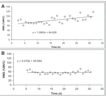
Comparing the neuromuscular efficiency indices, sig-nificant differences between the SG and CG were shown for the longissimus and iliocostalis muscles, but only on the right side for both groups (Table 1). The SG individuals demonstrated greater muscle activation while the fatigue protocol was being conducted on the right longissimus and right iliocostalis muscles, i.e., to maintain the same strength level, they showed a steeper straight-line inclination, with positive inclination coefficients (ϕ), thus indicating lower neuromuscular efficiency (Figure 2). The neuromuscular efficiency indices for the longissimus and iliocostalis mus-cles on the left side, did not differ significantly between the SG and CG.
Symmetry comparisons between the right and left sides of the trunk, using the fatigue and neuromuscular eiciency indi-ces, did not demonstrate any signiicant diferences between the two sides, for any of the four muscles, for either the SG or the CG. his result indicates that the neuromuscular activation level was similar for both sides, as was the fatigue level shown by the muscles, independent of the group to which the indivi-dual belonged.
Discussion
The purpose of this study was to investigate the poten-tial for using surface EMG to evaluate the neuromuscular efficiency and localized muscle fatigue of the lumbar exten-sors among individuals with scoliosis. The results showed that the individuals in the SG group presented significantly lower strength values than did the individuals in the CG.
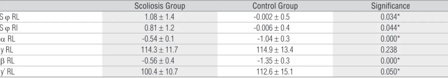
Scoliosis Group Control Group Significance
RMS ϕ RL 1.08 ± 1.4 -0.002 ± 0.5 0.034*
RMS ϕ RI 0.81 ± 1.2 -0.006 ± 0.4 0.044*
MF α RL -0.54 ± 0.1 -1.04 ± 0.3 0.000*
MF y RL 114.3 ± 11.7 114.9 ± 13.4 0.238
MF β RL -0.56 ± 0.4 -1.35 ± 0.3 0.000*
MF y’ RL 100.4 ± 10.7 112.6 ± 15.1 0.050*
Table 2. Mean and standard deviationvalues of the neuromuscular efficiency indices (ϕ) and localized muscle fatigue indices (α, y, β, y’), and p
values obtained via ANOVA, comparing the scoliosis and control groups (RL: right longissimus; RI: right iliocostalis).
17
One possible explanation for this result is that the SG indivi-duals had lumbar pain and, for this reason, they might have not used their maximum strength during the MVIC. It has been reported that individuals with pain tend to put a pain protection mechanism into action, which stops them from attaining maximum effort with the muscles in question25, 26.
Therefore, pain has an important role in body protection, because the changes in motor recruitment in an individual who suffers from pain may be due to some type of strategic control that the nervous system performs through a specific neural route27. The effects of this neural mechanism suggest
that there is a reduction in agonist activation and an incre-ase in antagonist activation27.
Some studies have presumed that, when people with pain perform an MVIC test, they attain submaximal efort because of their own pain situation, and that this has an inluence on the fatigue test result when based on a protocol of, for example, 80% MVIC28. he results from the present study go
along with that supposition, since the individuals in the SG presented strength values 42.6% lower than those of the CG, probably because they did not exert their maximum strength during the MVIC.
It has been documented that, in muscle fatigue situations like those occurring during sustained contractions, there is an increase in RMS and a decrease in MF of the EMG signal23,24.
hus, while performing muscle fatigue protocols, it is common to observe spectral compression towards the lower frequen-cies, thereby decreasing the MF. he reduction in MF that oc-curs because of decreased conduction velocity of the muscle iber action potential has been accepted by many researchers
as an indicator for the muscle fatigue that occurs during sustai-ned isometric contraction20,21,23,29. he results from the present
study showed this behavior, such that the SG reported signii-cantly lower muscle fatigue indices and therefore a straight line sloping less steeply, thereby demonstrating greater capacity to resist fatigue than did the individuals in the CG (Figure 1). Initially, it had been speculated that individuals with scoliosis would show a steeper slope on the MF straight line and would be more susceptible to muscle fatigue than would be the indi-viduals of the CG. he results showed an inverted relationship between fatigue and scoliosis, i.e., the right longissimus mus-cle, representing the concave side of the individuals’ curvature (contracted musculature), did not show early manifestations of muscle fatigue, as measured using fatigue indices. It can be un-derstood that this result was inluenced by the non-attainment of maximum strength during the MVIC, since the fatigue test depends on this result.
he results regarding neuromuscular eiciency showed sig-niicant diferences between the SG and CG, on the right side, for both the longissimus and the iliocostalis muscles. In other words, the individuals with scoliosis showed lower neuromus-cular eiciency indices and therefore greater neuromusneuromus-cular activation on the concave side. hese results partially corrobo-rate previous studies17,30 that suggested that, among individuals
with scoliosis, the trunk muscles on both the convex side and the concave side produce greater electrical activation than is seen among healthy individuals without scoliosis.
Since muscles cause movements and maintain tonus, they can be considered to produce skeletal deformities in situations of imbalance. In other words, situations of
y = -0.7526x + 97.854
0 20 40 60 80 100
0 5 10 15 20 25 30 35
Time (s)
MF
(%
MVC)
A
y = -1.4118x + 106.23
0 20 40 60 80 100
0 5 10 15 20 25 30 35
Time (s)
MF (%MVC)
B
Figure 1. Behavior of the median frequency (MF) of the right longissimus muscle from two representative individuals during the fatigue test: (a) from the scoliosis group and (b) from the control group.
y = 1.0993x + 64.828
0 20 40 60 80 100 120 140
0 5 10 15 20 25 30 35
Time (s) R M S ( %MVC) A
y = 0.019x + 55.684
0 20 40 60 80 100 120 140
0 5 10 15 20 25 30 35
Time (s)
RMS
(%MVC)
B
References
imbalance of the back muscles may be the causal factor for scoliosis31. For some authors, paravertebral muscles having
increased electrical signals on the convex side of the curve indicate that the convex side is stronger than the concave side. For other authors, it would be just the opposite, i.e., the electrical signals correspond to weak muscles that res-pond to the stimuli with all of their fibers and consequently produce stronger signals31. Although there have been
refe-rences in the literature to electrical activation asymmetry among individuals with scoliosis, independent of which side (concave or convex) is stronger, the results from the present study do not sustain any of these findings. One possible ex-planation for these results could be that the isometric con-traction of the trunk extensors performed during the fatigue test, as proposed in the present study, does not ensure that the muscle effort is symmetrical and therefore the activa-tion would also not be symmetrical. Therefore, one expla-nation for why the results did not indicate the asymmetries may relate to the fatigue protocol itself, not only because it was dependent on the MVIC (which, as already mentioned, did not correspond to the individuals’ maximum effort), but also because the trunk extension movement required in the protocol was not done unilaterally. Therefore, it may have
been more appropriate to make unilateral demands on the trunk extensors, as required in the Klapp exercises, thereby avoiding compensatory situations during the exertions of the fatigue test. These points constitute a limitation of the present study, in which the evaluation protocol was based on a situation that perhaps is not possible for individuals with scoliosis.
Conclusion
he results demonstrated that the individuals with sco-liosis demonstrated lower neuromuscular eiciency and strength values 42.6% lower than among the individuals in the control group. he results also showed that individuals with scoliosis showed neuromuscular activation symmetry between the right and left sides of the trunk and lower lo-calized muscle fatigue indices, thus contradicting the presu-ppositions previously established. he results suggest that surface EMG is an efective tool for functional assessments of scoliosis, from the point of view of neuromuscular eiciency, although the protocol established limited the participation of individuals with scoliosis.
1. Lopez F, Guille J, Bowen J. Rotation of the Spine in Congenital Scoliosis. J Pediatr Orthop. 1995;15(4):528-34.
2. Kisner C, Colby LA. Exercícios terapêuticos. Fundamentos e técnicas. São Paulo: Manole; 1998.
3. Meier MP, Klein MP, Krebs D, Grob D, Muntener M. Fiber transformations in multifidus muscle of young patients with idiopathic scoliosis. Spine. 1997;22(20):2357-64.
4. Hall C, Brody L, Koogan G. Exercício terapêutico na busca da função. Rio de Janeiro: Guanabara Koogan; 2001.
5. Tribastone F. Tratado de exercícios corretivos aplicados à reeducação motora postural. São Paulo: Manole; 2001.
6. Bradford DS, Lonstein JE, Moe JH, Ogilvie JW, Winter RB. Escoliose e outras deformidades da coluna. São Paulo: Santos; 1994.
7. Winter RB. Classification and terminology. In: Lonstein JE, Bradford DS, Winter RB, Ogilvie J. Moe’s textbook of scoliosis and other spinal deformities. 3rd ed. Philadelphia: W. B. Saunders Company; 1994. p. 39-44.
8. Bunnell WP. The natural history of idiopathic scoliosis before skeletal maturity. Spine. 1986;11:773-6.
9. Knoplich J. Enfermidades da coluna vertebral. 3a ed. São Paulo: Robe Editorial; 2003.
10. Figueiredo JD, Figueiredo UM. Incidência de escoliose no Maranhão. Rev Bras Ortop 1981;16(4):121-7.
11. Downie PA. Cash’s fisioterapia em ortopedia e reumatologia. São Paulo: Médica Panamericana; 1987.
12. Baraúna MA. Estudo comparativo entre a avaliação do equilíbrio estático de indivíduos amputados de coxa e não amputados. [Tese]. Lisboa: Universidade Técnica de Lisboa; 1997.
13. Bustamante JCF. Avaliação da convexidade torácica através da cifolordometria. [Dissertação]. Uberlândia: Centro Universitário do Triângulo; 2002.
14. Iunes DH, Castro FA, Salgado HS, Moura IC, Oliveira AS, Bevilaqua-Grossi D. Confiabilidade intra e interexaminadores e repetibilidade da avaliação
postural pela fotogrametria. Rev Bras Fisioter.2005;9(3):327-34.
15. Sizinio H, Xavier R, Pardini AG. Ortopedia e traumatologia. Princípios e
técnicas. 3a ed. Porto Alegre: Artmed; 2003.
16. Alexandre NMC, Moraes MAA. Modelo de avaliação físico-funcional da coluna vertebral. Rev Latinoam Enferm. 2001;9(2):67-75.
17. Gaudreault N, Arsenault AB, Larivière C, DeSerres SJ, Rivard CH. Assessment of the paraspinal muscles of subjects presenting an idiopathic scoliosis: an
19 18. Correia PP, Santos PMH, Veloso A. Electromiografia: fundamentação
fisiológica, métodos de recolha e processamento, aplicações cinesiológicas.
1a ed. Lisboa: Faculdade de Motricidade Humana; 1993.
19. Ricard, F. Las escoliosis. In: Ricard, F. Tratado de radiologia osteopática del raquis. Edición 2000. Madrid: Editorial Medica Panamericana; 1999. p. 269-77.
20. De Luca JC. Use of the surface EMG signal for performance evaluation of
back muscles. Muscle Nerve, 1993;16:210-6.
21. Merletti R. Standards for reporting EMG data. J Electromyogr Kinesiol. 2000;(7):1-2.
22. Basmajian JV, De Luca CJ. Muscles alive. Their functions revealed by eletromyography. Baltimore: Williams and Wilkins; 1985.
23. Roy SH, De Luca CJ, Snyder-Mackler L, Emley MS, Crenshaw RL, Lyons JP. Fatigue, recovery, and low back pain in varsity rowers. Med Sci Sports Exerc. 1990; 22(4):463-9.
24. Candotti CT, Guimarães ACS, Cardoso MFS. Detection of low-back pain in volleyball players and non-athletes using EMG. Braz J Biomech. 2000;1(1):15-9.
25. Larivière C, Arsenault AB, Gravel D, Gagnon D, Loisel P. Surface electromyography assessment of back muscle intrinsic properties. J Electromyogr Kinesiol. 2003;13:305-18.
26. Guyton AC. Tratado de fisiologia médica.9a ed. Rio de Janeiro: Guanabara
Koogan; 1997.
27. Dieën JH, Selen LPJ, Cholewichi J. Trunk muscle activation in low-back pain
patients, an analysis of the literature. J Electromyogr Kinesiol. 2003;13:333-51.
28. Elfving B, Dedering A, Németh G. Lumbar muscle fatigue and recovery in patients with long-term low-back trouble-electromyography and health-related factors. Clin Biomech. 2003;18(7):619-39.
29. Gonçalves M, Silva SRD. O efeito do uso de cinto pélvico em teste para determinação do limiar de fadiga eletromiográfico. Anais do IX Congresso Brasileiro de Biomecânica. Gramado. 2001;(1):311-6.
30. Reuber M, Schultz A, McNeill T, Spencer D. Trunk muscle myoelectric
activities in idiopathic scoliosis. Spine.1983;8(5):447-56.