Article
Synthesis of New Trimeric Lignin Model Compounds Containing 5-5’ and
β
-
O
-4’
Substructures, and their Characterization by 1D and 2D NMR Techniques
Vera L. Alvesa, Mariza G. Drumonda, Guglielmo M. Stefania, Chen-Loung Chenb and Dorila Piló-Velosoa*
a
Departamento de Química - ICEx - Universidade Federal de Minas Gerais, Belo Horizonte, MG, Brazil.
b
Department of Wood and Paper Science, North Carolina State University, Raleigh, NC, USA.
Os trímeros-modelo de lignina contendo as subestruturas bifenila (5-5’) e aril-éter (β-O-4’) foram sintetizados a partir dos derivados da desidrodivanilina e da α-bromo acetovanilona pela reação de Williamson. O estudo de RMN de 1H e de 13C destes trímeros foi feito utilizando as técnicas homo e heteronucleares. A atribuição dos sinais de RMN de 1H e de 13C e a conformação das moléculas são também discutidas neste artigo.
Trimeric lignin model compounds containing biphenyl (5-5’) and β-aryl ether (β-O-4’) were synthesized from dehydrodivanillin derivatives and α-bromo acetovanillone derivatives via Williamson’s reaction. The 1H and 13C NMR characteristics of the resulting trimers were studied using correspond-ing 1H and 13C NMR spectra as well as homo- and heteronuclear 2D NMR techniques. The results are discussed in terms of signal assignment and conformation of the molecules.
Keywords: trimeric lignin model compounds, biphenyl substructure, β-aryl ether substructure, synthesis, 2D NMR spectroscopy
Printed in Brazil 0103 - 5053 $6.00+0.00
Introduction
Lignin is an integral component of wood consisting approximately one-third of the woody material in vascular plants. In general, lignin is removed in wood based indus-tries, particularly in pulping processes in the pulp and pa-per industry, as by-products, usually burned in the recov-ery furnace to produce energy for mill operation. However, there is an increasing interest in its structural study because of its potential utilization as chemical feedstock in the chemical industry1,2.
In a previous study, Drumond et al.3 have shown that in milled wood lignin (MWL) from spruce wood, the frequency of 5-5’ is approximately 24-26 units per 100 C9-units (phenylpropane units)1.These 5-5’ units, consist mostly in the form of etherified 5-5’ units, approximately 75% of the total and the remainder in the form of nonetherified units. Recently, Karhunen et al.4-6 showed that most of the etheri-fied 5-5’ units are present in the form of dibenzo-2H,3H-1,4-dioxocin structure containing α-O-4’ and β-O-4’ sub-structures. However, Ede and Kilpeläinen7 determined the amounts of α-O-4’ substructures in a number of soft- and hardwood MWLs by 2D NMR using inverse detection tech-niques. They concluded that these MWLs contain a total
α-O-4’ unit of less than 0.3 units per 100 C9-units1,8.
The objectives of this series of investigation are, there-fore three-fold: (a) to synthesize trimeric lignin model com-pounds, (b) to study 1H and 13C NMR spectroscopic char-acteristics of this class of compounds, and finally (c) to compare these NMR spectroscopic characteristics to clarify the presence of these substructures in lignin.
In this work, we will report the synthesis and the NMR spectroscopic characteristics of new trimeric lignin model compounds containing both 5-5’ and β-O-4’ substructures1,3. These compounds include 4-O-acetyl-4’-O-[α -(3-methoxy-4-ethoxyphenyl)-α-oxoethyl]dehydrodivanillin (I), 4’-O-[α -(3-methoxy-4-ethoxyphenyl)-α-oxoethyl] dehydrodivanillin (II), 4’-O-[α-(3-methoxy-4-ethoxyphenyl)-α-oxo-β -hydroxymethylethyl]dehydrodivanillin (III), 4’-O-[α -(3-methoxy-4-ethoxyphenyl)-α-hydroxyethyl] dehydrodiva-nillyl alcohol (IV) (Figure 1).
Experimental
Melting points were determined on Mettler FT 80 melt-ing point apparatus and are uncorrected. Elemental analy-sis were obtained on a Perkin Elmer 2400 apparatus. All NMR spectra were recorded on Bruker DRX 400 AVANCE
spectrometer. Deuterated dimethyl-sulphoxide (DMSO-d6) was used as solvent, and tetramethylsilane as internal refer-ence. The pulse and acquisition conditions employed were: for 1H-1H-COSY45 - dwell time (DW) 128.800 µs (F1), ac-quisition time (AQ) 0.132 s, number of transient (NS) 32, recycle delay (RD) 2.000 s, time domain (TD) 1024 (F1),
TD 128 (F2); for PENDANT9 - DW 15.700 µs, AQ 1.029 s, NS 10240, RD 1.000 s, power level for composite pulse decoupling (PL-CPD) 28 dB, 1/4JXH (D4) 1.850 ms, 1/8JXH (D7) 4.620 ms; for DEPT-135 – DW 15.700 µs, AQ 1.029 s, NS 2048, RD 1.500 s, delay for antiphase magnetization (D2) 3.571 ms; for nOe differential - DW 60.800 µs, AQ 3.985 s, NS 180, RD 2.000 s, TD 65.536, power level for nOe buildup (PL14) 80.00 dB; HMBC – 83.400µs, AQ 0.342 s, NS 16, RD 2.000 s, delay for long range coupling (D6) 0.070 s, TD 4096 (F2) and 1024 (F1). 4-O -acetyldehydrodi-vanillin was prepared as described in the literature10, and so was α-bromo-3-methoxy-4-ethoxyacetophenone11.
i β α 6" 5" 4" 3" 2" 1" 6' 5' 4' 3' 2' 1' 6 5 4 3 2 C A B
ii iii iv
i K2CO3, acetone, reflux; ii piperidine, EtOH, reflux; iii para formaldehyde, DMF; iv NaBH4, EtOH CHO
OCCH3
O
CH3O
+
OCH2CH3 C=O CH2Br
OCH3 CHO OH OCH3 CHO O OCH3 CH2 C=O CHO OCCH3 O
CH3O
OCH2CH3 OCH3
OCH2CH3 OCH3 CHO
OH CH3O
CHO O OCH3 CH2 C=O 1 1 2 3 4 5 6 1' 2' 3' 4' 5' 6' A B β α 1" 2" 3" 4" 5" 6" C C 6" 5" 4" 3" 2" 1" α B A 56''
4' 3' 1
OCH2CH3 OCH3 CHO
OH CH3O
CHO
O
OCH3 CHCH2OH C=O 2 3 4 5 6 1' 2' β β 2' 1' 6 5 4 3 2 1
CH2OH
O
OCH3 CH2 CHOH CH2OH
OH CH3O
OCH2CH3 OCH3 B A C 3' 4' 5' 6' 1" 2" 3" 4" 5" 6" α I
II III IV
4-O-acetyl-4’-O-[α-(3-methoxy-4-ethoxyphenyl)-α-oxoethyl]
dehydrodivanillin (l)
To a solution of α -bromo-3-methoxy-4-ethoxyac-etophenone (0.19 g, 0.71 mmol) and 4-O -acetyldehydrodi-vanillin (0.29 g, 0.84 mmol) in 12 mL of dried acetone, anhydrous potassium carbonate ( 0.18 g, 1.3 mmol) was added. The mixture was refluxed under magnetic stirring for 1hr. The insolubles were filtered off and the solvent was reduced under vacuum to give a residue that was dissolved in chloroform and washed with 0.5 mol L-1 NaOH solution. The organic layer was dried over anhy-drous Na2SO4, and the solvent was removed under reduced pressure. The product was precipitated by addition of tetrahydrofuran-hexane mixture to the resulting solution to give a yellow solid (0.28 g, 73%), mp. 75-78 oC. Anal. Calcd for C29H28O10, C: 64.93, H: 5.22. Found: C: 64.80, H: 5.20. 1H NMR [(400 MHz, DMSO-d
6) δ 1.36 (t, J 6.95 Hz, 3 H), 2.10 (s, 3 H), 3.92 (s, 3 H), 3.82 (s, 3 H), 3.79 (s, 3 H), 4.10 (q, J 6.95 Hz, 2 H), 5.34 (s, 2 H), 7.57 (d, J 1.65 Hz,1 H), 7.60 (d, J 1.75 Hz,1 H), 7.33 (d, J 1.93 Hz, 1 H), 6.97 (d, J 8.45 Hz, 1 H), 7.55 (d, J 1.65 Hz, 1 H), 7.38 (d, J
1.75 Hz, 1 H), 7.47 (dd, J 1.93 and 8.45 Hz, 1 H), 9.94 (s,1 H), 9.92 (s, 1 H). 13C NMR (100 MHz, DMSO-d
6) δ 15.6, 21.0, 56.3, 57.0, 57.1, 64.8, 75.0, 111.1, 111.6, 113.3, 112.4, 123.3, 126.3, 126.6, 127.8, 130.0, 132.3, 132.9, 135.1, 143.0, 149.4, 150.0, 152.6, 153.4, 192.4, 192.7, 193.1.
4’-O-[α-(3-methoxy-4-ethoxyphenyl)-α-oxoethyl]
dehydrodivanillin (ll)
A mixture of I (0.076 g, 0.14 mmol), toluene (0.2 mL), piperidine (0.1 mL) and 95% EtOH (1 mL) was refluxed under magnetic stirring for 30 min. The mixture was neu-tralized with AcOH, and 5 mL H2O was added to the solu-tion. The resulting aqueous solution was extracted with CHCl3. The organic layer was dried over anhydrous Na2SO4 and the solvent was removed under reduced pressure. The product was precipitated by addition of petroleum ether to the resulting solution to give a yellow solid (0.039 g, 55%), mp. 133.6-136.8 oC). Anal. Calcd for C
27H26O9, C: 65.59, H: 5.26. Found: C: 65.39, H: 5.24. 1H NMR [(400 MHz, DMSO-d6) δ 1.34 (t, J 6.90 Hz, 3 H), 3.88 (s, 3 H), 3.82 (s, 3 H), 3.76 (s, 3 H), 4.10 (q, J 6.90 Hz, 2 H), 5.29 (s, 2 H), 7.39 (d, J 1.88 Hz, 1 H), 7.54 (d, J 1.92 Hz, 1 H), 7.30 (d, J 1.99 Hz, 1 H), 6.95 (d, J 8.28 Hz, 1 H), 7.36 (d, J 1.88 Hz, 1 H), 7.45 (d,
J 1.92 Hz, 1 H), 7.44 (dd, J 1.99 and 8.28 Hz, 1 H), 9.74 (s, 1 H), 9.92 (s, 1 H), 9.98 (1H, exchange with D2O). 13C NMR (100 MHz, DMSO-d6) δ 14.5, 55.4, 55.9, 56.0, 63.9, 74.2, 109.2, 110.1, 111.3, 111.4, 122.5, 124.4, 125.4, 126.6, 127.8, 128.0, 131.0, 131.6, 147.7, 148.0, 148.5, 150.2, 150.3, 152.5, 191.0, 191.7, 192.4.
4’-O-[α-(3-methoxy-4-ethoxyphenyl)-α-oxo-β-hydroxymethylethyl]
dehydrodivanillin (III)
A solution of I (0.10 g, 0.18 mmol), paraformaldehyde (0.026 g, 0.3 mmol) and NaOH (0.0076 g) in 5.5 mL of DMF was stirred on an oil bath at 60 oC under a N
2 atmo-sphere for 12 h. The reaction mixture was then poured into 10 mL of ice-cooled water. The precipitate was filtered off, and the aqueous layer was extracted with CHCl3, and the solvent was removed under reduced pressure. The product was precipitated by addition of ice-cooled water into the resulting solution to give a beige solid (0.010g, 10%), mp. 91-94 oC. Anal. Calcd for C28H28O10, C: 64.12, H: 5.34. Found: C: 63.91, H: 5.33. 1H NMR [(400 MHz, DMSO-d
6) δ 1.36 (t, J 7.00 Hz, 3 H), 3.88 (s, 3 H), 3.61 (s, 3 H), 3.75 (s, 3 H), 4.10 (q, J 7.00 Hz, 2 H), 4.81 (d, J 5.80 Hz, 2 H), 4.87 (s, 1 H, exchange with D2O), 5.54 (t, J 5.80 Hz, 2 H), 7.43 (d, J 2.00 Hz, 1 H), 7.46 (d, J 2.00 Hz, 1 H), 7.58 (d, J 1.90 Hz, 1 H), 7.30 (d, J 1.80 Hz, 1 H), 6.97 (d, J 8.20 Hz, 1 H), 7.33 (d, J 1.90 Hz, 1 H), 7.50 (dd, J 1.80 and 8.20 Hz,1 H), 9.73 (s, 1 H), 9.89 (s, 1 H), 9.96 (1H, exchange with D2O). 13C NMR (100 MHz, DMSO-d
6) δ 15.6, 21.0, 56.3, 57.0,
57.1, 64.8, 75.0, 109.2, 110.1, 111.3, 123.2, 124.0, 127.0, 127.6, 127.8, 128.6, 130.3, 131.0, 140.8, 148.3, 150.1, 150.2, 151.4, 152.3, 192.4, 192.7, 193.1.
4’-O-[α-(3-methoxy-4-ethoxyphenyl)-α-hydroxyethyl]
dehydrodivanillyl alcohol (IV)
To a solution of I (0.10g, 0.18 mmol) in 2 mL CHCl3 and 3 mL 95% EtOH, NaBH4 (0.051 g, 1.35 mmol) was added. The solution was stirred at room temp. for 24 h. The reaction mixture was then acidified and extracted with CH2Cl2. The organic layer was dried over anhydrous Na2SO4, and the solvent was removed under reduced pres-sure. The product was precipitated by addition of hexane to the resulting mixture to give a beige solid (0.047 g, 51%, mp. 70-74.1 oC). Anal. Calcd for C
27H32O9, C: 64.80, H: 6.40. Found: C: 64.59, H: 6.42. 1H NMR [(400 MHz, DMSO-d6) δ 1.26 (t, J 7.00 Hz, 3 H), 3.81 (s, 3 H), 3.82 (s, 3 H), 3.68 (s, 3 H), 3.89-3.95 (m, 4 H), 4.38-4.43 (m, 3 H), 4.90-5.30 (m, 3 H, exchange with D2O), 6.64-7.74 (m, 7 H), 9.60 (1H, exchange with D2O). 13C NMR (100 MHz, DMSO-d6) δ 14.7, 55.3, 55.7, 55.8, 62.6, 62.7, 63.7, 71.3, 78.0, 109.4, 110.1, 110.3, 113.1, 118.6, 121.0, 121.1, 125.5, 132.2, 132.5, 134.8, 137.3, 142.4, 144.5, 146.9, 147.5, 149.0, 151.8.
Results and Discussion
and confirmed by 1H/1H-COSY, HMBC and nOe differ-ence spectra. For example, for compound I: the 1H reso-nances at δ 7.33 (doublet, J 1.93Hz), 6.97 (doublet,
J 8.45Hz) and 7.47 (double doublet J 1.93 and 8.45 Hz) indicate the three spin system of ring C and were con-firmed by HMBC technique (Table 2) and nOe difference spectra (Table 3).
The HMBC data confirmed the aromatic and formyl hydrogen chemical shifts for rings A and B. Hence, Table 2 data show that methylene at δ 5.34 (β to B and C) as well as both H-2 at δ 7.60 and H-6 at δ 7.38 are correlated with C-4 at δ 152.6. These correlations confirm ring B hydrogen assignments. H-2 at δ 7.60 also correlates with formyl carbon at δ 192.4 and C-6 at δ 126.3. Once the hydrogen chemical shifts for rings B and C were assigned, the remaining ones were attributed to ring A. Finally, H-2 at δ 7.57 shows corre-lation with formyl group of ring A at δ 192.7.
The nOe difference spectra are useful in establishing molecular conformation and steric arrangement of sub-stituents. Therefore, spectra obtained by using these tech-niques were used to verify the conformation of I (Table 3).
Irradiation of OCH2CO hydrogens at δ 5.34 enhances the signals of H-2” at δ 7.33 and H-6” at δ 7.47, both aromatic hydrogens in the ring C of compound I, while the intensity of the signal at δ 3.82, corresponding to hydrogens of methoxy group in ring B, was not affected. This last observation shows that the C4’- O bond is not free to rotate. On the other hand, irradiation of the methoxyl group resonance at δ 3.82 only enhances the signal at δ 7.60 corresponding to H-2’ of ring B.
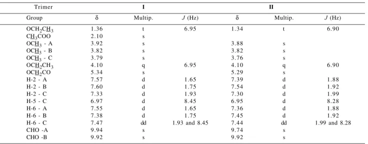
Table 1. 1H Chemical Shifts (δ) of trimers I and II
Trimer I II
Group δ Multip. J (Hz) δ Multip. J (Hz)
OCH2CH3 1.36 t 6.95 1.34 t 6.90
CH3COO 2.10 s
OCH3 - A 3.92 s 3.88 s
OCH3 - B 3.82 s 3.82 s
OCH3 - C 3.79 s 3.76 s
OCH2CH3 4.10 q 6.95 4.10 q 6.90
OCH2CO 5.34 s 5.29 s
H-2 - A 7.57 d 1.65 7.39 d 1.88
H-2 - B 7.60 d 1.75 7.54 d 1.92
H-2 - C 7.33 d 1.93 7.30 d 1.99
H-5 - C 6.97 d 8.45 6.95 d 8.28
H-6 - A 7.55 d 1.65 7.36 d 1.88
H-6 - B 7.38 d 1.75 7.45 d 1.92
H-6 - C 7.47 dd 1.93 and 8.45 7.44 dd 1.99 and 8.28
CHO -A 9.94 s 9.74 s
CHO -B 9.92 s 9.92 s
Multip.= Multiplicities (400 MHz, DMSO-d6)
Table 2. H/C correlations (3J
CH) for model compound I using
HMBC technique
δ1H H Ring C (δ13C)
9.94 CHO A C-2 (111.6) and C-6 (126.6)
7.57 H-2 A C-4 (143.0 ), C-6 (126.6 ) and CHO (192.7) 7.55 H-6 A C-2 (111.6 ) and C-4 (143.0 )
9.92 CHO B C-2 (113.3) and C-6 (126.3)
7.60 H-2 B C-6 (126.3 ), C-4 (152.6 ) and CHO (192.4) 7.38 H-6 B C-2 (113.3 ), C-4 (152.6 ) and CHO (192.4) 7.33 H-2 C C-4 (153.4 ), C-6 (123.3 ) and CO (193.1 ) 6.97 H-5 C C-3 (149.5 ) and C-1 (130.0 )
7.47 H-6 C C-2 (111.1 ) and C-4 (153.4 ) 5.34 CH2-β B/C C-4/B (152.6) and C-1/C (130.0)
Table 3. Interpretation of nOe difference spectra* of trimer I δ Irradiation (H/Ring) δ Observed Nuclear Overhauser
Enhancements (H/Ring)
9.92 (CHO/B) 7.38 and 7.60 (H-6 and H-2/B) 7.38 (H-6/B) 9.92 (CHO) 9.94 (CHO/A) 7.55 and 7.57 (H-6 and H-2/A) 3.92 (OCH3/A) 7.57 (H-2/A)
7.33 (H-2/C) 3.79 (OCH3/C) and 5.34 (OCH2CO) 4.10 (CH2CH3) 6.97 (H-5/C) and 1.36 (CH2CH3) 3.82 (OCH3/B) 7.60 (H-2/B)
5.34 (OCH2CO) 7.33 (H-2/C) and 7.47 (H-6/C) *at 400 MHz in DMSO-d6
β 2' 1' 6 5
4 3 2
CHO
OR CH3O
1
3'
4' 5' 6'
A B
α 1" 2" 3"
4" 5" 6" C
CHO
OCH3
H2C O
C=O
CH3O
CH3CH2O
These results show that the 3-methoxy-4-ethoxyphenacyl group is syn to ring A and also that ring C is rotating about Cα-C1’’ bond. In addition, the analysis of nOe dif-ference spectra has allowed to assign unambiguously the chemical shifts of hydrogens and methoxy groups of each aromatic ring (Table 3).
From broadband decoupled 13C NMR and DEPT spec-tra of compound I, the signals for aliphatic carbons at δ 15.6 (OCH2CH3), 21.0 (CH3COO), 56.3, 57.0 and 57.1 (OCH3), 64.8 (OCH2CH3), 75.0 (CH2-β), 192.4 and 192.7 (CHO), and 193.1 (CO) were assigned.
In 13C NMR spectrum of trimer II, obtained by PEN-DANT pulse sequence9 methyl carbons (CH3 and OCH3), aromatic CH and CHO show normaly phase positive sig-nals while secondary (>CH2) and quarternary (>C<) car-bons show phase inverted signals. On the basis of these results and the signal assignments of I, signals for aliphatic carbons were assigned at δ 14.5 (CH3), 55.4, 55.9, and 56.0 (OCH3), 63.9 (OCH2), 74.2 (CH2-β), 191.0 and 191.7 (CHO), and 192.4 (CO).
By combining the data obtained from 13C NMR, DEPT, and PENDANT spectra with those of 2D 1H/13C - COSY contour plot it was possible to assign the aromatic hydro-genated carbons for each A, B, and C rings of trimers I and
II. 1H/13C - COSY data are summarized in Table 4. 1H NMR spectra of trimers III and IV were analyzed using the 1HNMR spectra of both compounds I and II as a reference. This leads to assignments of hydrogens in tri-mers III and IV. The results are summarized in Table 5.
In addition, the presence of a hydroxymethyl group in trimer III was verified by comparing chemical shifts of trimer II (Table 1) with those of trimer III (Table 5). This group was characterized by signals at δ 4.87 (CH2OH) and
δ 4.81 (CH2OH). As for trimers I and II, the 13C chemical shift assignments for compounds III and IV were deduced from 13C NMR spectra with help of the corresponding DEPT spectra and 1H/13C - COSY.
The 13C spectrum of trimer III exhibited signals for aliphatic carbons at δ 14.6 (CH3CH2), 55.3, 55.7, and 55.9 (OCH3), 64.0 (CH3CH2O), 62.5 (OCHCH2OH), 83.8
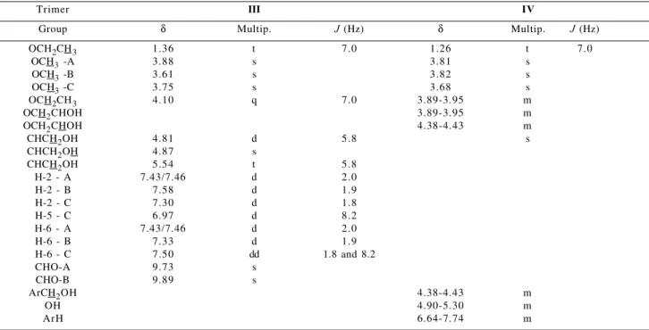
Table 5. 1H Chemical Shifts (δ) of trimers III and IV
Trimer III IV
Group δ Multip. J (Hz) δ Multip. J (Hz)
OCH2CH3 1.36 t 7.0 1.26 t 7.0
OCH3 -A 3.88 s 3.81 s
OCH3 -B 3.61 s 3.82 s
OCH3 -C 3.75 s 3.68 s
OCH2CH3 4.10 q 7.0 3.89-3.95 m
OCH2CHOH 3.89-3.95 m
OCH2CHOH 4.38-4.43 m
CHCH2OH 4.81 d 5.8 s
CHCH2OH 4.87 s
CHCH2OH 5.54 t 5.8
H-2 - A 7.43/7.46 d 2.0
H-2 - B 7.58 d 1.9
H-2 - C 7.30 d 1.8
H-5 - C 6.97 d 8.2
H-6 - A 7.43/7.46 d 2.0
H-6 - B 7.33 d 1.9
H-6 - C 7.50 dd 1.8 and 8.2
CHO-A 9.73 s
CHO-B 9.89 s
ArCH2OH 4.38-4.43 m
OH 4.90-5.30 m
ArH 6.64-7.74 m
Multip.= Multiplicities (400 MHz, DMSO-d6)
Table 4. Heteronuclear correlation (1H/13C-COSY)* for trimers I and II
Trimer Ring C-2 / H-2 C-5 / H-5 C-6 / H-6 CHO
A 111.6 / 7.57 126.6 / 7.55 192.7 / 9.94
I B 113.3 / 7.60 126.3 / 7.38 192.3 / 9.92
C 111.1 / 7.33 112.4 / 6.97 123.3 / 7.47
A 109.2 / 7.39 128.0 / 7.36 191.0 / 9.74
II B 111.3 / 7.54 126.6 / 7.45 191.7 / 9.92
C 110.1 / 7.30 111.4 / 6.95 122.5 / 7.44
(OCHCH2OH), 191.0 and 191.7 (CHO), and 194.1 (CO). The 13C spectrum of trimer IV presented signals for ali-phatic carbons at δ14.7 (CH3CH2O), 55.3, 55.7 and 55.8 (OCH3), 62.6 and 62.7 (CH2OH), 63.7 (CH3CH2O), 71.3 (OCH2CHOH), and 78.0 (OCH2CHOH).
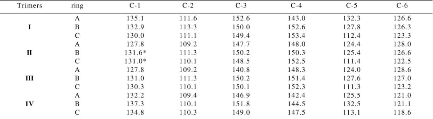
Table 6 summarizes the 13C NMR chemical shifts of aromatic carbons in trimers I to IV. The chemical shifts of aromatic carbons for compound III were assigned by com-parison with those of compound II, while those of aro-matic carbons in trimer IV were determined by compari-son with model compounds 1 and 2 (Figure 2)10,11.The chemical shift assignments for compoud IV were also de-duced from the 13C NMR spectrum with help of the corre-sponding DEPT spectra.
Table 7 shows the 13C NMR chemical shifts for these three compounds. From these data it may be verified that aromatic carbons of rings A and B of trimer IV as well as those of parent compound 1 present very close chemical shifts. Similarly, the chemical shifts assigned to aromatic carbons of ring C are closely related to those of compound 2.
Conclusions
Trimeric lignin model compounds containing 5-5’ and
β-O-4’ substructures were synthesized, successfuly, by way of acetyldehydrodivanillin and α -bromo-3-methoxy-4-ethoxyacetophenone. This paves the way for the synthe-sis of oligomeric lignin model compounds containing
biphenyl (5-5’) and β-aryl ether (β-O-4’) substructures through Williamson’s reaction.
The chemical shifts and coupling constants for I and II
were deduced from 1D NMR spectra, and confirmed by HMBC, 1H/1H-COSY and nOe difference spectra.
This last one was useful in establishing the conforma-tion of 4-O-acetyl-4’-O-[α
-(3-methoxy-4-ethoxyphenyl)-Table 6. 13C chemical shifts (δ) for aromatic carbons of trimers I - IV
Trimers ring C-1 C-2 C-3 C-4 C-5 C-6
A 135.1 111.6 152.6 143.0 132.3 126.6
I B 132.9 113.3 150.0 152.6 127.8 126.3
C 130.0 111.1 149.4 153.4 112.4 123.3
A 127.8 109.2 147.7 148.0 124.4 128.0
II B 131.6* 111.3 150.2 150.3 125.4 126.6
C 131.0* 110.1 148.5 152.5 111.4 122.5
A 127.8 109.2 140.8 148.3 124.0 128.6
III B 131.0 111.3 150.2 151.4 127.6 127.0
C 130.3 110.1 150.1 152.3 111.3 123.2
A 132.2 109.4 146.9 142.4 125.5 121.0
IV B 137.3 110.1 151.8 144.5 132.5 121.1
C 134.8 110.3 149.0 147.5 113.1 118.6
100MHz, DMSO-d6, *these values may be exchangeable.
Table 7.13C NMR chemical shifts (δ) of trimer IV and dimers 110 and 211
Compound Ring C-1 C-2 C-3 C-4 C-5 C-6
IV A 132.2 109.4 146.9 142.4 125.5 121.0
1 A 132.4 109.6 147.3 142.5 125.7 121.2
IV B 137.3 110.1 151.8 144.5 132.5 121.0
1 B 137.3 110.2 152.5 144.4 133.0 121.2
IV C 134.8 110.3 148.5 147.5 112.6 118.2
2 C 135.1 110.9 149.0 147.5 113.1 118.6
100MHz, DMSO-d6
β 2' 1' 6
5
4 3 2
1
CH2OH
O
OCH3 CH2 CHOH CH2OH
OH CH3O
OCH2CH3 OCH3 B
A
C 3'
4' 5' 6'
1" 2" 3"
4" 5" 6"
α
IV
6' 5'
4' 3'
A B
CH2OH
OH CH3O
CH2OH
OCH2CH3 OCH3 1
2
3
4 5 6
1' 2'
1
2
D
α
C
OCH2CH3 OCH3 COOH
O
OCH3 CH2 CHOH β
Figure 2. Model compounds related to IV.
α-oxoethyl]dehydrodivanillin (I). The results obtained show that the 3-methoxy-4-ethoxyphenacyl group is syn
to ring A and also that ring C is rotating about Cα-C1’’ bond with no restriction. In addition, the analysis of nOe difference spectra has allowed to assign unambiguously the chemical shifts of hydrogens and methoxyl groups of each aromatic ring.
1H NMR spectra of trimers III and IV were analyzed using the 1H NMR spectra of both compounds I and II as a reference. As for trimers I and II, the 13C chemical shift assignments for compouds III and IV were deduced from 13C NMR spectra with help of the corresponding DEPT, 1H/13C COSY and HMBC spectra. While the chemical shifts of aromatic carbons for compound III were assigned by comparison with those of compound II, those of aro-matic carbons in trimer IV were determined by compari-son with dimeric model compounds.
From the 1D and 2D 1H, 13C-NMR spectroscopic stud-ies of trimers I-IV, it is evident that the acetoxy group in trimer I undergoes base-catalyzed hydrolysis to give tri-mer II (Table 6). The later compound has a hydroxyl group at C-4 of ring A. This resulted in shielding (∆δ - 2.9 to ∆δ - 7.9) of C-1, C-2, C-3 and C-5 and deshielding of ∆δ + 5.0 and ∆δ + 1.4 for C-4 and C-6 in ring A, respectively. For ring B, all aromatic carbons are shielded (∆δ - 0.2 to 2.4). For ring C, C-1, C-2, C-4, and C-5 are shielded (∆δ - 0.9 to
∆δ - 1.0).
Furthermore, chemical structures of trimers II and IV, differ in the substituents at C-1 of rings A/B and at C-α of side chain in ring C. As compared to trimer II, C-4 and C-6 of ring A of trimer IV are shielded by ∆δ - 5.6 to - 7.0, while C-1 and C-5 are deshielded by ∆δ + 4.4 and + 1.1, respectively. For ring B of IV, C-2 and C-4 are shielded by
∆δ - 1.2 and - 5.8, respectively, in addition to deshielding
of C-1, C-3 and C-5 (∆δ + 1.6-5.7). For ring C of IV, C-4 and C-6 undergo shielding of ∆δ - 5.0 and - 3.9, respec-tively, and C-1 deshielding of ∆δ + 3.8.
Acknowledgments
This research project is supported by CNPq, FAPEMIG, and FINEP, for which the authors are grateful. This work constitutes part of the Doctoral Thesis of V.L. Alves.
References
1. Adler, E. Wood Science and Technol. 1977, 2, 169. 2. Lin, S. Y. Progress in Biomass Conversion 1983,
4, 31.
3. Drumond, M. G.; Aoyama, M.; Chen, C. -L.; Robert, D.
J. Wood Chem. Technol. 1989, 9, 421.
4. Karhunen, P.; Rummakko, P.; Sipilä, J.; Brunow, G.; Kilpeläinen, I. Tetrahedron Lett. 1995, 36, 169. 5. Karhunen, P.; Rummakko, P.; Sipilä, J.; Brunow, G.;
Kilpeläinen, I. Tetrahedron Lett. 1995,36, 4501. 6. Karhunen, P.; Rummakko, P.; Pajunen, A.; Brunow, G.
J. Chem. Soc. Perkin Trans. I 1996, 2303.
7. Ede, R. M.; Kilpeläinen, I. Res. Chem. Intermed. 1995,
21, 313.
8. Glasser, W. G.; Honeycutt, S. S.; Barnett, C. A.; Morohoshi, N. Tappi1979, 62, 111.
9. Homer, J.; Perry, M. C. J. Chem. Soc., Chem. Commun.
1994, 373.
10. Drumond, M. G.; Piló-Veloso, D.; Cota, S. D. S.; Morais, S. A. L.; Nascimento, E. A.; Chen., C. -L. Holzforschung 1992, 46, 127.
11. Hassi, H.Y.; Aoyama, M.; Tai, D.; Chen, C. -L.; Gratzl, J. S. J. Wood Chem. Technol. 1987,7, 551.