J. Braz. Chem. Soc., Vol. 16, No. 2, 210-213, 2005. Printed in Brazil - ©2005 Sociedade Brasileira de Química 0103 - 5053 $6.00+0.00
Article
* e-mail: [email protected]
Biotransformation of Steviol Derivatives by
Aspergillus niger
and
Fusarium moniliforme
Brás H. de Oliveira*,a José Dias S. Filhob and Paulo C. Leala
a
Universidade Federal do Paraná, Departamento de Química, CP19081, 81531-970 Curitiba - PR, Brazil
b
Universidade Federal de Minas Gerais, Departamento de Química, 31270-90 Belo Horizonte - MG, Brazil
Derivados do esteviol foram submetidos a biotransformações por fungos. O ent-11β ,13-diidróxi-15,16-epoxicauran-19-oato de metila foi hidroxilado em C-11 por Aspergillus niger enquanto que o ácido ent-16β-hidróxibaierano-19-óico foi hidroxilado em C-6 e C-7 pelo Fusarium moniliforme. A hidroxilação em posições não ativadas do esqueleto carbônico é discutida tendo em vista as propriedades de importantes diterpenóides descritos na literatura.
Steviol derivatives have been submitted to biotransformations by fungi. Methyl ent-11β ,13-dihydroxy-15,16-epoxikauran-19-oate was hydroxylated at C-11 by Aspergillus niger, whereas
ent-16β-hydroxybeyeran-19-oic acid was hydroxylated at C-6 and C-7 by Fusarium moniliforme. The hydroxylation at non-activated positions at the carbon skeleton is discussed in connection with the properties of important polyhydroxylated diterpenoids described in the literature.
Keywords: kaurene and beyerane diterpenes, steviol, biotransformation, Aspergillus niger, Fusarium moniliforme
Introduction
Stevioside (1),1 a natural glucoside extracted from
Stevia rebaudiana, is produced commercially in several countries. Besides its use as low caloric sweetener stevioside has also other important properties. Studies have shown that it has, for example, antihypertensive,
antihyperglycemic2 and cancer-chemopreventive3
activities. Stevioside aglucone, steviol (2), is a tetracyclic diterpenoid with kaurene skeleton and a number of important biological properties4 have also been attributed
to diterpenoids of this class.
Several bioactive polyhydroxylated diterpenoids have been described. Isodon5 and Rabdosia6 diterpenoids, for
example, are antimicrobial and antitumour compounds and their activity is dependent on the number and position of
hydroxyl groups in the molecule. Aphidicolin,7 an
anticancer and antileishmanial compound, is a tetrahydroxylated tetracyclic diterpenoid and the free hydroxyl groups are essential for its activity. Obtaining hydroxylated derivatives of available diterpenes may, therefore, lead to new or improved properties.
The use of biological methods for the transformation of organic compounds have gained increasing attention due to its inherent advantages.8 Diterpenoids have been
subject to this kind of transformation in a number of occasions9 and our previous results have shown that these
diterpenes are prone to hydroxylation by some fungi.10,11
Therefore this technique can be useful for obtaining new diterpenoid derivatives.
In our continuing work for obtaining new derivatives of steviol we now describe the biotransformation of ent-13-hydroxy-15,16-epoxikauran-19-oate (3) by Aspergillus niger and ent-16-hydroxybeyeran-19-oic acid (5) by Fusarium moniliforme.
Experimental
IR: KBr discs; NMR: 200 MHz (1H) and 50 MHz (13C);
Solvents were distilled before use.
Preparation of the substrates
The substrates were prepared as described previously.12
Briefly, stevioside (1) was hydrolysed under acidic
conditions and the crude mixture obtained was crystallized to produce isosteviol (5). It was then reduced with sodium borohydride producing the corresponding alcohol (6). From the mother liquor of the hydrolysis reaction a mixture of steviol and its ∆15 isomer was isolated by CC, esterified
211 Biotransformation of Steviol Derivatives by Aspergillus niger and Fusarium moniliforme
Vol. 16, No. 2, 2005
be separated by CC, leading to steviol epoxide methyl ester and its isomer, methyl ent-13-dihydroxy-15,16-epoxikauran-19-oate (3). The spectroscopic data for the compounds were in accordance with those from the literature.
Fungi and culture conditions
Stock cultures of A. niger and F. moniliforme were stored on potato dextrose agar slants, at 4 oC. Seed cultures were
obtained by transferring fungi from stock cultures to a liquid medium containing (g L-1) glucose (10.00), corn
steep liquor (8.00) and yeast extract (2.00).
The experiments were conducted with conical flasks (1000 mL) containing 200 mL of the same liquid medium. For each fungus two flasks were inoculated with the seed culture and shaken at 30 oC for 48 h. Then the substrate in
DMSO was added and the fermentation continued for seven more days. Then the mycelium was filtered off and washed with EtOAc. The broth was extracted with the same solvent and the extracts were combined and dried with anhydrous Na2SO4. After filtration the solvent was evaporated under reduced pressure and the crude extracts were fractionated by CC on silica gel.
Biotransformation of methyl ent-13-hydroxy-15,16-epoxikauran-19-oate (3) by A. niger
The epoxide ester (3) (350 mg) in DMSO (2.0 mL) was
evenly distributed among 4 flasks of a 48 h old culture and the fermentation continued for 7 more days. The crude extract obtained was fractionated on a silica gel column. Elution with CHCl3-EtOAc (2:1 v/v) produced a solid (80.0 mg) which was recrystalised from CHCl3-hexane. It was
characterized as methyl ent-11β
,13-dihydroxy-15,16-epoxikauran-19-oate (4). IR νmax/cm
-1: 3408, 1722; 1H
NMR (CDCl3) δ: 0.90 (3H, s, H-20), 1.19 (3H, s, H-18), 1.22 (3H, s, H-17), 3.40 (1H, bs, H-15β), 3.62 (3H, s, CO2Me), 4.25 (1H, m, H-11α).
Biotransformation of ent-16β-hydroxybeyeran-19-oic acid by F. moniliforme
The alcohol (6) 500 mg was distributed among 5 flasks and after 7 days fermentation a crude extract was obtained. It was esterified with ethereal diazomethane and fractionated by CC on silica gel. Elution with CHCl3 -EtOAc (2:1 v/v) gave a solid (17 mg) which was characterized as methyl ent-6α,16β -dihydroxybeyeran-19-oate (7); IR νmax /cm
-1: 3421, 1719; 1H NMR (CDCl 3) δ:
0.90 (3H, s, H-20), 0.94 (3H, s, H-17), 1.21 (3H, s, H-18), 2.50 (1H, bs, H-12α), 3.63 (3H, s, -OCH3), 3.85 (1H, dd, J 5.3 and 10.1 Hz, H-16β), 4.20 (1H, td, J 6.3 and 10.1 Hz, H-6α).
Further elution gave another solid (56 mg) which was characterized as methyl ent-7α,16β -dihydroxybeyeran-19-oate (8); IR νmax/cm
-1: 3527, 3437, 1720; 1H NMR
(CDCl3) δ: 0.73 (3H, s, H-20), 0.94 (3H, s, H-17), 1.16 (3H, s, H-18), 2.50 (1H, d, J 13.2 Hz, H-12α), 3.57 (1H, t, J 2.6 Hz, H-7α), 3.63 (3H, s, -OCH3), 3.88 (1H, dd, J 5.4 and 10.1 Hz, H-16β).
Results and Discussion
The biotransformation of methyl ent-13-hydroxy-15,16-epoxikauran-19-oate (3) by A. niger resulted in a more polar compound as determined by tlc. Its IR spectrum showed a strong band at 3408 cm-1,indicating the presence
of a hydroxyl group, as well as the ester carbonyl at 1722 cm-1. Its 1H NMR, compared to the parent compound,
showed a new peak at 4.25 ppm integrating for one proton, indicating a possible hydroxylation at a methylene carbon. That was confirmed by the 13C NMR spectra (Table 1) which
212 de Oliveira et al. J. Braz. Chem. Soc.
The stereochemistry at C-11 (Figure 1) was inferred from the multiplicity of the corresponding signal in the 1H NMR
spectrum (4.25 ppm). It was a broad singlet indicating a weak coupling with neighbouring hydrogens. The axial (β) orientation for the hydroxyl is, therefore, the most likely and a molecular model showed that, in that position, the dihedral angle between H-11α and the protons at C-12 and C-10 was aproximately 60o. The product is methyl
ent-11α,13-dihydroxy-15,16-epoxikauran-19-oate (4). Hydroxylation at C-11 of a kaurene skeleton has been described previously. Steviol methyl ester was transformed by Gibberella fujikuroi,13 indicating that different
microorganisms may have similar monooxigenases.
The biotransformation of the alcohol (6) by F.
moniliforme resulted in two more polar compounds as determined by TLC and they were analysed as their methyl esters. The first one produced a IR spectrum with a strong band at 3421 cm-1 indicating the presence of a hydroxyl
group as well as the ester carbonyl at 1719 cm-1. Its 1H
NMR spectrum, compared to the parent compound, showed a new peak at 4.20 ppm integrating for one hydrogen, indicating a possible hydroxylation at a
methylene carbon. That was confirmed by the 13C NMR
spectra (Table 1) which showed 8 methylene carbons (DEPT), instead of 9 of the parent compound, as well as extra methine signal. The signal corresponding to C-6 in the substrate, a methylene at 21.0 ppm, was missing and the extra CH appeared at 68.6 ppm. The hydroxylation at C-6 was confirmed by the paramagnetic shifts of neighbouring C-5 and C-7 (3 and 2.6 ppm respectively). The stereochemistry at C-6 (Figure 2) was deduced from the multiplicity of corresponding C-H signal in the 1H
NMR spectrum (4.20 ppm). It was a collapsed triplet of doublets showing a diaxial coupling (10.1 Hz) with neighbouring protons. A NOESY experiment also showed a strong interaction with the singlet at 0.73 (H-20). The equatorial (β) orientation of the hydroxyl is, therefore, the
most likely and the product is methyl ent-6α,16β
-dihydroxy-beyeran-19-oate (7). Hydroxylation at C-6 has been achieved by Aspergillus ochraceous on other beyerane diterpenes.14 This is another example of the
presence of similar monooxigenases in different microorganisms.
The second metabolite produced a ir spectrum with two hydroxyl bands at 3527 and 3437 cm-1 as well as the
ester carbonyl at 1720 cm-1. Its 1H NMR spectrum, compared
to the parent compound, showed a new peak at 3.57 ppm integrating for one proton, indicating a possible hydroxylation at a methylene carbon. That was confirmed
by the 13C NMR spectra (Table 1) which showed 8
methylenes (dept), instead of 9 of the parent compound, as well as extra methine signal. The signal corresponding to C-7 in the substrate, a methylene at 21.0 ppm, was missing and the extra methyne appeared at 76.8 ppm. The hydroxylation at C-7 was confirmed by the paramagnetic shifts of neighbouring C-6 and C-8 (6 and 3 ppm respectively). Furthermore the signals of C-5 and C-9, bearing axial protons, undergone significant paramagnetic shifts (8.6 and 10.1 ppm respectively) indicating 1,3 diaxial interactions with an axial hydroxyl at C-7. The stereochemistry at C-7 (Figure 3) was confirmed from the multiplicity of corresponding C-H signal in the 1H NMR Table 1. 13C NMR of methyl ent
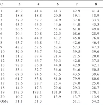
-13-hydroxy-15,16-epoxikauran-19-oate (3), methyl ent -11α,13-dihydroxy-15,16-epoxikauran-19-oate (4), ent-16β-hydroxy-beyeran-19-oic acid (6), methyl ent -6α,16β-dihydroxy-beyeran-19-oate (7) and methyl ent -7α,16β-dihydroxy-beyeran-19-oate (8). Solvent: CDCl3 except for (6) (CD3OD)
C 3 4 6 7 8
1 40.7 41.4 41.3 42.9 41.4
2 18.8 18.8 20.0 19.1 18.9
3 37.9 37.7 34.9 37.8 33.5
4 43.5 43.5 44.6 44.0 43.3
5 56.5 56.7 58.4 61.0 49.8
6 20.4 20.8 22.3 68.6 28.9
7 38.4 44.9 43.2 45.8 76.8
8 43.7 46.8 43.1 42.6 46.4
9 48.2 57.5 57.4 57.3 47.3
1 0 39.0 36.7 39.2 39.5 39.6
1 1 21.2 87.4 21.4 21.5 20.0
1 2 35.7 46.7 39.3 42.0 37.8
1 3 78.8 86.0 44.0 42.9 42.1
1 4 33.4 32.7 56.6 54.6 50.5
1 5 67.0 74.5 43.5 43.5 39.6
1 6 61.7 83.8 81.0 79.9 80.0
1 7 28.6 28.7 25.4 24.0 25.0
1 8 14.9 17.3 29.6 29.3 28.7
1 9 178.0 178.1 181.9 178.1 178.1
2 0 10.7 14.7 13.9 13.7 13.9
OMe 51.1 51.3 51.1 54.2
Figure 2.
213 Biotransformation of Steviol Derivatives by Aspergillus niger and Fusarium moniliforme
Vol. 16, No. 2, 2005
spectrum (3.56 ppm). It was a broad triplet with weak coupling with neighbouring protons, compatible with an axial (β) position for the hydroxyl group. The product therefore is methyl ent-7α,16β -dihydroxy-beyeran-19-oate (8). This compound has been described previously, resulting from the biotransformation of the same substrate by Gibberella fujikuroi.15 The hydroxylation at C-7 of
kaurenes and beyerenes by Fusarium sp has been reported in a number of occasions. It indicates that the monooxygenase responsible for that transformation is not too specific and accepts a broad range of compounds of those classes.
The results shown above indicate that the fungi used are able to increase the polarity of the parent compounds. Hydroxylating non-activated positions using microbiological methods can, therefore, be used as a viable procedure for obtaining diterpenoid derivatives with potentially useful biological properties, like those of Isodon5 and Rabdosia6 diterpenoids previously mentioned.
Acknowledgements
The financial support of FINEP and CNPq (Brazil) are greatly acknowledged. We are also thankful to LAREMAR, UFMG, for the NMR spectra.
Figure 3.
References
1. Geuns, J.M.C.; Phytochemistry2003,64, 913.
2. Jeppesen, P.B.; Gregersen, S.; Rolfsen, S.E.D.; Jepsen, M.; Colombo, M.; Agger, A.; Xiao, J.; Kruhoffer, M.; Orntoft, T.; Hermansen, K.; Metab. Clin. Exp.2003,52, 372.
3. Konoshima, T.; Takasaki, M.; Pure Appl. Chem. 2002,74,
1309.
4. Ghisalberti, E.L.; Fitoterapia1993, 68, 303.
5. Fujita, E.; Nagao, Y.; Node, M.; Kaneko, K.; Nakazawa, S.; Kuroda, H.; Experientia1976,32, 203.
6. Fujita, E.; Node, M.; Fortschr. Chem. Org. Naturst.1984,46,
77.
7. Kayser, O.; Kiderlen, A.F.; Bertels, S.; Siems, K.; Antimicrob.
Agents Chemother.2001,45, 288.
8. Schmid, A.; Dordick, J.S.; Hauer, B.; Kiener, A.; Wubbolts, M.; Witholt, B.; Nature2001,409, 258.
9. Hanson, J.R.; Nat. Prod. Rep.1992,9, 139.
10. de Oliveira, B.H.; Strapasson, R.A.; Phytochemistry1996,43,
393.
11. de Oliveira, B.H.; dos Santos, M.C.; Leal, P.C.; Phytochemistry 1999,51, 737.
12. Avent, A.G.; Hanson, J.R.; de Oliveira, B.H.; Phytochemistry 1990,29, 2712.
13. Shigematsu, Y.; Murofushi, N.; Takahashi, N.; Agric. Biol. Chem.1982,46, 2313.
14. Beilby, J.P.; Ghisalbe, E.L.; Jefferie, P.R.; Sefton, M.A.; Sheppard, P.N.; Tetrahedron Lett.1973, 2589.
15. Ali, M.S.; Hanson, J.R.; de Oliveira, B.H.; Phytochemistry 1992,31, 507.
Received: January 4, 2004