Spectroscopic Study of the Interaction of Nd
3+with Amino Acids:
Phenomenological 4f-4f Intensity Parameters
Soraya Jericóa, Célia R. Carubellia, Ana M.G. Massabnia,
Elizabeth B.Stucchia, Sergio R. de A. Leitea*, and Oscar Maltab
a
Instituto de Química, Universidade Estadual Paulista C.P. 355,
14801-970 Araraquara -- SP, Brazil b
Departamento de Química Fundamental, Universidade Federal de Pernambuco,
50670-901 Recife -- PE, Brazil
Received: June 1, 1998
Estudamos o comportamento dos parâmetros fenomenológicos de intensidade das transições 4f-4f em compostos de Nd3+ com glicina, ácido L-aspártico, ácido L-glutâmico, L-histidina, ácido DL-málico e Aspartame®em solução aquosa como função dos valoresde pK e das cargas parciais sobre os átomos de oxigênio dos grupos carboxilatos dessas moléculas. Os resultados são discutidos e interpretados qualitativamente em termos dos mecanismos das intensidades 4f-4f por dipolo elétrico forçado e acoplamento dinâmico, indicando como dominante o mecanismo de dipolo elétrico forçado.
We have studied the bevahior of the phenomenological 4f-4f intensity parameters in compounds of the Nd3+ ion with glycine, L-aspartic acid, L-glutamic acid, L-histidine, DL-malic acid and AspartameTM in aqueous solution, as a function of the pK values and partial charges on the oxygens of the carboxylate groups of these molecules. The results are discussed and qualitatively interpreted in terms of the forced electric dipole and dynamic coupling mechanisms of the 4f-4f intensities, thus indicating that the forced electric dipole mechanism is dominant.
Keywords:neodymium, amino acids, transition intensity parameters
Introduction
The study of the chemical bonding between trivalent lanthanide ions (Ln3+) and amino acids or peptides has its
origin in the interest in using these ions as structural probes in biological systems, particularly in those systems which contain Ca2+ in their structure1,2. The Ca2+ ion is optically
inactive and is, therefore, not suitable for providing infor-mation, through optical spectroscopic measurements, about the chemical environment in which it is embedded. On the other hand, almost all Ln3+ ions are know to exhibit
rich optical spectra, either in absorption or emission, and it occurs that, due to the similarity between ionic radii, they may substitute Ca2+ ions in the chemical structures.
There is strong evidence that the bonding between Ln3+
ions and amino acids is made with the oxygens of the carboxylate group, and that the bonding via the nitrogen of
the amino group is unlikely to occur at least in a range of pH values up to 5.63,4. In this paper we examine the relation between the basicity of the carboxylate groups of glycine (Gly), L-aspartic acid (Asp), L-glutamic acid (Glu), L-his-tidine (His), DL-malic acid (Mal) and AspartameTM (APM) (Table 1), and the intensity parameters of 4f-4f transitions in their compounds with Nd3+ ion. The intensity parameters are qualitatively interpreted in terms of the forced electric dipole and dynamic coupling mechanisms of 4f-4f intensi-ties5-10. Both mechanisms are dependent on the chemical environment around the Ln3+ ion. The basicity of the car-boxylate groups is considered according to pK values and partial atomic charges on the oxygens which were calcu-lated from molecular mechanics and the semi-empirical PM3 quantum chemical method.
Article
* Correspondence should be addressed at: Instituto de Química - UNESP, C.P. 355, 14801-970 Araraquara -- SP, Brazil.
Experimental
The samples were prepared from aqueous solutions of Nd(ClO4)3 and the amino acids glycine, L-aspartic acid,
L-glutamic acid and L-histidine, the dipeptide Aspar-tameTM or the DL-malic acid. The absorption spectra were measured in a Carl Zeiss M-40 UV-visible spectro-photometer between 11000 cm-1 and 30000 cm-1. Solutions of Nd(ClO4)3 with pH 5.0-5.5 were prepared from Nd2O3
(Aldrich, 99.99%) and standardized by EDTA / xylenol orange titration. Solutions of the ligands were standardized by titration with NaOH / phenolphtalein or by potentiomet-ric titration. The concentration of all solutions were around 5.00 x 10⁻² mol L-1.
We have firstly examined the behavior of the
4I
9/2 →4G5/2, 2G7/2 hypersensitive transitions of Nd3+,
be-tween 16600 cm-1 and 18200 cm-1, as a function of the molar ratio Nd3+: Ligand and the pH of the solution, which was varied from 1 to 5.5 for each ratio. The solutions of the Nd(ClO4)3 and the ligand were mixed in a quartz cell of
1.00 cm optical pathway which was coupled to a quartz bulb with 20 mL capacity. Volumes were measured with calibrated pipetes. The molar ratio Nd3+ / Ligand was varied from 1:1 to 1:10. The pH was adjusted by addition of acid or base and measured directly in the cell with a combined glass microelectrode. Concentrations were cor-rected for the volume. Wavenumber scan was made with variable slits and constant energy beam at the photodetector and the absorbance values were read to the fourth decimal place in a digital display. All the measurements were made
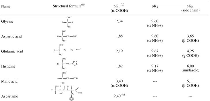
at 25 ± 1 °C. Doubly destilated water was used as reference. The samples with the ratio Nd3+ / Ligand and pH that gave the highest absorption in the region of hypersensitivity were used to obtain the entire spectra from which the spectroscopic parameters were obtained. The best ratio Nd3+ / Ligand is 1/4 except for the Nd3+ / Aspartame, in which case the experimental oscilator strength always rises with increasing of the quantity of the ligand. Figure 1 shows the curves of Pexpvs. pH for several samples using histidine
as ligand. As we see, Pexp decreases as the ratio goes to
higher values than 1/4.
As we observed nine groups of transitions in the entire spectral region under investigation, we obtained nine equa-tions for P (Eq. 2), where the following values were intro-duced for the calculation of τλ: experimental P values as obtained in the Eq. 1, experimental σ values as the baricen-ter of a group of transitions obtained from the area meas-urements under the absorption curve, U(λ) values as obtained of the medium value for the U(λ) of the transitions in each group11. A program in BASIC was developed to treat these nine equations by a least square method to obtain the τλ parameters. The least square method reduces a sys-tem of n equations with 3 unknown quantities to a syssys-tem of 3 equations with 3 unknowns, which are the pheno-menological τλ parameters.
Results and Discussion
The experimental oscillator strengths are obtained through the expression
Table 1. Covalent structures of the ligands.
Name Structural formula(a) pK1(b)
(α-COOH)
pK2 pKR
(side chain)
Glycine 2,34 9,60
(α-NH3+)
Aspartic acid 1,88 9,60
(α-NH3+)
3,65 (β-COOH)
Glutamic acid 2,19 9,67
(α-NH3+)
4,25 (γ-COOH)
Histidine 1,82 9,17
(α-NH3+)
6,00 (imidazole)
Malic acid 3,40
(α-COOH)
--- 5,11
(β-COOH)
Aspartame 2,40 (c) ---
---(a) Ionic forms predominating at pH 7,0; (b) The pKa values from the CRC Handbook of Chemistry and Physics21; (c) Estimated value from titration curves in this work.
C COO -NH3 + H H H
NH3+
COO
-C CH2 COO
-H
OH COO
-C CH2 COO
-C COO
-NH3 +
H CH2 CH2 COO
-C COO
-H H C
H
NH3+
C C C O N H O OCH3 CH2 H C COO
-NH3+
H CH2
Pexp= 4.318 x 10⁻⁹
∫
ε(σ) dσ (1)were ε(σ) is the molar extintion coefficient at wavenumber
σ (cm-1) and the integral in Eq. 1 is directly proportional to the area under the absorption curve.
According to the theory of 4f-4f intensities5, the oscil-lator strength of a transition between two manifolds, with respective total angular momenta J and J’, of a given 4fN
electronic configuration is given by:
P =
∑
λ= 2,4,6
στλ<(4 f
N)ψ ’J’ U(λ)(4 fN)ψ J >2
(2J + 1) (2)
where σ is the baricenter of the transition energy (in wavenumbers), U(λ) is a unit tensor operator of rank λ and the τλ are the so-called intensity parameters which depend on the chemical environment, radial integrals and interconfigurational energy differences in the lanthanide ion. The reduced matrix elements of U(λ) in Eq. 2 have been calculated, in the intermediate coupling scheme, for the whole series of the trivalent lanthanides12.
An alternative way of expressing the theoretical oscil-lator strength has been of common use in the literature, in terms of the Ωλ intensity parameters which are related to the τλ parameters by Ωλ = τλ/1.085 x 1011 χ cm-1, where χ = (η2 + 2)2/9η, η being the index of refraction of the
medium13. For the sake of comparison with the results of
previous studies on 4f-4f intensities in compounds of triva-lent lanthanides with amino acids14,15, the expression of the
theoretical oscillator strength in terms of the τλ intensity parameters as in Eq. 2 is used in the present work.
In principle these parameters can be calculated from theoretical models provided structural data around the
lan-thanide ion are available5,9. However, a common procedure is to treat them as adjustable parameters to reproduce the observed oscillator strengths. The τλ thus obtained are refered to as phenomenological intensity parameters. In this procedure the least square method is commonly used in which the input data are the values of the measured oscillator strengths, the squared reduced matrix elements of U(λ) and the transition energies σ. Table 2 presents the values of the oscilator strengths and transition energies, corresponding to the transitions observed in the absorption spectrum of the complex with L-aspartic acid, as a function
Figure 1. Oscillator strength vs. pH. Nd3+: L-histidine system.
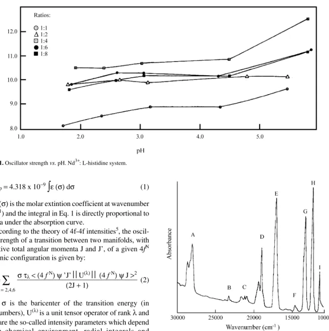
Figure 2. Absorption spectrum of Nd(ClO4)3: L-aspartic acid in aqueous solutions.
Transitions from the 4I9/2 to: A) 2L15/2, 4D1/2, 4D5/2, 2I11/2, 4D3/2 B) 2P1/2
C) 4G11/2 (2D,2P)3/2, 2G9/2, 2K15/2 D) 4G9/2, 4G7/2, 2K13/2 E) 4G5/2, 2G7/2 F)
4F
Table 2. Spectral transitions of Nd(III) complexed by L-aspartic acid: average wavenumber (spectral band baricenter), experimental and calculated oscillator strength at some pH values.
Transition from 4I9/2 to: pH ν (cm-1) Pexp (x 109 ) Pcalc. (x 109)
1.6 28415 8.90 10.0
2L
15/2, 4D1/2, 4D5/2, 2I11/2, 4D3/2 2.4 28415 8.80 10.0
3.5 28390 8.45 10.0
4.2 28365 9.05 10.2
5.4 28365 8.74 9.30
1.6 23360 0.40 0.400
2P
1/2 2.4 23355 0.400 0.400
3.5 23345 0.400 0.400
4.2 23355 0.300 0.400
5.4 23340 0.300 0.200
1.6 21350 1.70 1.50
4G
11/2, (2D, 2P)3/2, 2G9/2, 2K15/2 2.4 21320 1.70 1.50
3.5 21355 1.60 1.50
4.2 21350 1.66 1.50
5.4 21345 1.51 1.40
1.6 19250 5.40 5.70
4
G9/2, 4G7/2 2.4 19235 6.50 5.70
3.5 19240 6.75 5.80
2K
13/2 4.2 19240 6.90 5.90
5.4 19240 6.44 5.70
1.6 17365 8.90 10.6
4G
5/2, 2G7/2 2.4 17350 9.10 11.1
3.5 17315 11.0 11.8
4.2 17305 11.7 11.8
5.4 17305 12.0 12.7
1.6 14700 0.500 0.900
4F
9/2 2.4 14685 0.500 0.900
3.5 14690 0.495 0.900
4.2 14710 0.565 0.900
5.4 14700 0.528 0.900
1.6 13490 8.22 12.5
4S
3/2, 4F7/2 2.4 13485 8.30 12.4
3.5 13480 8.40 12.4
4.2 13470 8.31 12.4
5.4 13470 8.30 12.4
1.6 12540 8.60 10.6
2H
9/2, 4F5/2 2.4 12548 8.10 10.6
3.5 12560 8.10 10.3
4.2 12532 8.10 10.4
5.4 12533 8.10 10.6
1.6 11533 2.00 2.10
4
F3/2 2.4 11550 2.10 2.10
3.5 11540 2.20 2.10
4.2 11543 2.10 2.20
of the pH. It may be noted that the intensity of the hyper-sensitive 4I9/2→4G5/2,2G7/2 transitions increases as the pH
increases up to aproximately 5.4 value. For pH values above 5.5 the Nd3+ ion hydrolyses.
In the fitting procedure to obtain the phenomenological
τλ parameters, the squared reduced matrix elements of U(λ)
for the transitions separated by groups, as indicated in Table 2, were summed together. The results are presented in Table 3.
In the case of the glycine ligand, the τλ values presented in Table 3 in the range of pH above the pK1 agree with the
values obtained by Legendziewicz et al.14,15 for the com-pounds in the crystalline phase. The same agreement is not observed when the ligand is glutamic acid, where τ4 and τ6
are discrepant from the solid state values. This fact can be explained if we consider that the two carboxylate groups of the glutamic acid may or may not be involved in the coordination at the pH value used in these measurements.
Among the τλ parameters, in general τ2 is the most
sensitive to the coordination geometry and the charac-teristics of the ligands13. We have examined the behavior
of τ2 with the ligand’s pK; τ2 has varied linearly with pK1
provided the monocarboxylic and dicarboxylic species were considered separately (Fig. 3). We have also exam-ined the behavior of τ2 with the average value <pk> = (pK1
+ pK2) / 2, since at pH ~5 both carboxylic groups are
expected to be equally deprotonated. In this case, τ2 has
increased with <pk>, but not linearly (Fig. 4). Note that in this plot, <pk> = pK1 for the monocarboxylic species.
An interesting correlation is also obtained between τ2
and the average partial charges on the carboxylate oxygens. The molecular amino acid modelling was performed by empirical calculation methods. Firstly, the geometry opti-mization was made by a method of molecular mechanics, using a modified MM2 force field16,17,18 named MM+, and the Polak-Ribiere minimum energy search procedure19. Secondly, a single point calculation was performed by the quantum mechanical semi-empirical PM3 method20. This
Table 3. Observed values of the τλ parameters (x 109 cm-1).
Ligand PH τ2 τ4 τ6
Glycine 5.08 3.22 4.63 10.8
4.04 3.26 4.33 10.5 3.02 3.27 4.36 10.5 2.06 2.41 5.44 10.8 1.07 2.10 5.35 10.7 L-Aspartic acid 5.43 3.60 4.82 9.50 4.22 3.28 5.07 9.55 3.50 3.05 4.74 9.69 3.40 1.82 4.90 9.64 1.62 1.70 4.85 9.65 L-Glutamic acid 5.42 4.06 7.17 5.59 L-Histidine 5.45 3.17 4.46 11.1 4.17 2.83 4.52 11.1 3.68 2.74 4.32 10.9 2.84 2.30 5.12 10.6 1.72 2.10 5.20 10.7 DL-Malic acid 5.45 4.92 7.38 13.3 AspartameTM 5.05 3.24 5.16 11.6 4.10 2.91 3.73 11.1 3.61 2.86 4.91 10.8 2.88 2.56 4.92 10.8 1.81 2.19 4.93 10.6
Figure 4. Judd-Ofelt τ2 parameter versus the carboxylic acid average ionization constants <pK>= (pK1 + pK2)/2. For monocarboxylic species,
<pK> values are merely the pK1. From left to right: His, Gly, APM, Asp,
Glu and Mal.
Figure 3. Judd-Ofelt parameter τ2 versus first ionization acid constant
pK1. From left to right:
leads to the partial atomic charges in the ligands. The idea is not to get the most reliable set of partial atomic charges, but rather to follow the trends involving partial charges on the oxygens, the pK values, and consequently the basicity of the oxygens, and τ2. The results are summarized in Table
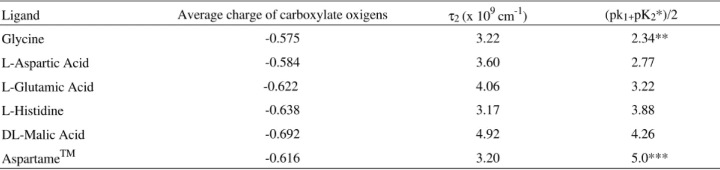
4. Figure 5 shows a plot of τ2vs. the average charges on the
carboxylate oxygens. As in the case of Fig. 4 an increasing behavior of τ2 is also observed.
Figure 6 indicates the partial charges on the concerned atoms of the ligand molecules.
The oxygen charges in Table 4 and Fig. 5 correspond in each case to the average value between the charges on the oxygens of the carboxylate groups.
From the point of view of the ligand field theory, the larger the negative charge on the oxygens, the greater is the ionic interaction between the ligand and the lanthanide ion.
Figure 5. Judd-Ofelt parameter τ2vs. average carboxylic oxygen partial charges, in units of electron charge.
Table 4. Average atomic charges on carboxylate oxygens, as obtained from a PM3 calculation. Comparison with τ2 values and average pK.
Ligand Average charge of carboxylate oxigens τ2 (x 109 cm-1) (pk1+pK2*)/2
Glycine -0.575 3.22 2.34**
L-Aspartic Acid -0.584 3.60 2.77
L-Glutamic Acid -0.622 4.06 3.22
L-Histidine -0.638 3.17 3.88
DL-Malic Acid -0.692 4.92 4.26
AspartameTM -0.616 3.20 5.0***
* For L-histidine and AspartameTM, pK2 is the constant for the 2nd acid group ionized at the concerned pH, which in these cases is not a carboxylate
group; ** Only pK1; *** Estimate from a titration plot.
-0.541
-0.609
-0.574
-0.596
-0.628
-0.628
-0.584
-0.592
-0.051
-0.600 -0.677
-0.699 -0.654 -0.352
-0.712 -0.704
-0.490 -0.172 -0.380
-0.605 -0.628
Figure 6. Optimized structures of ligand molecules with Mülliken partial atomic charges, calculated by a PM3 semiempirical quantum mechanical
Theoretically, it has been accepted that there are two domi-nating mechanisms contributing to the τλ parameters. These are the forced electric dipole and dynamic coupling mechanisms8,9. The τ
λ can be expressed as:
τλ= C
∑
t,p
Bλ,t,p
2
(2t + 1) (3)
where C is a constant and the quantities Bλ,t,p are the
so-called intensity parameters for 4f-4f transitions between individual Stark levels. The Bλ,t,p are expressed as a sum of
the two contributions:
Bλ,t,p= Bλ,t,p(forced electric dipole)+
+ Bλ,t,p(dynamic coupling) (4)
It has been shown that these two contributions have opposite signs10. A strong ligand field tends to favor the forced electric dipole mechanism. Further, the dynamic coupling Hamiltonian is directly proportional to the oxygen polarizability, which decreases as the localized charge on the oxygens increases. Thus, the observed increasing be-havior of τ2 with the pK and with the partial charges on the
carboxylate oxygens suggests that, in the present com-pounds with the Nd3+ ion, the forced electric dipole mecha-nism is dominant. On the other hand, it is not obvious why this increasing behavior of τ2 is approximately linear. This
is a point which deserves a more detailed theoretical analy-sis and is beyond the scope of this paper.
Finally, we notice that in the range of pH with accept-able τλ values, there is a predominant complex species that allows a comparative analysis of the results in the scope of this work.
Acknowledgments
The authors acknowledge the Conselho Nacional de Desenvolvimento Científico e Tecnológico (CNPq), the Fundação de Amparo à Pesquisa do Estado de São Paulo (FAPESP) and the Fundação para o Desenvolvimento da UNESP (FUNDUNESP) for financial support. FUNDU-NESP also helped in meeting the publication costs of this article.
We are also very grateful to Prof. Romeu Magnani (IQ-UNESP) for the development of the computational program to calculate the τλ parameters.
References
1. Nieboer, E. Structure and Bonding, 1975.
2. Martin, R.B.; Richardson, F.S. Quart. Rev. Biophys.
1979, 12, 181.
3. Prados, R.; Stadtherr, L.G.; Donato Jr., H.; Martin, R.B. J. Inorg. Nucl. Chem. 1974,36, 689.
4. Legendziewicz, J.; Huskowska, E.; Kozlowski, H.; Jezowska-Trzebiatowska, B. Inorg. Nucl. Chem. Let-ters1979,15, 349.
5. Judd, B.R. Phys. Rev. 1962,127, 750. 6. Ofelt, G.S. J. Chem. Phys. 1962,37, 511.
7. Mason, S.F.; Peacock, R.D.; Stewart, B. Chem. Phys. Letters1974, 29, 149.
8. Judd, B.R. J. Chem. Phys. 1979,70, 4830.
9. Malta, O.L.; Ribeiro, S.J.L.; Faucher, M.; Porcher, P.
J. Phys. Chem. Solids 1991,52, 587.
10. Xia, S.-D.; Reid, M.F. J. Phys. Chem. Solids 1993, 54, 777.
11. Carnall, W.T.; Fields, P.R.; Rajnak, K. J. Chem. Phys. 1968, 49, 4424.
12. Carnall, W.T.; Crosswhite, H.; Crosswhite, H.M. In
Energy Level Structure and Transition Probabilities of the Trivalent Lanthanides in LaF3, Argonne
Na-tional Laboratory, Report, 1977.
13. Peacock, R.D. Structure and Bonding 1975, 22,83. 14. Legendziewicz, J.; Huskowka, E.; Strek, W.;
Je-zowsk-Trzebiatowska, B. J. Luminescence 1981,
24/25, 819.
15. Legendziewicz, J.; Huskowska, E.; Argay, Gy.; Waskowska, A. Inorg. Chim.Acta. 1984, 95, 57. 16. Allinger, N.L. J. Am. Chem. Chem. Soc. 1977, 99,
8127.
17. Allinger, N.L.; Yuh, Y.H. In Quantum Chemistry Program Exchange, Bloomington, IN Program # 395. 18. Burkert, U.; Allinger, N.L. In Molecular Mechanics,
ACS Monograph 177, 1982.
19. HyperChemTM In Computational Chemistry, Auto-desk, Sausalito, CA, 1993.
20. Stewart, J.J.P. J. Comp. Chem. 1991,12, 320. 21. Lide, D.R., editor; ‘‘CRC Handbook of Chemistry and
Physics’’, 72nd ed., 1991-1992.