Article
J. Braz. Chem. Soc., Vol. 26, No. 11, 2307-2312, 2015. Printed in Brazil - ©2015 Sociedade Brasileira de Química 0103 - 5053 $6.00+0.00
A
*e-mail: [email protected]
Chemical Constituents of the Seeds of
Raphanus sativus
and their Biological Activity
Ki Hyun Kim,a Eunjung Moon,b Seoung Rak Lee,a Kyoung Jin Park,a Sun Yeou Kim,c Sang Un Choid and Kang Ro Lee*,a
aSchool of Pharmacy, Sungkyunkwan University, 440-746 Suwon, Republic of Korea bCharmzone R&D center, Charmzone Co. LTD., 220-962 Wonju, Republic of Korea
cCollege of Pharmacy, Gachon University, 406-799 Incheon, Republic of Korea dKorea Research Institute of Chemical Technology, 305-600 Deajeon, Republic of Korea
As part of our ongoing search for bioactive constituents of natural Korean medicinal resources, a bioassay-guided fractionation and chemical investigation of the MeOH extract of Raphanus sativus (Brassicaceae)seeds resulted in the isolation and identification of fifteen compounds, including a new phenolic compound. The structure of the new compound was determined by extensive spectroscopic analysis and the Mosher’s method. One of the compounds has been recently reported as a synthetic product. Some compoundsshowed moderate antiproliferative activities against the tumor cell lines A549, SK-OV-3, SK-MEL-2, and HCT-15 with IC50 values in the
range of 5.62 to 28.88 µM. Moreover, the anti-neuroinflammatory activities of the isolates were determined by measuring the nitric oxide (NO) levels in the medium using murine microglia BV-2 cells. With exception of one specific compound, all the others inhibited the lipopolysaccharide (LPS)-stimulated NO production (IC50 values < 200 µM).
Keywords:Raphanus sativus, Brassicaceae, cytotoxicity, anti-inflammation, Mosher’s method
Introduction
The edible root vegetable Raphanus sativus L. (Brassicaceae), commonly known as radish, is one of the most widely grown and consumed vegetables throughout the world. In China, it has been used as a traditional herbal medicine for over 1400 years, recorded in ‘Tang Materia Medica’, the first Chinese pharmacopoeia.1 Different parts
of R. sativus, including roots, seeds, and leaves, are known to possess a variety of medicinal properties.2 In particular,
the seeds of R. sativus, also known as Raphani Semen, have been used as a traditional Korean medicine since ancient times, as carminative, diuretic, expectorant, laxative, and stomachic agents, and have also been used as anti-cancer and/or anti-inflammatory agents.3-5 These effects have
been supported by previous phytochemical investigations reporting pharmacologically active glucosinolates responsible for cancer-chemoprotective properties.1,6-8
In a previous work,9,10 we found that the MeOH extract
of Raphani Semen exhibits significant cytotoxic activity against the human tumor cell lines, A549, SK-OV-3,
SK-MEL-2, and HCT-15, in addition to inhibitory effects on nitric oxide (NO) production in lipopolysaccharide (LPS)-stimulated BV-2 microglial cells. This observation led to the isolation and identification of 4-methylthio-butanyl derivatives and phenylpropanoid sucrosides that correlated with the cytotoxic and anti-inflammatory activities.9,10 Using a bioassay-guided methodology, we
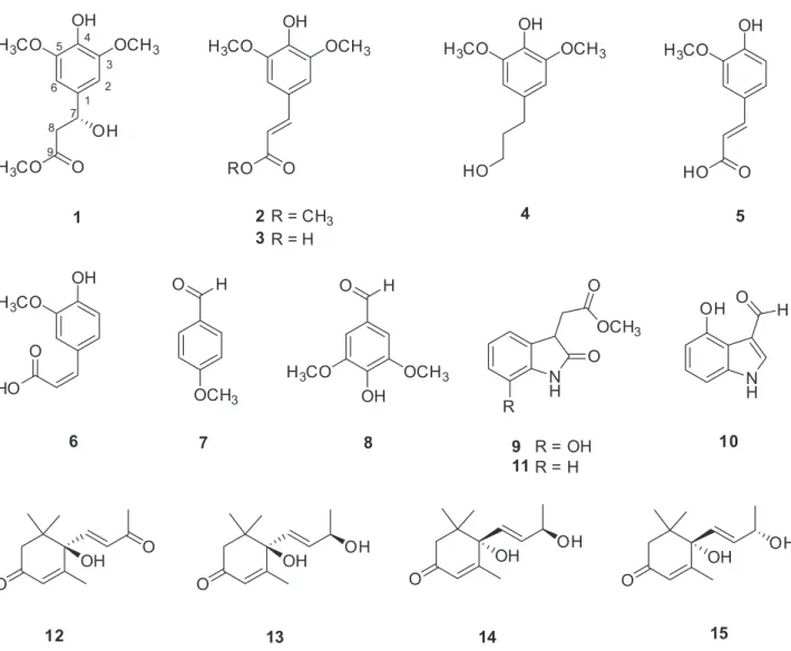
are now reporting the isolation of fifteen compounds (1-15), including a new phenolic (1), from the most active CHCl3-soluble fraction of the MeOH extract (Figure 1).
In addition, their antiproliferative activities against A549, SK-OV-3, SK-MEL-2, and HCT-15 cell lines, and their inhibitory effects on NO production in a LPS-activated BV2 cell line, have been evaluated.
Experimental
General experimental procedures
spectra were recorded with a Shimadzu 1601 UV-Visible spectrophotometer (Shimadzu, Tokyo, Japan). High-resolution electrospray ionisation mass spectrometry (HR-ESIMS) and ESIMS spectra were recorded on a Micromass QTOF2-MS (MicroMass, Waters, Milford, MA, USA). Nuclear magnetic resonance (NMR) spectra, including 1H-1H COSY, HMQC, and HMBC experiments,
were recorded on a Varian UNITY INOVA 500 NMR spectrometer (Varian, Palo Alto, CA, USA) operating at 500 MHz (1H) and 125 MHz (13C), with chemical shifts
given in ppm (d). Semi-preparative high performance liquid chromatography (HPLC) was conducted using a Gilson 306 pump (Gilson, Middleton, WI) with Shodex refractive index detector (Shodex, New York, NY). Silica gel 60 and RP-C18 silica gel (230-400 mesh,
Merck, Darmstadt, Germany) were used for column chromatography. Low-pressure liquid chromatography (LPLC) was carried out over a LiChroprep Lobar-A Si 60 column (240 mm × 10 mm i.d.; Merck) with a FMI QSY-0 pump. The packing material for molecular sieve column
chromatography was Sephadex LH-20 (Pharmacia, Uppsala, Sweden). Merck precoated silica gel F254 plates
and RP-18 F254s plates (Merck, Darmstadt, Germany)
were used for thin layer chromatography (TLC). Spots were detected on TLC under UV light or by heating after spraying with 10% H2SO4 in C2H5OH (v/v).
Plant material
The seeds of R. sativus were purchased at the Kyungdong herbal market, Seoul, Korea, in January 2010, and identified by one of the authors (K. R. Lee). A voucher specimen (SKKU-2010-01) has been deposited in the herbarium of the School of Pharmacy, Sungkyunkwan University, Suwon, Korea.
Extraction and isolation
The dried seeds of R. sativus (3.6 kg) were ground and extracted twice with 80% aqueous MeOH at room OH
H3CO OCH3
O H3CO
OH
1
OH
H3CO OCH3
O RO
2 R = CH3 3 R = H
OH
H3CO OCH3
HO
4
OH
H3CO
O HO
OH
H3CO
O
HO
OH
OCH3 H3CO
O H
OCH3 O H
N H
H O OH
N H
O O
OCH3
R
5
6 7 8 9 R = OH
11R = H
10
O
OH OH
O
OH OH
O
OH OH
O
O OH
12 13 14 15
1 2 3 4 5
6
7 8
9
temperature, and subsequently filtered. The filtrate was evaporated in vacuo to obtain a crude extract (325 g), which was fractionated by sequential liquid-liquid partitioning of H2O with n-hexane, CHCl3, and n-BuOH to yield 12,
8, and 30 g of residues, respectively. Each fraction was evaluated for cytotoxicity against A549, OV-3, SK-MEL-2, and HCT-15 cell lines using a sulforhodamine B (SRB) assay. The CHCl3-soluble fraction exhibited the
highest cytotoxic activity against the tested tumor cell lines. It also showed potent inhibition of NO production in LPS-stimulated BV-2 cells. The CHCl3-soluble fraction
(8 g) was separated by column chromatography on a reversed-phase C18 column using a gradient of increasing MeOH in H2O [40% MeOH (1.5 L), 60% MeOH (1.5 L),
and 100% MeOH (2.0 L)] to give 13 fractions [C1-C13; C1-C3 (each 0.5 L of 40% MeOH), C4-C8 (each 0.3 L of 60% MeOH), and C9-C13 (each 0.4 L of 100% MeOH)], whose compositions were monitored by TLC. Fraction C4 (180 mg) was subjected to semi-preparative reversed-phase HPLC using a 250 mm × 10 mm i.d., 10 µm, EconosilRP-18 column (Alltech, Nicholasville, KY, USA), with MeOH-H2O (2:3) as eluent, to yield fraction C41.
Fraction C41 (55 mg) was reapplied to semi-preparative normal-phase HPLC using a 250 mm × 10 mm i.d., 5 µm, Apollo Silica column (Alltech), with CHCl3-MeOH
(30:1) as eluent, to yield compounds 1 (8 mg) and 4 (6 mg). Fraction C5 (530 mg) was applied to LPLC on a 240 mm × 10 mm i.d., 40-63 µm, LiChroprep Lobar-A Si 60 column (Merck), with n-hexane-EtOAc (1:1, 900 mL) as eluent, to give three fractions [C51-C53; C51 (200 mL), C52 (200 mL), and C53 (500 mL)]. Fraction C51 (182 mg) was purified using semi-preparative normal-phase HPLC with n-hexane-CHCl3-EtOAc (1:1:2) as eluent, to yield
compounds 7 (5 mg), 8 (15 mg), 12 (7 mg), 13 (6 mg), and 14 (8 mg). Fraction C52 (162 mg) was also purified using semi-preparative normal-phase HPLC with CHCl3-MeOH
(30:1) as eluent, to yield compound 15 (9 mg). Fraction C7 (400 mg) was subjected to a Sephadex LH-20 column using CH2Cl2-MeOH (1:1) as eluent, to yield two fractions
(C71 and C72). Fraction C72 (150 mg) was then applied to semi-preparative normal-phase HPLC with n-hexane-CHCl3-EtOAc (1:0.1:1) as eluent, to afford compounds 5
(5 mg), 6 (5 mg), 9 (6 mg), and 11 (6 mg). Fraction C8 (350 mg) was separated over a Sephadex LH-20 column using CH2Cl2-MeOH (1:1) as eluent, to yield compound 10
(7 mg). Fraction C10 (280 mg) was also applied to a Sephadex LH-20 column using the same eluent, to give two fractions (C101 and C102). Fraction C101 (75 mg) was further purified using semi-preparative normal-phase HPLC with n-hexane-CHCl3-EtOAc (1:0.1:1) as eluent, to
yield compounds 2 (33 mg) and 3 (9 mg).
Methyl (3R)-hydroxy-3-(4-hydroxy-3,5-dimethoxyphenyl)
propanoate (1)
Colorless gum; [α]D25 +17.3 (c 0.40, MeOH); UV
(MeOH) λmax / nm (log ε) 270 (1.2), 238 (2.8), 216 (3.9);
IR (KBr) νmax / cm−1 3357, 2946, 2832, 1720, 1451, 1116,
1032, 674; 1H (500 MHz) and 13C (125 MHz) NMR, see
Table 1; ESIMS (positive-ion mode) m/z 279 [M + Na]+;
HRESIMS (negative-ion mode) m/z 255.0877 [M − H]+
(calcd. for C12H15O6, 255.0869).
Preparation of (R)- and (S)-MTPA ester derivatives of 1
4-(Dimethylamino)pyridine (DMAP, 1 mg) and (S)-(+)-α-methoxy-α-(trifluoromethyl)phenylacetyl chloride (MTPA-Cl, 10 µL) were added to a stirred solution of 1 (2.0 mg) in pyridine (400 µL), and the mixture was further stirred at room temperature for 16 h. The reaction mixture was then passed through a silica gel Waters Sep-Pak Vac 6cc and eluted with n-hexane-EtOAc (15:1) to give the (R)-Mosher ester 1r. Treatment of 1 (2.0 mg) with (R)-MTPA-Cl (10 µL) as described above, yielded the corresponding (S)-MTPA ester 1s.
Cytotoxicity assays
All tumor cell cultures were maintained using RPMI1640 cell growth medium (Gibco, Carlsbad, CA), supplemented with 5% fetal bovine serum (FBS) (Gibco), 100 units mL−1 penicillin and 100 µg mL−1 streptomycin.
The human tumor cell lines, A549 (non-small cell lung carcinoma), SK-OV-3 (ovarian malignant ascites), SK-MEL-2 (skin melanoma), and HCT-15 (colon adenocarcinoma) were provided by the National Cancer Institute (NCI). The cytotoxicity of the isolates against cultured human tumor cell lines was evaluated by the sulforhodamine B (SRB) method. Each tumor cell line was inoculated into standard 96-well flat-bottom microplates and incubated for 24 h at 37 °C in a humidified atmosphere of 5% CO2. The attached cells were then incubated with
(purity ≥ 98%; Sigma) was used as a positive control. Tested compounds were demonstrated to be pure as evidenced by NMR and HPLC analysis (purity ≥ 95%).
Measurement of NO production
The murine microglial BV2 cell line was generously provided by PhD E. Choi from Korea University (Seoul, Korea), and maintained in Dulbecco’s modified Eagle (DMEM) medium supplemented with 5% FBS, 100 units mL−1 penicillin, and 100 µg mL−1 streptomycin.
All cells were incubated at 37 °C in a humidified incubator with 5% CO2. BV-2 cells were plated into a 96-well plate
(3 × 10⁴ cells well−1). Following culture for 24 h, cells
were pretreated with the compounds for 30 min, and subsequently stimulated with 100 ng mL−1 of LPS for a
further 24 h. Control cultures received the carrier solvent (0.1% dimethyl sulfoxide). Nitrite, a soluble oxidation product of NO, was measured in the culture media using the Griess reaction. The supernatant (50 µL) was harvested and mixed with an equal volume of Griess reagent (1% sulfanilamide, 0.1% N-1-napthylethylenediamine dihydrochloride in 5% phosphoric acid). After 10 min, the absorbance at 570 nm was measured using a microplate reader. Sodium nitrite was used as a standard to calculate the NO2− concentration. NG-monomethyl-L-arginine (NMMA,
Sigma, St. Louis, MO, USA), a well-known NO synthase inhibitor, was tested as a positive control.
Results and Discussion
Compound 1 was isolated as a colorless gum. The molecular formula of 1 was determined to be C12H16O6 by
the negative mode HRESIMS at m/z 255.0877 [M − H]+
(calcd. for C12H15O6, 255.0869). The IR spectrum displayed
absorption bands associated with hydroxyl (3357 cm−1)
and carbonyl ester (1720 cm−1) groups. The 1H NMR
spectrum of 1 (Table 1) showed characteristic signals attributable to a 1,3,4,5-tetrasubstituted aromatic ring at dH 6.54 (2H, s, H-2 and H-6), one downfield shifted
methine at dH 5.00 (1H, ddd, J 9.0, 4.0, 3.0 Hz, H-7),
one methylene adjacent to a carbonyl group at dH 2.70
(1H, dd, J 16.0, 9.0 Hz, H-8a) and 2.62 (1H, dd, J 16.0, 4.0 Hz, H-8b), and three methoxy groups, two that were overlapping at dH 3.83 (6H, s) and another at dH 3.66 (3H,
s). The 13C NMR spectrum of 1 (Table 1) exhibited a
total of 12 carbons including six aromatic carbons in the range dC 147.1-102.3, one oxygenated carbon at dC 70.5,
one methylene carbon at dC 43.4, one carbonyl carbon at
dC 172.8, and three methoxy carbons at dC 56.3 (× 2) and
51.9. Interpretation of the 2D NMR spectra correlations,
including 1H-1H correlated spectroscopy (COSY),
heteronuclear multiple quantum correlation (HMQC), and heteronuclear multiple bond correlation (HMBC), revealed compound 1 to be the sinapic acid derivative, methyl 3-hydroxy-3-(4-hydroxy-3,5-dimethoxyphenyl) propanoate (Figure 2). The absolute configuration of C-7 of 1 was established on the basis of the modified Mosher’s method.11 Treatment of 1 with (S)-(+)-α-methoxy-α
-(trifluoromethyl)phenylacetyl chloride [(S)-MTPA-Cl] and DMAP in pyridine gave the (R)-MTPA ester 1r. Similar treatment of 1 with (R)-(−)-MTPA-Cl afforded the (S)-MTPA ester 1s. Analysis of the 1H NMR chemical
shift differences (∆dS-R) [see Supporting Information (SI) section] of the two MTPA esters allowed the assignment of the absolute configuration of C-7 as R (Figure 3). Thus, the structure of 1 was elucidated as methyl (3R)-hydroxy-3-(4-hydroxy-3,5-dimethoxyphenyl) propanoate, which appears to be the epimer of methyl (3S)-hydroxy-3-(4-hydroxy-3,5-dimethoxyphenyl) propanoate, previously reported in Ruta graveolens L.12 The spectroscopic data of the known
epimer is similar to those of 1 (Table 1), except for the splitting pattern of H-7,12 however, its optical rotation value
([α]D25−1.03 in MeOH) was easily distinguishable from
that of 1 ([α]D25 +17.3 in MeOH).12
The known compounds were identified as sinapic acid methyl ester (2),13 sinapic acid (3),14 dihydrosinapic alcohol
(4),15 (E)-ferulic acid (5),16 (Z)-ferulic acid (6),16 p-anisic
aldehyde (7),17 and syringic aldehyde (8),18 by comparison of
their spectroscopic data with previously reported literature
Table 1.1H and 13C NMR data of compound 1 inCDCl 3a
Position 1
dH / ppm dC / ppm
1 − 133.7
2 6.54 (s) 102.3
3 − 147.1
4 − 134.2
5 − 147.1
6 6.54 (s) 102.3
7 5.00 (ddd, 9.0, 4.0, 3.0 Hz) 70.5
8 2.70 (dd, 16.0, 9.0 Hz) 2.62 (dd, 16.0, 4.0 Hz)
43.4
9 − 172.8
4-OH 5.41 (s) −
7-OH 3.06 (d, 3.0 Hz) −
3,5-OCH3 3.83 (s) 56.3
9-OCH3 3.66 (s) 51.9
values. Furthermore, three indole alkaloids were identified as methyl 7-hydroxyoxindole-3-acetate (9),19
4-hydroxy-3-indolecarbaldehyde (10),20 and methyl oxindole-3-acetate
(11),21 by comparison with previously published data. On
the other hand, methyl 7-hydroxyoxindole-3-acetate (9) has been recently reported as a synthetic product.19 Compound
9 appears to be an artifact of extraction with MeOH, since its free acid precursor has been reported to be present in Zea mays.19,22 Other known megastigmane derivatives were
also identified as (−)-dihydrovomifoliol (12),23
(6R,9R)-vomifoliol (13),24 (6S,9R)-vomifoliol (14),24 and
(6S,9S)-vomifoliol (15),24 by comparison of their spectroscopic
data with previously reported values. The absolute configurations of compounds 12-15 were established on the basis of their NMR data, optical rotation values, and circular dichroism (CD) data.23,24
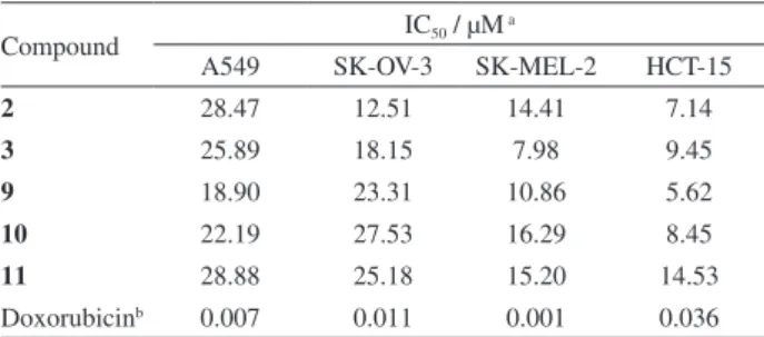
The antiproliferative activities of compounds 1-15 were evaluated by determining their inhibitory effects on four human tumor cell lines, namely A549, SK-OV-3, SK-MEL-2, and HCT-15, using the SRB bioassay.25 The
results (Table 2) showed that compounds 2, 3, and 9-11 displayed moderate antiproliferative activities against all the tumor cell lines. However, the other compounds were inactive (IC50 > 30 µM) in all four cell lines. In particular,
among the isolates, indole alkaloids 9-11 showed good antiproliferative effects against all the tumor cells tested, with IC50 values ranging from 5.62 to 28.88 µM.
Compounds 1-15 were also examined for their anti-neuroinflammatory activities by measuring the NO levels produced in LPS-activated BV-2 cells, a microglial cell
line. The results (Table 3) showed that all the compounds, with the exception of compound 6,inhibited NO production (IC50 values < 200 µM). These compounds had no effect on
cell viability in LPS-treated BV-2 cells at their respective IC50 values (data not shown). Among these, compounds 2,
4, and 12 significantly inhibited NO levels with IC50 values
of 18.99, 18.69, and 15.60 µM, respectively. In particular, compound 12 showed potent inhibition of NO in LPS-stimulated NO production with an IC50 of 15.60 µM, which
is a lower concentration than that displayed by the positive control NG-nonomethyl-L-arginine (IC
50 19.15 µM).
Excess production of NO by activated microglia induces neuronal cell death,26,27 which consequently leads to various
neurodegeneration disorders of the CNS. Therefore, the active compounds 2, 4, and 12 could be considered potential candidates in the prevention of the progressive damage resulting from neurodegenerative diseases.
Conclusions
The bioassay-guided fractionation and chemical investigation of the MeOH extract of R. sativus seeds resulted in the isolation and identification of fifteen OH
H3CO OCH3
O H3CO
OH 1 2
3 4 5
6
7 8
9
Figure 2. The 1H-1H COSY correlations (bond) and key HMBC correlations (H→C) of 1.
OH
H3CO OCH3
O
H3CO
OMT PA
+0.023 +0.017 +0.020
-0.0 59 -0.0 07
-0.0 01 -0.0 07
-0.0 01
Figure 3.∆d Values (dS – dR) in ppm of the two MTPA esters of 1.
Table 3. Inhibitory effects of compounds 1-15 on NO production in LPS-activated BV-2 cells
Compound IC50 / µMa Compound IC50 / µMa
1 58.74 9 166.71
2 18.99 10 36.91
3 24.05 11 27.15
4 18.69 12 15.60
5 41.25 13 76.50
6 > 200 14 39.24
7 44.76 15 53.21
8 31.41 NMMAb 19.15
aIC
50 value of each compound was defined as the concentration (µM) that caused 50% inhibition of NO production in LPS-activated BV-2 cells; bNMMA as positive control.
Table 2. Antiproliferative activities of compounds 2, 3, and 9-11 isolated from R. sativus
Compound IC50 / µM
a
A549 SK-OV-3 SK-MEL-2 HCT-15
2 28.47 12.51 14.41 7.14
3 25.89 18.15 7.98 9.45
9 18.90 23.31 10.86 5.62
10 22.19 27.53 16.29 8.45
11 28.88 25.18 15.20 14.53
Doxorubicinb 0.007 0.011 0.001 0.036
aIC
50 value of compounds against each cancer cell line. The IC50 value was defined as the concentration (µM) causing 50% inhibition of cell growth
compounds (1-15), including a new phenolic compound, methyl (3R)-hydroxy-3-(4-hydroxy-3,5-dimethoxyphenyl) propanoate (1). Among the isolates, three indole alkaloids (9-11) showed good antiproliferative effects against all the tumor cells tested, with IC50 values ranging from
5.62 to 28.88 µM. In addition, compounds 2, 4, and 12 significantly inhibited LPS-stimulated NO production in murine microglia BV-2 cells with IC50 values of 18.99,
18.69, and 15.60 µM, respectively. These results suggest that the above mentioned active compounds with cytotoxic or anti-neuroinflammatory activities, could be considered as lead molecules for drug development related to various cancers or neurodegenerative diseases.
Supplementary Information
Supplementary data are available free of charge at http://jbcs.sbq.org.br as a PDF file.
Acknowledgments
This research was supported by Basic Science Research Program through the National Research Foundation of Korea (NRF) funded by the Ministry of Science, ICT & Future Planning (2015R1C1A1A02037383). We thank Ja Phil Ku and Chai Yeon Kang at the Sungkyunkwan University Cooperative Center for Research Facilities for their aid in the NMR spectra measurements.
References
1. Duan, L. X.; Feng, B. M.; Wang, W.; Chen, F.; Cai, G. M.; Pei, Y. H.; Wang, Y. Q.; Helv. Chim. Acta2006, 89, 2953. 2. Nadkarni, K. M.; Indian Materia Medica; Popular Prakashan:
Bombay, 1976.
3. Yeung, H. C.; Handbook of Chinese Herbs and Formulas; Institute of Chinese Medicine: Los Angeles, 1985.
4. Duke, J. A.; Ayensu, E. S.; Medicinal Plants of China; Reference Publ., Inc.: Michigan, 1985.
5. Chopra, R. N.; Nayar, S. L.; Chopra, I. C.; Glossary of Indian Medicinal Plants (Including the Supplement), Council of
Scientific and Industrial Research: New Delhi, 1986. 6. Barillari, J.; Cervellati, R.; Paolini, M.; Tatibout, A.; Rollin, P.;
Iori, R.; J. Agric. Food Chem. 2005, 53, 9890.
7. Daxenbichler, M. E.; Spencer, G. F.; Carlson, D. G.; Rose, G. B.; Brinker, A. M.; Powell, R. G.; Phytochemistry1991, 30, 2623. 8. Nastruzzi, C.; Cortesi, R.; Esposito, E.; Menegatti, E.; Leoni, O.;
Iori, R.; Palmieri, S.; J. Agric. Food Chem. 1996, 44, 1014. 9. Kim, K. H.; Kim, C. S.; Park, Y. J.; Moon, E.; Choi, S. U.; Lee,
J. H.; Kim, S. Y.; Lee, K. R.; Bioorg. Med. Chem. Lett. 2015,
25, 96.
10. Kim, K. H.; Moon, E.; Kim, S. Y.; Choi, S. U.; Lee, J. H.; Lee, K. R.; J. Ethnopharmacol. 2014, 151, 503.
11. Kim, K. H.; Choi, S. U.; Kim, Y. C.; Lee, K. R.; J. Nat. Prod.
2011, 74, 54.
12. Rollinger, J. M.; Schuster, D.; Danzl, B.; Schwaiger, S.; Markt, P.; Schmidtke, M.; Gertsch, J.; Raduner, S.; Wolber, G.; Langer, T.; Stuppner, H.; Planta Med.2009, 75, 195. 13. Yao, C.-S.; Lin, M.; Wang, L.; Chem. Pharm. Bull. 2006, 54,
1053.
14. Sun, K.; Li, X.; Shenyang Yaoke Daxue Xuebao2003, 20, 419. 15. Hungerford, N. L.; Sands, D. P. A.; Kitching, W.; Aust. J. Chem.
1998, 51, 1103.
16. Salum, M. L.; Robles, C. J.; Erra-Balsells, R.; Org. Lett. 2010,
12, 4808.
17. Iinuma, M.; Moriyama, K.; Togo, H.; Tetrahedron2013, 69, 2961.
18. Kim, H.; Ralph, J.; Lu, F.; Ralph, S. A.; Boudet, A. M.; MacKay, J. J.; Sederoff, R. R.; Ito, T.; Kawai, S.; Ohashi, H.; Higuchi, T.;
Org. Biomol. Chem. 2003, 1, 268.
19. Homer, J. A.; Sperry, J.; Tetrahedron Lett.2014, 55, 5798. 20. Zhou, H. F.; Jian, R. J.; Kang, J.; Huang, X. L.; Li, Y.; Zhuang,
C. L.; Yang, F.; Zhang, L. L.; Fan, X.; Wu, T.; Wu, X.; J. Agric. Food Chem.2010, 58, 12717.
21. Kinashi, H.; Suzuki, Y.; Takeuchi, S.; Kawarada, A.; Agric. Biol. Chem. 1976, 40, 2465.
22. Lewer, P.; Bandurski, R. S.; Phytochemistry1987, 26, 1247. 23. Mori, K.; Tetrahedron Lett.1973, 28, 2635.
24. Yamano, Y.; Ito, M.; Chem. Pharm. Bull.2005, 53, 541. 25. Song, J. H.; Choi, H. J.; Song, H. H.; Hong, E. H.; Lee, B. R.;
Oh, S. R.; Choi, K.; Yeo, S. G.; Lee, Y. P.; Cho, S.; Ko, H. J.;
J. Ginseng Res.2014, 38, 173.
26. Minghetti, L.; Levi, G.; Prog. Neurobiol.1998, 54, 99. 27. Boje, L. M.; Arora, P. K.; Brain Res.1992, 587, 250.
Submitted: July 23, 2015